Abstract
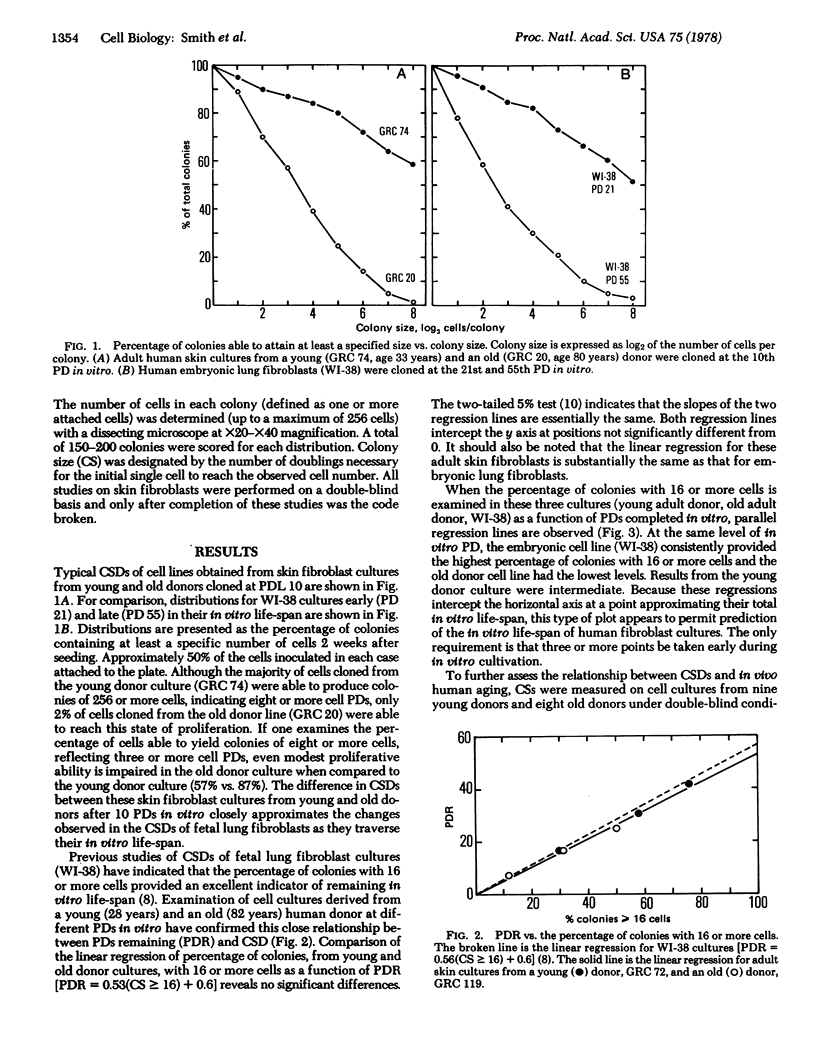
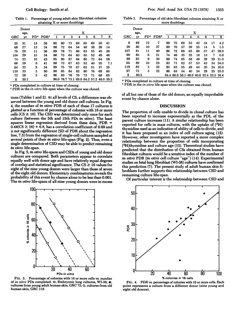
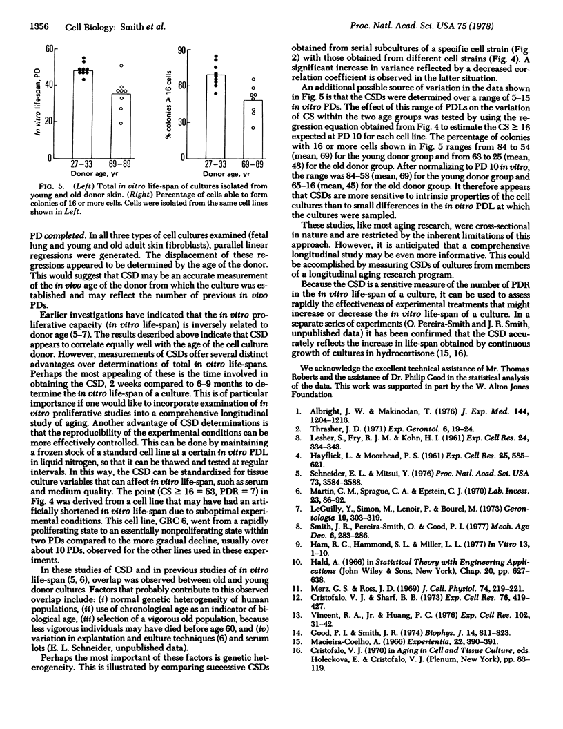
Individual human diploid cells plated at low cell density and incubated for 2 weeks develop into colonies ranging in size from one cell to several thousand cells. The resultant colony size distribution is an accurate indicator of the number of subsequent in vitro population doublings that can be attained by the parent culture. This relationship holds for both human fetal lung and adult skin fibroblast cultures. In addition, the colony size distributions obtained from fetal, young adult, and old adult human cell cultures at the same low level of in vitro passage are indicative of the in vivo age of the level of in vitro passage are indicative of the in vivo age of the cell culture donor. Cell cultures of fetal origin give rise to the highest percentage of colonies with significant proliferative abilities, whereas cultures from old adults give rise to the lowest percentage of large colonies. Therefore, colony size distributions appear to be good indicators of both in vitro and in vivo human cellular aging.
Full text
PDF


Selected References
These references are in PubMed. This may not be the complete list of references from this article.
- Albright J. W., Makinodan T. Decline in the growth potential of spleen-colonizing bone marrow stem cells of long-lived aging mice. J Exp Med. 1976 Nov 2;144(5):1204–1213. doi: 10.1084/jem.144.5.1204. [DOI] [PMC free article] [PubMed] [Google Scholar]
- Cristofalo V. J., Sharf B. B. Cellular senescence and DNA synthesis. Thymidine incorporation as a measure of population age in human diploid cells. Exp Cell Res. 1973 Feb;76(2):419–427. doi: 10.1016/0014-4827(73)90394-7. [DOI] [PubMed] [Google Scholar]
- Good P. I., Smith J. R. Age distribution of human diploid fibroblasts. A stochastic model for in vitro aging. Biophys J. 1974 Nov;14(11):811–823. doi: 10.1016/S0006-3495(74)85951-5. [DOI] [PMC free article] [PubMed] [Google Scholar]
- Ham R. G., Hammond S. L., Miller L. L. Critical adjustment of cysteine and glutamine concentrations for improved clonal growth of WI-38 cells. In Vitro. 1977 Jan;13(1):1–10. doi: 10.1007/BF02615497. [DOI] [PubMed] [Google Scholar]
- LESHER S., FRY R. J., KOHN H. I. Age and the generation time of the mouse duodenal epithelial cell. Exp Cell Res. 1961 Aug;24:334–343. doi: 10.1016/0014-4827(61)90436-0. [DOI] [PubMed] [Google Scholar]
- Le Guilly Y., Simon M., Lenoir P., Bourel M. Long-term culture of human adult liver cells: morphological changes related to in vitro senescence and effect of donor's age on growth potential. Gerontologia. 1973;19(5):303–313. doi: 10.1159/000211984. [DOI] [PubMed] [Google Scholar]
- Macieira-Coelho A. Action of cortisone on human fibroblasts in vitro. Experientia. 1966 Jun 15;22(6):390–391. doi: 10.1007/BF01901156. [DOI] [PubMed] [Google Scholar]
- Martin G. M., Sprague C. A., Epstein C. J. Replicative life-span of cultivated human cells. Effects of donor's age, tissue, and genotype. Lab Invest. 1970 Jul;23(1):86–92. [PubMed] [Google Scholar]
- Merz G. S., Jr, Ross J. D. Viability of human diploid cells as a function of in vitro age. J Cell Physiol. 1969 Dec;74(3):219–222. doi: 10.1002/jcp.1040740302. [DOI] [PubMed] [Google Scholar]
- Schneider E. L., Mitsui Y. The relationship between in vitro cellular aging and in vivo human age. Proc Natl Acad Sci U S A. 1976 Oct;73(10):3584–3588. doi: 10.1073/pnas.73.10.3584. [DOI] [PMC free article] [PubMed] [Google Scholar]
- Smith J. R., Pereira-Smith O., Good P. I. Colony size distribution as a measure of age in cultured human cells. A brief note. Mech Ageing Dev. 1977 Jul-Aug;6(4):283–286. doi: 10.1016/0047-6374(77)90029-x. [DOI] [PubMed] [Google Scholar]
- Thrasher J. D. Age and the cell cycle of the mouse esophageal epithelium. Exp Gerontol. 1971 Feb 1;6(1):19–24. doi: 10.1016/0531-5565(71)90044-1. [DOI] [PubMed] [Google Scholar]
- Vincent R. A., Jr, Huang P. C. The proportion of cells labeled with tritiated thymidine as a function of population doubling level in cultures of fetal, adult, mutant, and tumor origin. Exp Cell Res. 1976 Oct 1;102(1):31–42. doi: 10.1016/0014-4827(76)90296-2. [DOI] [PubMed] [Google Scholar]


