

Abstract
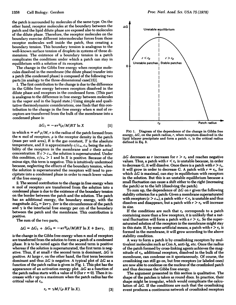
Crosslinking of membrane surface receptors may lead to their segregation into patches and then into a single large aggregate at one pole of the cell. This process is called capping. Here, a novel explanation of such a process is presented in which the membrane is viewed as a supersaturated solution of receptors in the lipid bilayer and the adjacent two aqueous layers. Without a crosslinking agent, a patch of receptors that is less than a certain size cannot stay in equilibrium with the solution and thus should dissolve. Patches greater than a certain size are stable and can, in principle, grow by the precipitation of the remaining dissolved receptors from the supersaturated solution. The task of the crosslinking molecules is to form such stable patches. These considerations are based on a qualitative thermodynamic calculation that takes into account the existence of a boundary tension in a patch (in analogy to the surface tension of a droplet). Thermodynamically, these systems should cap spontaneously after the patches have reached a certain size. But, in practice, such a process can be very slow. A suspension of patches may stay practically stable. The ways in which a cell may abolish this metastable equilibrium and thus achieve capping are considered and possible effects of capping inhibitors are discussed.
Keywords: cap formation, cell surface, cell membrane proteins
Full text
PDF
Selected References
These references are in PubMed. This may not be the complete list of references from this article.
- Bourguignon L. Y., Singer S. J. Transmembrane interactions and the mechanism of capping of surface receptors by their specific ligands. Proc Natl Acad Sci U S A. 1977 Nov;74(11):5031–5035. doi: 10.1073/pnas.74.11.5031. [DOI] [PMC free article] [PubMed] [Google Scholar]
- Bretscher M. S. Directed lipid flow in cell membranes. Nature. 1976 Mar 4;260(5546):21–23. doi: 10.1038/260021a0. [DOI] [PubMed] [Google Scholar]
- Edelman G. M. Surface modulation in cell recognition and cell growth. Science. 1976 Apr 16;192(4236):218–226. doi: 10.1126/science.769162. [DOI] [PubMed] [Google Scholar]
- Edidin M., Weiss A. Antigen cap formation in cultured fibroblasts: a reflection of membrane fluidity and of cell motility. Proc Natl Acad Sci U S A. 1972 Sep;69(9):2456–2459. doi: 10.1073/pnas.69.9.2456. [DOI] [PMC free article] [PubMed] [Google Scholar]
- Frye L. D., Edidin M. The rapid intermixing of cell surface antigens after formation of mouse-human heterokaryons. J Cell Sci. 1970 Sep;7(2):319–335. doi: 10.1242/jcs.7.2.319. [DOI] [PubMed] [Google Scholar]
- Harris A. K. Recycling of dissolved plasma membrane components as an explanation of the capping phenomenon. Nature. 1976 Oct 28;263(5580):781–783. doi: 10.1038/263781a0. [DOI] [PubMed] [Google Scholar]
- Karnovsky M. J., Unanue E. R. Mapping and migration of lymphocyte surface macromolecules. Fed Proc. 1973 Jan;32(1):55–59. [PubMed] [Google Scholar]
- Loor F., Forni L., Pernis B. The dynamic state of the lymphocyte membrane. Factors affecting the distribution and turnover of surface immunoglobulins. Eur J Immunol. 1972 Jun;2(3):203–212. doi: 10.1002/eji.1830020304. [DOI] [PubMed] [Google Scholar]
- Nicolson G. L. Transmembrane control of the receptors on normal and tumor cells. I. Cytoplasmic influence over surface components. Biochim Biophys Acta. 1976 Apr 13;457(1):57–108. doi: 10.1016/0304-4157(76)90014-9. [DOI] [PubMed] [Google Scholar]
- Schreiner G. F., Fujiwara K., Pollard T. D., Unanue E. R. Redistribution of myosin accompanying capping of surface Ig. J Exp Med. 1977 May 1;145(5):1393–1398. doi: 10.1084/jem.145.5.1393. [DOI] [PMC free article] [PubMed] [Google Scholar]
- Singer S. J., Nicolson G. L. The fluid mosaic model of the structure of cell membranes. Science. 1972 Feb 18;175(4023):720–731. doi: 10.1126/science.175.4023.720. [DOI] [PubMed] [Google Scholar]
- Stackpole C. W., DeMilio L. T., Jacobson J. B., Hämmerling U., Lardis M. P. A comparison of ligand-induced redistribution of surface immunoglobulins, alloantigens, and concanavalin A receptors on mouse lymphoid cells. J Cell Physiol. 1974 Jun;83(3):441–447. doi: 10.1002/jcp.1040830315. [DOI] [PubMed] [Google Scholar]
- Taupin C., Dvolaitzky M., Sauterey C. Osmotic pressure induced pores in phospholipid vesicles. Biochemistry. 1975 Oct 21;14(21):4771–4775. doi: 10.1021/bi00692a032. [DOI] [PubMed] [Google Scholar]
- Unanue E. R., Karnovsky M. J., Engers H. D. Ligand-induced movement of lymphocyte membrane macromolecules. 3. Relationship between the formation and fate of anti-Ig-surface Ig complexes and cell metabolism. J Exp Med. 1973 Mar 1;137(3):675–689. doi: 10.1084/jem.137.3.675. [DOI] [PMC free article] [PubMed] [Google Scholar]
- Unanue E. R., Perkins W. D., Karnovsky M. J. Ligand-induced movement of lymphocyte membrane macromolecules. I. Analysis by immunofluorescence and ultrastructural radioautography. J Exp Med. 1972 Oct 1;136(4):885–906. doi: 10.1084/jem.136.4.885. [DOI] [PMC free article] [PubMed] [Google Scholar]
- Wolf D. E., Schlessinger J., Elson E. L., Webb W. W., Blumenthal R., Henkart P. Diffusion and patching of macromolecules on planar lipid bilayer membranes. Biochemistry. 1977 Jul 26;16(15):3476–3483. doi: 10.1021/bi00634a029. [DOI] [PubMed] [Google Scholar]
- de Petris S., Raff M. C. Distribution of immunoglobulin on the surface of mouse lymphoid cells as determined by immunoferritin electron microscopy. Antibody-induced, temperature-dependent redistribution and its implications for membrane structure. Eur J Immunol. 1972 Dec;2(6):523–535. doi: 10.1002/eji.1830020611. [DOI] [PubMed] [Google Scholar]