Abstract
Background:
The etiology of spontaneous rupture of the extensor pollicis longus tendon includes systemic or local steroid injections, wrist fracture, tenosynovitis, synovitis, rheumatoid arthritis, and repetitive wrist motions.
Case Report:
We encountered a case of extensor pollicis longus tendon rupture with an unusual etiology, cow milking. In this case, transfer of the extensor indicis proprius tendon was performed successfully. At 1 year after surgery, extension of the thumb was sufficient.
Conclusion:
It appears that patients with occupations involving repetitive motions are at a high risk of closed tendon ruptures.
Keywords: EPL, repetitive motion, rupture, spontaneous, tendon
The etiology of spontaneous rupture of the extensor pollicis longus tendon (SREPL) includes systemic or local steroid injections, wrist fracture, tenosynovitis, synovitis, rheumatoid arthritis, and repetitive wrist motions (1). This report presents a SREPL in a patient who was a housewife with no immediately apparent predisposing factors. She had a history of milking a cow on average every day for 3 years.
We believe that, in this case, the prolonged repetitive activity associated with cow milking caused the extensor pollicis longus (EPL) tendon to rupture. We review the relevant literature and present this unusual case with the mechanism underlying the occurrence.
CASE PRESENTATION
A 40-year-old female was admitted to our clinic complaining of pain in her left wrist and inability to extend her left thumb. The complaint started 1 month before admission. She had no history of local or systemic steroid injection, RA, or trauma. She cited her occupation as housewife and stated that she had usually milked a cow every day for the preceding 3 years.
Upon physical examination, tenderness and swelling were discovered on the third extensor compartment. Loss of thumb extension was identified at the interphalangeal joint. Rheumatoid factor was negative. Both C-reactive protein levels (0.05 mg/dL) and erythrocyte sedimentation rate (4 mm/hour) were within the normal range (<0.08 mg/dL and <9 mm/hour, respectively). Radiographic analysis revealed no clinical signs such as fractures or RA. The clinical findings clearly indicated a rupture of the EPL.
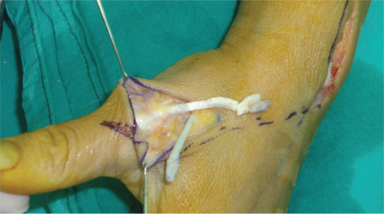
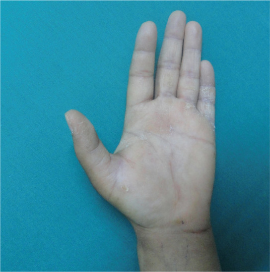
Upon surgical exploration, the distal stump of the EPL, which was degenerated, was found between the third compartment and zone five of the thumb (Figure 1). The proximal stump could not be identified because of retraction toward the forearm. After the atrophic and degenerative areas were surgically removed, the extensor indicis proprius (EIP) tendon was transferred to the distal stump of the EPL by using Pulvertaft’s technique. After 1 year of follow-up, the patient was fully capable of actively extending of her left thumb (Figure 2).
FIG. 1.

Degeneration and atrophy in the distal stump of the EPL and the EIP tendon is prepared for suture
FIG. 2.

At 1-year follow-up, extension of the thumb was satisfactory
DISCUSSION
The most common reasons for SREPL are steroid medication, RA, tenosynovitis, and synovitis. Other reasons include ankylosing spondylitis, tophaceous gout infiltration, Madelung’s deformity, non-metastatic distal radius or scaphoid fractures, wrist fractures, prolonged non-union of the scaphoid, misplaced fixators or plates, and subluxation of the distal ulna (1, 2).
The nutrition impairment theory of Engkvist et al. (3) is the main theory describing the cause of spontaneous tendon ruptures, and states that increased pressure on Lister’s tubercle leads to ischaemia and subsequent SREPL. Their angiographic study has shown that EPL has an avascular zone, which is supplied via diffusion from the synovial fluid around Lister’s tubercle. The other mechanisms are nutrition impairment, pressure caused by necrosis, a sharp fragment of bone that erodes the tendon, crushing injury, non-union of Lister’s tubercle, a roughened area of the radius caused by attrition of the tendon, repetitive activities that cause tenosynovitis, or a combination of these mechanisms (4).
Zvijac et al. (5) reported that attrition around Lister’s tubercle resulted in SREPL regardless of age and predisposing factors. Lloyd et al. (6) reported a spontaneous EPL rupture in a kickboxer and posited that this was caused by excessive and repetitive use of the wrist. There is a limitation in SREPL reports due to occupational activity, with the absence of other predisposing factors in the literature (2, 7).
In our case, the patient was a housewife with no other predisposing factors. She had a history of regularly milking a cow and such an action requires repetitive flexion and extension of the wrist, which can then cause the EPL to stretch and mechanical attrition to occur. Thus, excessive use of the wrist may cause rupture, due to the fact that the EPL tendon has a sharp turn around Lister’s tubercle.
We suggest that repetitive motion may cause attrition of the tendon and predispose it to rupture, and that several occupations may be at an increased risk of closed tendon rupture. Therefore, occupational groups at increased risk of tendon rupture should be advised to try and utilise the elbow instead of the wrist in tasks that require frequent flexion and extension motions.
Footnotes
This report was presented as a poster at the 34th Congress of Turkish Plastic Reconstructive and Aesthetic Surgery Association, 31 October– 4 November 2012, Antalya, Turkey and 10th Congress of Northern Cyprus Plastic Reconstructive and Aesthetic Surgery Association, 12–16 September 2012, Girne, Cyprus
Ethics Committee Approval: N/A
Informed Consent: Written informed consent was obtained from the participants of this study.
Peer-review: Externally peer-reviewed.
Author contributions: Concept – T.S., B.S., B.E.; Design – T.S., B.S., B.E.; Supervision – T.S., B.S., B.E.; Resource – T.S., B.S., B.E.; Materials – T.S., B.S., B.E.; Data Collection&/or Processing – T.S., B.S., B.E.; Analysis&/or Interpretation – T.S., B.S., B.E.; Literature Search – T.S., B.S., B.E.; Writing – T.S., B.S., B.E.; Critical Reviews – T.S., B.S., B.E.
Conflict of Interest: No conflict of interest was declared by the authors.
Financial Disclosure: The authors declared that this study received no financial support.
REFERENCES
- 1.Bjorkman A, Jorgsholm P. Rupture of the extensor pollicis longus tendon:a study of aetiological factors. Scand J Plast Reconstr Surg Hand Surg. 2004;38:32–5. doi: 10.1080/02844310310013046. [DOI] [PubMed] [Google Scholar]
- 2.Choi JC, Kim WS, Na HY, Lee YS, Song WS, Kim DH, et al. Spontaneous rupture of the extensor pollicis longus tendon in a tailor. Clin Orthop Surg. 2011;3:167–9. doi: 10.4055/cios.2011.3.2.167. [DOI] [PMC free article] [PubMed] [Google Scholar]
- 3.Engkvist O, Lundborg G. Rupture of the extensor pollicis longus tendon after fracture of the lower end of the radius - A clinical and microangio-graphic study. Hand. 1979;11:76–86. doi: 10.1016/s0072-968x(79)80015-7. [DOI] [PubMed] [Google Scholar]
- 4.Fujita N, Doita M, Yoshikawa M, Fujioka H, Sha N, Yoshiya S. Spontaneous rupture of the extensor pollicis longus tendon in a professional skier. Knee Surg Sports Traumatol Arthrosc. 2005;13:489–91. doi: 10.1007/s00167-004-0539-z. [DOI] [PubMed] [Google Scholar]
- 5.Zvijac JE, Janecki CJ, Supple KM. Non-traumatic spontaneous rupture of the extensor pollicis longus tendon. Orthopedics. 1993;16:1347–50. doi: 10.3928/0147-7447-19931201-10. [DOI] [PubMed] [Google Scholar]
- 6.Lloyd TW, Tyler MP, Roberts AH. Spontaneous rupture of extensor pollicis longus tendon in a kick boxer. Br J Sports Med. 1998;32:178–9. doi: 10.1136/bjsm.32.2.178. [DOI] [PMC free article] [PubMed] [Google Scholar]
- 7.Kim CH. Spontaneous rupture of the extensor pollicis longus tendon. Arch Plast Surg. 2012;39:680–2. doi: 10.5999/aps.2012.39.6.680. [DOI] [PMC free article] [PubMed] [Google Scholar]


