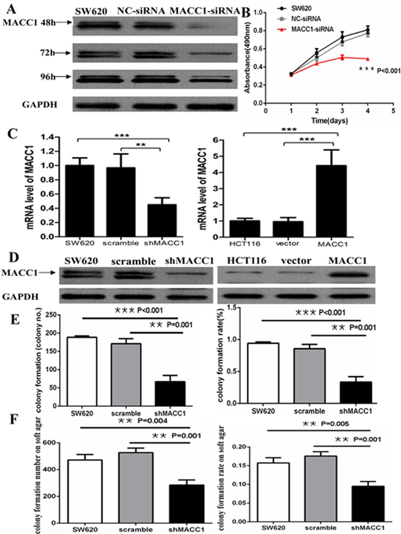
Figure 3.
MACC1 protein expression reduced in SW620 cells transfected with MACC1-siRNA at 48, 72, and 96 hours. GAPDH was used as loading control (A); MACC1 knockdown dramatically suppressed cell proliferation of SW620 at 48, 72, and 96 hours compared with the scramble siRNA(NC-siRNA) and untreated SW620 group by MTT analysis, respectively (B); MACC1 mRNA and protein expression was suppressed in SW620 cells (left) stably transfected shMACC1 compared with the scramble and untreated control groups by real-time PCR and western blot analysis, respectively. MACC1 mRNA and protein expression was elevated in HCT116 cells (right) stably transfected with MACC1 expression plasmid compared with empty vector and untreated control groups, respectively. **P<0.01, ***P<0.001(C-D); MACC1 knockdown significantly inhibited colony formation (E) and soft agar colony formation (F) of SW620 cells, respectively. The histogram showed the relative mean number of colony number (left) and the colony formation rate (right) in SW620 cells.

