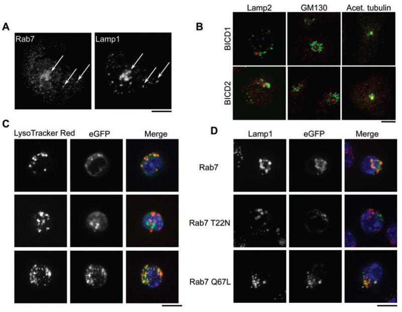
Figure 2. Intracellular localization of Rab7 and BICD1 in CTL.
A) hCTL labeled with antibodies against Rab7 and Lamp1. Arrows indicate colocalization. B) hCTL labeled with antibodies against BICD1 or BICD2 (red) and Lamp2, GM130 or acetylated tubulin (green), as indicated. C) Representative images of live OT-1 mCTL nucleofected with GFP-tagged human Rab7 constructs (as indicated) imaged with Lysotracker (lysosomes, red) and Hoechst (nuclei, blue) or (D) fixed after 24 h and stained with anti-Lamp1. Scale bars: (A) 5 μm; (B) 3 μm; (C,D) 10 μm.

