Abstract
Primitive neuroectodermal tumours (PNET) are aggressive childhood malignancies and offer a significant challenge to treatment. A two years old female child presented with weakness both lower limbs. Preoperative MRI of the spine and paravertebral regionIso – hyper intense posterior placed extradural lesion, non contrast enhancing from D11-L2 levels with cord compression D9 to L3 laminectomy done. Granulation tissue found from D11 to L2. with cord compression. The granulation tissue removed in toto. The pathological findings were consistent with PNET. Post operative neurological improvement was minimal. Cranial screening ruled out any intracranialtumour. Hence a diagnosis of primary spinal PNET was made. A review of the literature shows that only 19 cases of primary intraspinal PNETs have been reported to date and the present case extradural in location. Primary intraspinal PNETs are rare tumors and carry a poor prognosis.
Keywords: Spinal cord, Primitive neuroectodermal tumor (PNET)
Introduction
(PNETs) term coined by Hart and Earle is a group of malignant neoplasms derived from the primitive neural crest are highly malignant and mainly exist in the central nervous system (CNS), chest wall, lower extremities, trunk, kidney, and orbit but rarely in the spine. Though multidisciplinary treatments have been proposed the standard therapy for PNETs, is the complete excision followed by craniospinal irradiation.1
Case report
2 years old female child from Andhra Pradesh, INDIA apparently normal before 2 months presented with progressive weakness of both lower limbs. Mother noticed child not perceiving sensation below the level of umbilicus when she nurses the child. The weakness was getting worse day by day and child had difficulty in rolling over in bed and sitting up. Mother also noticed continuous dribbling of urine.
Child had no history of seizures, vomiting or weight loss. Child was alert with an active cry and normal feeding. Examination of Cranial nerves was essentially normal. Bulk was normal in all limbs. We noticed hypotonia of both lower limbs. On examination of power child was not moving both lowerlimbs spontaneously or to painful stimuli.
The superficial relexes in the lower abdomen and plantar was not elicitable and deep tendon reflexes were absent in the lower limbs.
Radiological Investigations
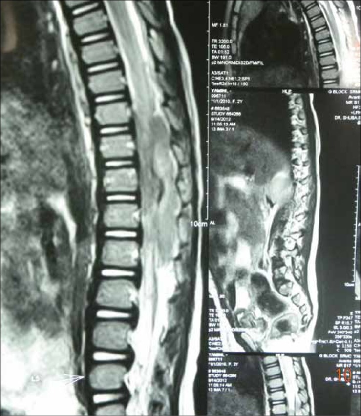
The child underwent MRI of the spine which revealed an iso – hyper intense posterior placed extradural lesion, non contrast enhancing from D11-L2 levels with cord compression. The cord was pushed anterolaterally. Considering the acute presentation and radiological findings we suspected either hemorrhage or an inflammatory mass Figure 1.
Fig. 1:
MRI of the spine which revealing an iso – hyper intense posterior placed extradural lesion , non contrast enhancing from D11-L2 levels with cord compression.
Surgery
Child was taken up for surgery on emergency basis. D9 to L3 open door laminotomy was done. Pinkish soft friable granulation likemass found from D11 to L2 with cord compression. Gross total removal of the mass was done. Tissue sent for HPE. Wound closed in layers after hemostasis.
Pathology
Tissue specimen represented acellular neoplasm composed of small round cells with reticular nuclei and finely developed chromatin cells arranged in dense sheets peritheliomatous and papillary pattern, areas of extensive hemorrhage and necrosis. Areas of micro calcification. Diagnosis of a small round cell tumour consistent with PNET was arrived.
Postoperative status
Postoperative status was uneventful. Wound healed well and sutures removed on the tenth day. The child showed little improvement of the power in the form of flickering movements and treated with antibiotics and steroids, Physiotherapy was done regularly. The child was evaluated for any intracranial lesion. MRI of the brain showed no intracranial lesion. We discussed with radiation oncology, considering the detrimental effects of radiation on the growing spine the child was not offered radiotherapy and the child was planned for chemotherapy.
Discussion
PNETs was first described by Bailey and Cushing in 1925, and were also called spongioblastomacerebelli. PNETs can occur outside the brain and throughout the body, as peripheral neuroblastomas and ewing sarcomas.
PNETs eventhough found in both children and adults are more common in children. The mean age is between 5 and 77 years and 80% of tumours occur in less than 15 years. There is male predominance, it is 1.4 to 4.8 times more common in males than in females. Usage of maternal folate, iron, and multivitamin supplementation reduces incidence of medulloblastomas.2–4
Several syndromes are associated with a familial increased incidence of medulloblastoma. They are Gorlin’s syndrome, Li-Fraumeni syndrome, and Turcot’ssyndrome. Intramedullary spinal cord tumors (IMSCTs) are usually rare. The onset of symptoms occurs in months. Frequently identified after a trivial trauma.5 Diagnosed by radiographic techniques. Majority of the tumors are benign in nature and have insidious growth pattern.
Tumours of the thoracic spine have a more insidious onset. Usually manifests with pain and progressive scoliosis. Examinationmay show paraspinal spasm and evidence of myelopathy. Sensory findings and bowel and bladder dysfunction occurs late.6
MRI is the investigation of choice. MRI provides excellent soft tissue imaging within the spinal column and any intramedullary lesions, edema and cysts can be visualized. Also the extent of the solid portion of the lesion to differentiate tumoral cysts from nontumoral cysts. Computed tomography and plain radiography are reserved for the evaluation of associated spinal deformities/instabilities.5
T1-weighted sequence, with and without gadolinium enhancement, and a T2-weighted sequence in the axial and sagittal planes are studied. A gradient echo sequence and a fluid-attenuated inversion recovery (FLAIR) gives information about hemosiderin deposits (gradient echo) or subtle intramedullary lesions (FLAIR). In children, a mass lesion within the spinal cord is probably an astrocytoma, ganglioglioma/glial neuronal tumor, or ependymoma. A hemangioblastoma or primitive neuroectodermal tumor is a rare possibility that can be considered when an unusual image or clinical state is present.7
Due to the aggressive behavior of the neoplasm and its great potential to metastasize, treatment should be multimodal, involving radical surgical resection, radiotherapy, and chemotherapy. Initial management of these tumours is almost always surgical since tissue biopsy is required. 80% resection or more gives a 5-year event-free survival rate higher than 70%. Aggressive resection has been shown to improve survival in children with SPNETs.
These tumours need radiation therapy but that there is a risk of injury to the growing spine and the risk of induction of second malignancies always exist. Radiation therapy is a critical component in the management of newly diagnosed SPNETs. The possibility of primitive neuro ectodermal tumour to disseminate along the neuraxis supports prophylactic craniospinal irradiation with an involved boost to the primary site.7-9
The utility of chemotherapy in the management of PNET remains unclear. The usual agents used includ ifosfamide, vincristine, methotrexate, cisplatin and lomustine. This combination gives 49.3% 3-year progression-free survival rate with optimal surgery followed by chemoradiation. The use of temozolomide has yet to meet significant success.10
References
- 1.Cabral GA, Nunes CF, Melo JO et al. Peripheral primitive neuro-ectodermal tumor of the cervical spine. SurgNeurol Int. 2012;3:91. doi: 10.4103/2152-7806.99938. [DOI] [PMC free article] [PubMed] [Google Scholar]
- 2.Akgun B, Ates D, Kaplan M. Ganglioneuroblastoma of the thoracic spinal cord: a very rare case report. ActaMedica (Hradec Kralove) 2012;55(1):50–52. doi: 10.14712/18059694.2015.76. [DOI] [PubMed] [Google Scholar]
- 3.Coumans JV, Walcott BP, Nahed BV et al. Multimodal therapy of an intramedullary cervical primitiveneuroectodermal tumor in an adult. J ClinOncol. 2012;30(2): doi: 10.1200/JCO.2011.38.6474. [DOI] [PubMed] [Google Scholar]
- 4.Parikh D, Short M, Eshmawy M et al. Surgical outcome analysis of paediatric thoracic and cervical neuroblastoma., Brown R. Eur J Cardiothorac Surg. 2012;41(3):630–634. doi: 10.1093/ejcts/ezr005. [DOI] [PubMed] [Google Scholar]
- 5.Gollard RP, Rosen L, Anson J et al. Intramedullary PNET of the spine: long-term survival after combined modality therapy and subsequent relapse. J PediatrHematolOncol. 2011;33(2):107–112. doi: 10.1097/MPH.0b013e3181f84b7f. [DOI] [PubMed] [Google Scholar]
- 6.Ellis JA, Rothrock RJ, Moise G et al. Primitive neuroectodermal tumors of the spine: a comprehensive review with illustrative clinical cases. Neurosurg Focus. 2011;30(1): doi: 10.3171/2010.10.FOCUS10217. [DOI] [PubMed] [Google Scholar]
- 7.Chang SI, Tsai MC, Tsai MD. An unusual primitive neuroectodermal tumor in the thoracic epidural space. J ClinNeurosci. 2010;17(2):261–3. doi: 10.1016/j.jocn.2009.05.018. [DOI] [PubMed] [Google Scholar]
- 8.Alexander HS, Koleda C, Hunn MK. Peripheral Primitive NeuroectodermalTumour (pPNET) in the cervical spine. J ClinNeurosci. 2010;17(2):259–261. doi: 10.1016/j.jocn.2009.05.020. [DOI] [PubMed] [Google Scholar]
- 9.Kiatsoontorn K, Takami T, Ichinose T et al. Primary epidural peripheralprimitive neuroectodermal tumor of the thoracic spine. Neurol Med Chir. 2009;49(11):542–545. doi: 10.2176/nmc.49.542. [DOI] [PubMed] [Google Scholar]
- 10.Hrabálek L, Kalita O, Svebisova H et al. Dumbbell-shaped peripheralprimitive neuroectodermal tumor of the spine-case report and review of the literature. J Neurooncol. 2009;92(2):211–217. doi: 10.1007/s11060-008-9744-9. [DOI] [PubMed] [Google Scholar]


