Abstract
Background
Acetabular revision THA with use of a large (jumbo) cup is an effective treatment for many cavitary and segmental peripheral bone defects. However, hip center elevation may occur with use of a jumbo cup owing to reaming superiorly and/or because of the increased diameter of the jumbo cup compared with the native acetabulum.
Questions/purposes
In our jumbo cup revision THAs, we attempted to avoid hip center elevation by placing the inferior edge of the cup at the inferior acetabulum. In this study, we asked (1) how much of an elevation in the hip center is observed radiographically with use of jumbo cups, and (2) how effective was our technique in minimizing hip center elevation during revision THA in clinical practice?
Methods
We retrospectively reviewed radiographic data for all patients, from one surgeon’s practice, who received a jumbo cup (defined as cup size ≥ 66 mm in men, ≥ 62 mm in women) during an acetabular revision between 1998 and 2012 and who had an anatomically placed THA or no THA on the contralateral side (so as to be able to make comparisons); 98 patients were identified and included. The height of the revised hip center was measured relative to the contralateral normal hip. Cup elevation resulting from superior reaming was determined by measuring the distance from the inferior cup edge to the interteardrop line. The mean hip center elevation and cup position relative to the interteardrop line in male and female patients were compared using unpaired t-tests.
Results
Radiographic analysis showed a mean hip center elevation of 11 mm. On average, 1 mm of the measured hip center elevation was the result of cup placement superior to its planned position at the interteardop line.
Conclusions
Our results indicate that revision THA with a jumbo cup is associated with hip center elevation despite placement of the cup at the inferior acetabulum. An increase in femoral head length may be needed to compensate for hip center elevation with use of a jumbo cup.
Level of Evidence
Level III, therapeutic study. See the Instructions for Authors for a complete description of levels of evidence.
Introduction
Revision THA with a cementless porous-coated jumbo cup and screw fixation is an effective technique to treat most failed acetabular components with cavitary or small segmental bone defects [1, 11, 13]. The acetabular bone is reamed into a larger hemispheric shape than the native acetabulum and cavitary defects are filled with morselized bone graft. The revision component is supported on host bone superiorly, posteriorly, and anteriorly. However, if reaming is directed superiorly, this also may raise the hip center [7]. A high hip center has been associated with altered hip biomechanics and hip instability [6]. Leg length discrepancy is not uncommon after revision THA. However, unlike primary THA, limb shortening is more common than lengthening during revision THA, which can be related to cup positioning [4].
To minimize the amount of hip center elevation with a jumbo cup technique in our revision THAs, we direct the reamer inferiorly to the inferior acetabular rim. However, elevation of the hip center may occur from directing the reamer superiorly and/or because the increased diameter of the jumbo cup shifts the hip center owing to the increase in the geometric radius of the oversized cup. In a prior computer simulation of jumbo cup reaming to span the superior-to-inferior dimensions of an acetabulum with a posterosuperior bone defect, we found that hip center elevation of 0.27 mm occurred for each millimeter of increase in reamer size [10].
In the current study, which is a clinical validation of the computer simulation study, we therefore asked (1) how much of an elevation in the hip center is observed radiographically with use of jumbo cups, and (2) how effective was our technique in minimizing hip center elevation during revision THA in clinical practice?
Materials and Methods
We conducted a retrospective radiographic review of all cementless jumbo cup acetabular revision THAs in the senior author’s (MDR) practice between January 1, 1998, and March 30, 2012. A jumbo acetabular cup was considered to be an implant of 62 mm or larger in diameter in women and 66 mm or larger in men, which is approximately 10 mm larger in diameter than the native acetabulum and consistent with the definition of a jumbo cup used in prior clinical studies [11, 13]. Cases with revision acetabular components smaller than 62 mm in diameter in women or smaller than 66 mm in men and those with structural bone grafts or augments, cages, and pelvic discontinuity were excluded. Ninety-eight patients were analyzed (57 men and 41 women). The mean cup diameter was 69.1 mm in men and 61.9 mm in women, with a median cup size of 68 mm in men and 64 mm in women (Table 1). Thirty-six patients had a well-positioned contralateral primary THA, ie, a cup fixed in approximately 45° abduction, between 20° and 30°, and with the inferior cup margin adjacent to the tear drop, and the remaining 62 patients had a native contralateral hip.
Table 1.
Patient characteristics and hip center measurements
| Characteristic | Measurements | p value |
|---|---|---|
| Number of patients | Total 98 | NA |
| Men 57 (58%) | ||
| Women 41 (42%) | ||
| Age (years) (mean ± SD, range) | Total (62.4 ± 12.2; 40–89) | p < 0.05 |
| Men (60 ± 12.1; 40–89) | ||
| Women (65.7 ± 11.8; 43–89) | ||
| Cup size (mm) | Total (67.3 ± 3.7; 62–78) | p < 0.05 |
| Men (69.1 ± 3.3; 66–78) | ||
| Women (64.9 ± 2.8; 62–72) | ||
| Contralateral hip center (a) mm | Total (21.7 ± 8; 6.3–52.5) | p = 0.91 |
| Men (21.6 ± 7.8; 9.8–52.5) | ||
| Women (21.8 ± 8.3; 6.3–41.9) | ||
| Jumbo hip center (a′) mm | Total (32.6 ± 9.6; 12.6–63.5) | p = 0.09 |
| Men (33.9 ± 10.3; 12.6–63.5) | ||
| Women (30.7 ± 8.3; 14.3–50.2) | ||
| Hip center difference (a′ − a) mm | Total (10.9 ± 10; −20.8 to 35) | p = 0.08 |
| Men (12.3 ± 10.8; −20.8 to 35) | ||
| Women (8.9 ± 8.3; −6.4 to 33.6) | ||
| Superior cup placement (d) mm | Total (1.2 ± 9.9; −26.5 to 28.6) | p = 0.07 |
| Men (2.6 ± 11.2; −26.5 to 28.6) | ||
| Women (−0.8 ± 7.4; −15.9 to 16.5) |
NA = not applicable; a′ = hip center height on the side with the jumbo cup; a = hip center height of the contralateral side; (a′ − a) = difference in hip center heights between both sides; d = amount of hip center elevation attributable to superior placement of the acetabular cup.
With our surgical technique we sought to direct acetabular reaming inferiorly to the level of the obturator foramen in an effort to place the inferior edge of the cup at the inferior acetabulum. The obturator foramen is easily palpated during revision THA, and can be used as an anatomic landmark to identify the inferior acetabulum. Sequentially increasing reamer sizes are used until a reamer engages the superior host bone. Reaming also may be directed medially through the medial wall to gain better superior coverage [3, 5]. However, with increasing reamer diameters to span the superior-to-inferior dimension of the acetabular bone defect, the anterior and/or posterior walls could be reamed away. To avoid reaming away the anterior wall by increased reamer size, the reamer may be shifted superiorly or an augment used above a smaller cup. If a reamer size is required that will compromise the anterior wall, we prefer to use a superior augment above a smaller cup rather than raise the hip center. A cup size was selected that contacted host bone superiorly to ensure a stable fit of the implant on host bone. Additional screw fixation was used as necessary. Forty-eight patients received a Reflection® InterfitTM (Smith & Nephew, Memphis, TN, USA), 38 patients received Reflection® Peripheral Hole (Smith & Nephew), five patients received Trident TritaniumTM (Stryker, Mahwah, NJ, USA), three received Constrained Tripolar (Stryker), and two patients each received Omnifit® PSL (Stryker) and R3TM Multi-hole (Smith & Nephew) acetabular implants. At the time of the revision procedure, the average age of the patients was 62.4 years (range, 40–83 years).
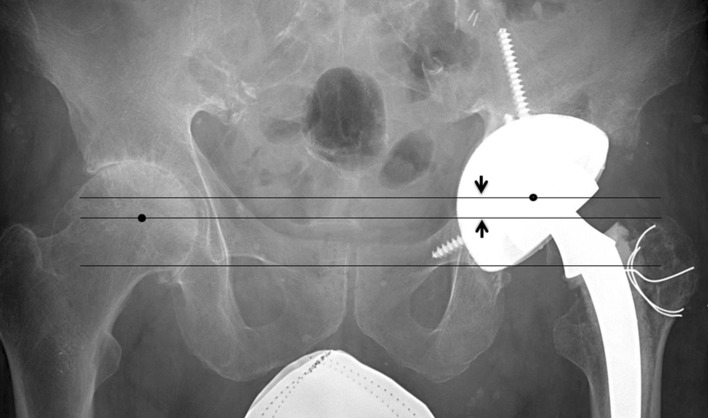
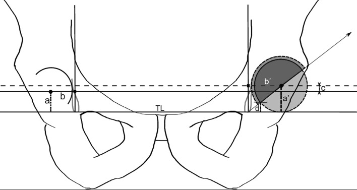
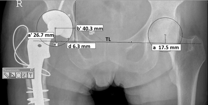
All radiographic measurements were made by one observer (CDN) and were taken from AP pelvis radiographs obtained at the 6-week followup using Philips iSiteTM PACS version 3.6 software (Koninklijke Philips Electronics NV, Andover, MA, USA). The primary outcome of interest was the vertical difference in the hip center of the revised hip compared with the contralateral hip (Fig. 1). To determine the vertical position of the hip center, first, a circle was made around the jumbo and the contralateral acetabular surfaces. The perimeter of these circles was aligned with the acetabular margins. The center of this circle was assumed to correspond to the hip center. The height of the hip center was estimated by measuring the height of a perpendicular line arising from the interteardrop line and ending at the hip center (Fig. 2). This was done for the hip with the jumbo cup (a′) and the contralateral hip (a), and the difference between the vertical positions of both hip centers was the measure of hip center elevation (Fig. 3). The acetabular teardrop was used as a landmark for measurements because the teardrop is a discrete anatomic structure, and its vertical position is not affected substantially by rotation of the pelvis.
Fig. 1.
An AP pelvic radiograph obtained after a left revision THA with a jumbo cup shows the revision cup is larger than the contralateral acetabulum. The interteardrop (lower) line intersects the inferior edge of the acetabular component indicating that the jumbo revision cup was placed at the planned position with the inferior edge of the cup at the inferior acetabulum. A middle line is parallel to the interteardrop line and intersects the right hip center. An upper line parallel to the interteardrop line intersects the center of the left jumbo cup. The distance between the two lines (arrows) indicates the amount of head center elevation that has occurred from the use of a jumbo cup.
Fig. 2.
A diagram shows the radiographic measurements used to determine relative hip center elevation and superior cup placement. TL = the interteardrop line; a′ = hip center height of the side with the jumbo cup; a = hip center height of the contralateral side; b′ = horizontal distance between a perpendicular line through the teardrop and the jumbo cup hip center; b = horizontal distance between a perpendicular line through the teardrop and the contralateral hip center; c = hip center elevation (a′ − a); d = amount of hip center elevation attributable to superior placement of the acetabular cup.
Fig. 3.
The radiographic lines and measurements made using the Philips iSiteTM PACS software are shown. TL = interteardrop line; a′ = hip center of the side with the jumbo cup; a = hip center of the contralateral side; a′ − a = hip center elevation; b′ = horizontal distance between a perpendicular line through the tear drop and the jumbo cup hip center; d = amount of hip center elevation attributable to superior placement of the acetabular cup.
Elevation of the hip center may be caused by superior reaming and placement of the cup above the interteardrop line, elevation resulting from a geometric increase in cup radius, or both.
To determine how much of the vertical change in the revision hip center was secondary to superior placement of the component above the interteardrop line, a measurement also was made of the vertical distance from the inferior edge of the jumbo cup to the interteardrop line. This was considered to be the hip center elevation caused by superior placement of the cup above its planned placement position. In the event that the inferior lip of the jumbo cup was below the interteardrop line, a negative value was assigned. Because the position of the inferior edge of the cup also is influenced by abduction angle, the cup position was normalized to 45° abduction. If the cup was not at 45° abduction, it was templated to 45° using a digital hemisphere to determine the appropriate measurement of relative cup elevation from the inferior edge of the templated cup position to the interteardrop line.
To account for the magnification of skeletal images on radiographs, the diameter of the acetabular cup was measured on the AP radiograph and divided by the actual diameter of the implanted cup. This yielded a mean magnification ratio of 1.2 (range, 1.1–1.35). All radiographic measurements were multiplied by this magnification factor to yield the true values. The mean hip center elevation and cup position relative to the interteardrop line in males and females was compared using unpaired t-tests.
Results
The radiographic analysis showed that our jumbo cup technique resulted in elevation of the hip center. The mean hip center height of the jumbo cup was 32.6 mm (male = 33.9 mm, female = 30.7 mm); and the mean hip center of the contralateral hip was 21.7 mm (male = 21.6 mm, female = 21.8 mm). This yielded a mean hip center difference of 10.9 mm, greater in men (n = 12.2 mm, range, −21 mm to 35.3 mm) than in women (n = 8.9 mm, range, −6.5 mm to 33.8 mm). There was no difference between either group (p = 0.085).
An average elevation of 1.2 mm was found to be the result of superior cup placement above its planned position. Superior cup placement accounted for 2.6 mm (range, −26.5 mm to 28.6 mm) of the hip center elevation in males and for −0.8 mm (range, −15.9 mm to 16.9 mm) in females. There was no measurable difference between either group (p = 0.076).
Discussion
Jumbo cups offer several advantages over other methods of acetabular revision. They provide a large area of contact between host bone and the porous implant surface facilitating reliable biological fixation, and they allow for a relatively straightforward and reproducible surgical technique of hemispheric reaming and screw fixation of the implant [1, 8, 11, 13]. The large size of the jumbo shell also can compensate for areas of bone loss, often eliminating the need for bulk allograft or metal augments. However, the jumbo cup technique requires preparation of the acetabular surface to fit a hemispheric implant that is larger in diameter than the native acetabulum. This may lead to elevation of the hip center secondary to reaming in a superior direction and placement of the cup above its planned position and/or resulting from a geometric increase in the radius of the cup. We questioned how much of an elevation in the hip center was observed radiographically with use of jumbo cups and how effective our jumbo cup technique was in minimizing hip center elevation during revision THA in clinical practice. We found that there was an elevation of approximately 1 cm in the hip center on average, primarily secondary to an increase in cup size, and much less so from reaming superiorly.
There are some limitations to the design of our study. Measurement errors can occur in a radiographic study. The measurements were done by one observer (CDN) using specific anatomic markers in each patient with a digital method in an effort to minimize these errors. However, neither intraobserver nor interobserver repeatability testing were included in the study so the reliability of the measurement technique cannot be accurately determined. Additionally, we included 36 patients with a contralateral THA. Given that we use the contralateral hip center as a reference point, this creates the possibility that changes in the contralateral hip center from the primary THA can influence our hip center elevation measurements. All 36 hips, however, were well-positioned, so any effects on measurements should be minimal. Another limitation of this study is that all the surgeries were performed by one surgeon (MDR), thus the results of this study may not be applicable to other surgeons whose revision THA technique may be different. We also did not quantitate acetabular bone loss using an established classification system in this series of patients.
Our radiographic analysis showed an average hip center elevation of 11 mm. The hip center elevation was 12 mm in men and 9 mm in women; however this difference was not significant. Since the average jumbo cup size was larger in men than women, but approximately 10 mm larger than the native acetabulum for males and females, our observation that the hip center elevation in both groups was similar may indicate that the amount of hip center elevation is more related to the amount of cup oversizing relative to the native acetabulum than the absolute cup diameter. The radiographic hip center elevation we observed correlates with, but exceeds the amount of hip center elevation predicted in a prior in vitro computer simulation [10]. In the simulation study, the hip center shifted superiorly by 0.27 mm for every millimeter increase in reamer size, so if we assume a presurgery diameter of 54 mm for the male acetabulum and revise it to a 69 mm jumbo cup (our average male jumbo cup diameter), this would predict a hip center elevation of 4.2 mm. The difference between the radiographic and computer simulation results may be partly attributable to the magnification of anatomic images that occur during radiography of the hip and to errors that arise from measuring three-dimensional relationships on two-dimensional planes.
Reaming in a superior direction may result in placement of the cup above the anatomic acetabulum and result in elevation of the hip center. Our results indicate that a surgical technique to place the inferior edge of the cup at the inferior acetabulum and avoid superior cup placement was relatively successful; our data show that on average approximately 1 mm of the total hip center elevation was attributable to placement of the cup above the interteardrop line.
A vertical hip center shift during revision THA may result in leg length discrepancies, altered hip biomechanics, and soft tissue laxity. A retrospective study involving 79 THA revisions by Dou et al. [4] showed that acetabular cup placement contributes to hip center elevation and leg length discrepancies after revision THA. In addition to its role in leg length changes, an elevated hip center can result in suboptimum biomechanics of the hip [2, 12]. Delp et al. [2] showed that superolateral placement of the hip center (2 cm superior and 2 cm lateral) decreases the moment arms of the hip abductors by an average of 28%, thus reducing force-generating capacity. Lachiewicz and Soileau [9] reported a dislocation rate of 10% after jumbo cup revision THA, which was the most common complication observed in their series. Hip center elevation in jumbo cup revision THA, as we observed, may be one of several factors that contribute to soft tissue laxity and hip instability. To compensate for an elevated hip center, appropriate adjustments on the femoral side can be made to increase leg length and/or offset.
Our results indicate that reaming inferiorly to the level of the obturator foramen was effective in maintaining the inferior edge of the cup at or near its planned placement position at the level of the acetabular teardrop. However, based on our radiographic analysis, the jumbo cup technique resulted in an average hip center elevation of approximately 1 cm. This suggests that an increase in femoral head length may be helpful to avoid limb shortening with use of a jumbo cup technique in revision THA.
Footnotes
One of the authors certifies that he (MDR), or a member of his or her immediate family, has or may receive payments or benefits, during the study period, an amount of USD 10,000–USD 100,000, from Smith & Nephew, Memphis, TN, USA.
All ICMJE Conflict of Interest Forms for authors and Clinical Orthopaedics and Related Research editors and board members are on file with the publication and can be viewed on request.
Each author certifies that his or her institution approved the human protocol for this investigation, that all investigations were conducted in conformity with ethical principles of research, and that informed consent for participation in the study was obtained.
Clinical Orthopaedics and Related Research neither advocates nor endorses the use of any treatment, drug, or device. Readers are encouraged to always seek additional information, including FDA-approval status, of any drug or device prior to clinical use.
This work was performed at the School of Medicine, University of California San Francisco, San Francisco, CA, USA.
References
- 1.Dearborn JT, Harris WH. Acetabular revision arthroplasty using so-called jumbo cementless components: an average 7-year follow-up study. J Arthroplasty. 2000;15:8–15. doi: 10.1016/S0883-5403(00)90999-9. [DOI] [PubMed] [Google Scholar]
- 2.Delp SL, Wixson RL, Komattu AV, Kocmond JH. How superior placement of the joint center in hip arthroplasty affects the abductor muscles. Clin Orthop Relat Res. 1996;328:137–146. doi: 10.1097/00003086-199607000-00022. [DOI] [PubMed] [Google Scholar]
- 3.Dorr LD, Tawakkol S, Moorthy M, Long W. Wan Z Medial protrusio technique for placement of a porous-coated hemispherical acetabular component without cement in total hip arthroplasty patients who have acetabular dysplasia. J Bone Joint Surg Am. 1999;81:83–92. doi: 10.2106/00004623-199901000-00012. [DOI] [PubMed] [Google Scholar]
- 4.Dou Y, Zhou Y, Tang Q, Yang D, Liu J. Leg-length discrepancy after revision hip arthroplasty: are modular stems superior? J Arthroplasty. 2013;28:676–679. doi: 10.1016/j.arth.2012.08.009. [DOI] [PubMed] [Google Scholar]
- 5.Fabi D, Gonzalez M, Goldstein W, Ahmed M. Acetabular cup revision with the use of the medial protrusio technique at an average follow-up of 6.6 years. J Arthroplasty. 2010;25:197–202. doi: 10.1016/j.arth.2009.01.019. [DOI] [PubMed] [Google Scholar]
- 6.Gustke KA. Jumbo cup or high hip center: is bigger better? J Arthroplasty. 2004;19(4 suppl 1):120–123. doi: 10.1016/j.arth.2004.02.012. [DOI] [PubMed] [Google Scholar]
- 7.Hendricks KJ, Harris WH. High placement of noncemented acetabular components in revision total hip arthroplasty: a concise follow-up, at a minimum of fifteen years, of a previous report. J Bone Joint Surg Am. 2006;88:2231–2236. doi: 10.2106/JBJS.E.00247. [DOI] [PubMed] [Google Scholar]
- 8.Jasty M. Jumbo cups and morsalized graft. Orthop Clin North Am. 1998;29:249–254. doi: 10.1016/S0030-5898(05)70323-0. [DOI] [PubMed] [Google Scholar]
- 9.Lachiewicz PF, Soileau ES. Fixation, survival, and dislocation of jumbo acetabular components in revision hip arthroplasty. J Bone Joint Surg Am. 2013;95:543–548. doi: 10.2106/JBJS.L.00758. [DOI] [PubMed] [Google Scholar]
- 10.Nwankwo C, Dong NN, Heffernan CD, Ries MD. Do jumbo cups cause hip center elevation in revision THA? A computer simulation. Clin Orthop Relat Res. 2014;472:572–576. doi: 10.1007/s11999-013-3169-2. [DOI] [PMC free article] [PubMed] [Google Scholar]
- 11.Patel JV, Masonis JL, Bourne RB, Rorabeck CH. The fate of cementless jumbo cups in revision hip arthroplasty. J Arthroplasty. 2003;18:129–133. doi: 10.1054/arth.2003.50038. [DOI] [PubMed] [Google Scholar]
- 12.Vasavada AN, Delp SL, Maloney WJ, Schurman DJ, Zajac FE. Compensating for changes in muscle length in total hip arthroplasty: effects on the moment generating capacity of the muscles. Clin Orthop Relat Res. 1994;302:121–133. [PubMed] [Google Scholar]
- 13.Whaley AL, Berry DJ, Harmsen WS. Extra-large uncemented hemispherical acetabular components for revision total hip arthroplasty. J Bone Joint Surg Am. 2001;83:1352–1357. doi: 10.2106/00004623-200109000-00010. [DOI] [PubMed] [Google Scholar]


