Abstract
A patient with zoster in multiple dermatomes, severe headache, and normal magnetic resonance angiography (MRA) and 4-vessel digital subtraction angiogram (DSA) developed 9 anterior circulation aneurysms 2 months later. Antiviral treatment resulted in clinical improvement, size reduction of most aneurysms, and complete resolution of the 2 largest aneurysms.
A patient with zoster in multiple dermatomes, severe headache, and normal magnetic resonance angiography (MRA) and 4-vessel digital subtraction angiogram (DSA) developed 9 anterior circulation aneurysms 2 months later. Antiviral treatment resulted in clinical improvement, size reduction of most aneurysms, and complete resolution of the 2 largest aneurysms.
Case report.
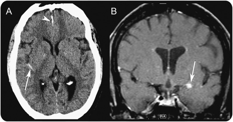
A 41-year-old woman with systemic lupus erythematosus (SLE) taking methotrexate 25 mg once weekly and oral prednisone 15 mg alternating with 10 mg daily was hospitalized for fever and right-sided headache of 2 days' duration. On the second day, she developed bilateral mandibular-distribution zoster and right-sided geniculate zoster on the hard palate. Brain MRI showed 7 areas of sulcal enhancement, several involving cerebral arteries. On day 3, she was started with IV acyclovir, 1 g q 8 h, for 21 days. On day 4, CSF opening pressure was 23 cm H2O, CSF contained 105 leukocytes/μL (91% mononuclear), CSF protein was 123 mg/dL and glucose 36 mg/dL, and PCR revealed 215,509 copies of varicella-zoster virus (VZV) DNA/mL CSF. Fluorescent antibody staining of facial rash swab identified VZV antigen. Severe headache continued. On day 5, brain CT revealed subarachnoid hemorrhage (SAH) in both sylvian fissures and orbitofrontal regions and an intra-axial hematoma in left gyrus rectus, and MRI showed areas of sulcal enhancement involving the blood vessels, but no aneurysms (figure 1); 4-vessel DSA was normal. Headache resolved on day 7. At discharge, she continued taking oral prednisone.
Figure 1. Subarachnoid and subdural hemorrhage with vasculitic inflammation.

At day 5 of illness, CT shows right sylvian subarachnoid hemorrhage and parafalcine subdural hematoma (A, arrow and arrowhead, respectively). Postgadolinium T1 coronal MRI at that time demonstrates focal nodular subarachnoid enhancement in several locations, including a lesion overlying the left middle cerebral artery M1 segment (B, arrow), consistent with intense vasculitic inflammation involving the middle cerebral artery M1 lenticulostriate perforators.
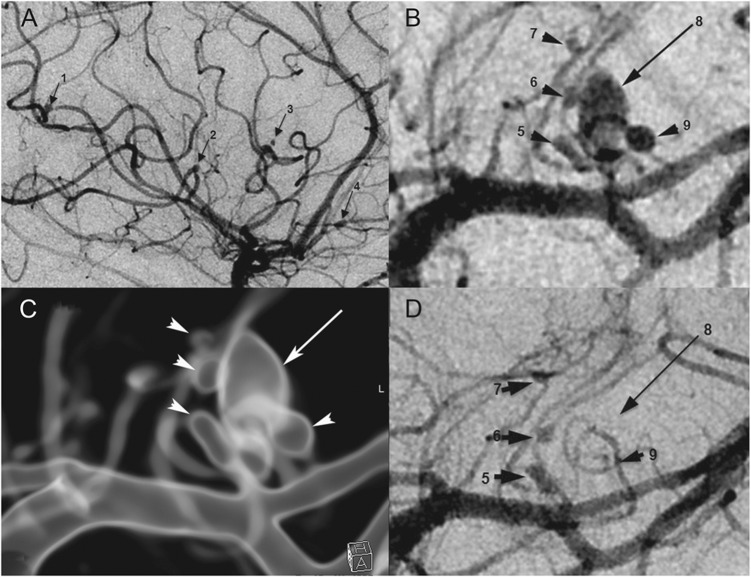
Two months later, severe headache recurred. Neurologic examination revealed nuchal rigidity. Brain CT detected new SAH in the left suprasellar cistern extending into sylvian fissure and multiple sulci of the left frontal and temporal lobes. Four-vessel DSA revealed 9 anterior circulation aneurysms with a cluster of left middle cerebral artery lenticulostriate perforator aneurysms surrounding a dominant 4 × 7-mm aneurysm (figure 2, A and B). Blood pressure was controlled in the low-normal range. The patient was treated with IV acyclovir 1.25 g q 8 h for 3 weeks. On day 2, she was dysarthric with right central facial weakness and right hemiplegia. Brain MRI showed left lenticulostriate-distribution restricted diffusion. She was treated with IV methylprednisolone 1 g daily for 5 days.
Figure 2. Nine aneurysms in varicella-zoster virus vasculopathy.
Digital subtraction angiogram (DSA) demonstrates a total of 9 mycotic aneurysms. Four aneurysms arise from the right middle cerebral artery (MCA) M3 segments (A, 1–4, arrows) in the sylvian fissure and a cluster of left MCA lateral lenticulostriate perforator aneurysms, configured as a dominant oval aneurysm (B, 8, arrow) surrounded by 4 smaller lesions (B, 5–7 and 9, arrowheads). A translucent rendered model from an angiographic 3D spin acquisition (C) further elucidates the complex arrangement of the left lateral lenticulostriate aneurysms. Repeat DSA after antiviral treatment and corona radiata perforator infarction shows interval thrombosis of the dominant aneurysm (D, 8, long arrow) and one of the satellite lesions (D, 9, arrowhead) that are no longer visible.
Nine days later, CSF was xanthochromic and contained 48,000–65,000 erythrocytes/μL and 25 leukocytes/μL (80% neutrophils, 20% mononuclear). CSF protein was 38 mg/dL and glucose 44 mg/dL; PCR revealed no VZV DNA. Eight days after treatment with IV acyclovir, DSA confirmed complete resolution of the dominant aneurysm (figure 2D, arrow 8), the smaller lenticulostriate aneurysm (figure 2D, arrow 9), and decreased size of the original aneurysms (figure 2, C and D). The patient continued treatment with oral valacyclovir 500 mg bid and prednisone as above. Acetylsalicylic acid 81 mg daily was started because of increased anticardiolipin antibodies associated with SLE. After 3–4 months, she remained headache-free without new symptoms but with residual right hemiplegia.
Discussion.
Our literature search back to 1935 using the terms “multiple” and “intracranial” and “aneurysms” identified 308 articles, only 5 of which reported the existence of 9 or more aneurysms in adults: a 45-year-old woman with 13 cerebral aneurysms and atrial myxoma1; a healthy 49-year-old woman with 13 anterior and posterior circulation aneurysms2 who might have had VZV vasculopathy without rash; a 46-year-old woman with long-standing asthma who developed 18 aneurysms during high-dose corticosteroid treatment, a known risk factor for VZV vasculopathy,3 with postmortem examination revealing inflammation in the wall of aneurysms,4 as manifest in VZV-infected arteries; and a 69-year-old woman with 10 anterior circulation aneurysms of unknown etiology,5 resembling our patient with 9 anterior circulation aneurysms. Although the exact reason for development of aneurysms in our patient is unknown, 1 month earlier, focal arteritis and intracranial hemorrhage (figure 1) developed, as found in patients with intracerebral and subarachnoid hemorrhage after zoster6 and varicella.7 The robust inflammatory response likely damaged the vessel wall, predisposing it to development of aneurysm, a notion supported by clinical improvement and radiographic resolution of aneurysms after antiviral and anti-inflammatory treatment. Overall, VZV vasculopathy should be considered in patients with rapid development of multiple cerebral aneurysms, particularly since one-third of VZV vasculopathy cases occur in the absence of rash and antiviral therapy may lead to dramatic improvement of vasculopathy and aneurysm regression.
Acknowledgments
Acknowledgment: The authors thank Marina Hoffman for reviewing the manuscript for errors in grammar, punctuation, spelling, readability, clarity, and accuracy.
Footnotes
Author contributions: Drs. Liberman, Hurley, Caprio, Bernstein, Nagel, and Gilden all contributed to drafting/revising the manuscript for content, study concept/design, analysis or interpretation of data, vital reagents/tools/patents, and acquisition of data.
Study funding: Supported in part by NIH grants AG006127 and AG032958 to D.G. and NS067070 to M.A.N.
Disclosure: The authors report no disclosures relevant to the manuscript. Go to Neurology.org for full disclosures.
References
- 1.George KJ, Rennie A, Saxena A. Multiple cerebral aneurysms secondary to cardiac myxoma. Br J Neurosurg 2012;26:409–411 [DOI] [PubMed] [Google Scholar]
- 2.Zacks DJ. Multiple intracranial aneurysms. Am J Roentgenol 1978;130:180–182 [DOI] [PubMed] [Google Scholar]
- 3.Gilden D, Cohrs RJ, Mahalingam R, Nagel MA. VZV vasculopathies: diverse clinical manifestations, laboratory features, pathogenesis and treatment. Lancet Neurol 2009;8:731–740 [DOI] [PMC free article] [PubMed] [Google Scholar]
- 4.Moriyama S, Saito K, Yokoyama T. Multiple intracranial aneurysms of inflammatory origin with subarachnoid hemorrhage. Acta Pathol Jpn 1980;30:815–823 [DOI] [PubMed] [Google Scholar]
- 5.Yamaki T, Takeda M, Takayama H, Nakagaki Y. Treatment of multiple intracranial aneurysms in the anterior circulation: case report. Neurol Med Chir 1990;30:47–50 [DOI] [PubMed] [Google Scholar]
- 6.Jain R, Deveikis J, Hickenbottom S, Mukherji SK. Varicella-zoster vasculitis presenting with intracranial hemorrhage. Am J Neuroradiol 2003;24:971–974 [PMC free article] [PubMed] [Google Scholar]
- 7.Danchaivijitr N, Miravet E, Saunders DE, Cox T, Ganesan V. Post-varicella intracranial haemorrhage in a child. Dev Med Child Neurol 2006;48:139–142 [DOI] [PubMed] [Google Scholar]


