Abstract
Tumors with similar grade and morphology often respond differently to the same treatment because of variations in molecular profiling. To account for this diversity, personalized medicine is developed for silencing malignancy associated genes. Nano drugs fit these needs by targeting tumor and delivering antisense oligonucleotides for silencing of genes. As drugs for the treatment are often administered repeatedly, absence of toxicity and negligible immune response are desirable. In the example presented here, a nano medicine is synthesized from the biodegradable, non-toxic and non-immunogenic platform polymalic acid by controlled chemical ligation of antisense oligonucleotides and tumor targeting molecules. The synthesis and treatment is exemplified for human Her2-positive breast cancer using an experimental mouse model. The case can be translated towards synthesis and treatment of other tumors.
Keywords: Chemistry, Issue 88, Cancer treatment, personalized medicine, polymalic acid, nanodrug, biopolymer, targeting, host compatibility, biodegradability
Introduction
In the post-genomic era when genomes of cancer have been unraveled (National Center for Biotechnology and The Cancer Genome Atlas), future treatment of cancer will account for genetic diversity of tumors often within the same tumor1-4. Bioinformatics and fast, not expensive DNA sequencing allows acquisition of malignant genes/mutations on a personal level2,4,5. Once the genes have been identified, patients will be treated with personalized medicine to modify or silence their expression of malignant genes6. The need to target cancer cells and deliver drugs into these cells calls for polyfunctional delivery systems. Obviously, nano drugs can meet this requirement7.
In a surging wave of discoveries nanoparticles have proven suitable to bring payloads of chemotherapeutic drugs, proteins and/or genetically active material to cancer cells. However, adverse effects remain to be addressed. One of them related to the absence of biodegradability may cause deposition of material in healthy tissue and organs with the likelihood to provoke diseases. To minimize deposition, we introduced non-toxic and non-immunogenic polymalic acid, which is of microbial origin and biodegradable to H2O and CO28. We use the polymer to synthesize an all-in-one covalent type of nano drug. It contains chemically attached chemotherapeutics such as Temozolomide, Doxorubicin, or antisense oligonucleotides and functional groups serving extravasation, tissue targeting, endosomolytic delivery. The drugs are intrinsically cleaved from the nano platform when they arrived in the targeted tumor cell, thereby regenerating their full pharmaceutical activity.
We describe the method of microbial production of the polymer nano platform, its purification, and the chemical synthesis of a nano drug that contains Trastuzumab (Herceptin) for cancer targeting and an antisense oligonucleotide for the inhibition of HER2 overproduction. In applying the nano drug to xenogeneic human Her2-positive breast cancer on nude mice, we demonstrate high efficacy of the cancer treatment. The principles of tumor targeting and gene silencing introduced here for polymalic acid nano drugs can be applied in the treatment of other cases of cancer.
Protocol
All experiments comply with official animal regulations including surgical and nonsurgical procedures performed in vivo and are in full accordance with IACUC protocols.
1. Bioproduction of Polymalic Acid
Grow a seed culture from spherules on agar and transfer plasmodia to 100 ml culture medium using a 25 °C thermo stated incubator and basal culture medium8,9.
Produce an amount of gravity-packed 500 ml micro plasmodia under the exclusion of light in order to avoid sporulation.
Prepare 80 g CaCO3 suspended in 8 L basal medium in 10 L bioreactor vessel. Transfer the 500 ml packed micro plasmodia into the mounted reaction vessel and allow the bioprocess for 75 hr at 25 °C, 10 L/min flow of filtered air and 150 rpm stirring by a segment stirrer.
Terminate when the culture broth has pH 4.8 indicating the end of production of PMLA and measure PMLA content by the hydroxamate/Fe(III) assay.
Cool the broth to 17 °C and allow cells to settle by gravity.
Cool to 4 °C and adjust to pH 7.5, use 2 M NaOH. Pump the supernatant through a column filled with 1.5 L of DEAE-cellulose in the direction bottom to top (coarse grain DEAE, equilibrated with 20 mM Tris-HCl pH 7.5 at 5 °C).
Wash with 3 columns of buffer containing 0.3 M NaCl after changing the direction from top to bottom and elute PMLA in the presence of 0.7 M NaCl.
Adjust to 0.1 M CaCl2 and precipitate PMLA-Calcium with 80% ice cold ethanol. Fractionate over Sephadex G25 into PMLA-calcium of 80 – 300 kDa, 50 – 80 kDa, and 10 – 50 kDa, use water in the absence of buffer and salt.
Pass each fraction over an Amberlite IR 120H+ column, and immediately freeze the flow-through polymalic acid in liquid nitrogen for lyophilization.
Dissolve in dry acetone. After filtration, remove solvent in dry air stream, lyophilize and store at minus 20 °C.
2. Synthesis of Polymalic Acid-based Nano Drug
Activate carboxyl groups of PMLA (P) by mixing 116 mg (1 mmol malyl units) of PMLA-H in 1 ml of anhydrous acetone and 1 mmol N-hydroxysuccinimide and 1 mmol dicyclohexyl carbodimide in 2 ml dimethyl formamide (DMF). Stir at 20 °C for 3 hr. Add 0.05 mmol of mPEG5000-NH2 (0.05 mol% of malyl units) in 1 ml DMF followed by 0.05 mmol triethylamine (TEA).
Add drop wise dissolved in DMF 0.4 mmol leucine ethyl ester (LOEt) (40 mol% of malyl units), 0.1 mmol 2-thiol-1-ethylamine (2-MEA) (10 mol% of malyl units) and 0.5 mmol TEA, all dissolved in DMF. After each addition, allow 1 hr and check for reaction completion by negative ninhydrin response (thin layer-chromatography, TLC).
Do remove leftover NHS-ester by spontaneous hydrolysis with phosphate-buffered saline (30 min, pH 6.8). Desalt over PD-10 column, obtain “preconjugate” as a white powder by lyophilization. Store dry at minus 20 °C.
Dissolve 30 mg antibody (mAb, IgG2a-κ) in 4.5 ml of 100 mM sodium phosphate, 150 mM NaCl, pH 5.5.
Reduce disulfide bonds with 5 mM tris(2-carboxy ethyl)phosphine hydrochloride for 30 min at 20 °C and purify over PD-10 column.
Dissolve Mal-PEG3400-Mal in 2 ml of the same buffer and add the reduced mAb at final volume of 10 ml and stir overnight at 4 °C. Concentrate to 2.5 ml over Vivaspin 20, exclusion 30 kD, and purify over Sephadex G75. Verify product over sec-HPLC and measure amount photometrically at 280 nm wavelength.
Attach Mal-PEG-Mal-mAb to “preconjugate” thiols obtaining P/PEG5000(5%)/ LOEt(40%)/mAb(0.2%)/2-MEA(10%). Do this by mixing 5 ml of 200 nmol mAb-PEG3400-Mal to 50 mg (P/mPEG5000/LOEt/2-MEA) in the above buffer pH 5.5, adjust concentration of sulfhydryl to 2 mM and incubate at 20 °C for 3 hr (yield 98%).
Solubilize 3’-H2N-AON in DMF/PBS (pH 7.2) and react with N-succinimidyl-3-(2-pyridyldithio)propionate (SPDP) in (1:1 v/v) DMF/methanol (ninhydrin test) and purify AON-PDP over Sephadex-LH20 in methanol, then in water and lyophilize.
Incubate 800 nmol activated AONs in the 5.5 pH buffer with the above prepared Mal-PEG3400-Mal-mAb-preconjugate overnight until the disulfide exchange reaction is terminated.
Incorporate fluorescent Alexa Fluor 680-maleimide forming thioether with polymer-2-MEA-SH. Block excess sulfhydryls with 3-pyridyldithio-propionate (PDP) and purify over Sephadex G75. Store at 4 °C or snap frozen at -80 °C until further use.
3. Assays and Properties
Perform tests for purity and stability in PBS and serum at 4 °C and 37 °C by sec-HPLC analysis. Use polystyrene sulfonate as molecular weight standards. Measure nano size distribution and zeta-potentials. Use commercial systems.
Add to 320 μl sample 160 μl of 10% (w/v) hydroxylammonium chloride and 160 μl of 10% (w/v) NaOH. After 10-15 min, mix with 160 μl of 5% (w/v) Fe(III)Cl3 in 12% (v/v) HCl and read A540 of hydroxamate/Fe(III). Calculate PMLA assuming 1 mg/ml PMLA corresponds to 2.5 A540.
Hydrolyze the nano drug overnight in 2 M HCl at 110 °C and assay malic acid on reversed phase chromatography with reference to samples spiked with malic acid standards. Cleave the nano drug with 100 mM dithiothreitol and measure Morpholino-AON by quantitative reversed phase HPLC against Morpholino AON standards.
Take nano drug without cleavage and measure antibody using a protein assay kit. Quantify mPEG by allowing its reaction with ammonium ferrothiocyanate, then extract with chloroform and read absorbance at 510 nm wavelength10.
Perform ELISA with 5 μl of nano drug (1-10 μg mAb) per well testing antibody activity and colligation. Use 0.5 μg/well transferrin receptor and HER2, respectively, as plate-coated antigens and peroxidase-coupled secondary antibodies.
Calculate achieved % for AON, mPEG and mAb with regard to malic acid (100%) and compare with the % intended by design (see examples Table 1 and Table 2).
4. In vitro Testing
Incubate the nano drug with cultured cancer cells and measure surviving cells over time by their activity to reduce yellow reagent [3-(4,5-dimethylthiazol- 2-yl)-5-(3-carboxymethoxyphenyl)-2-(4-sulfophenyl)-2H-tetrazolium, inner salt] (MTS) to a blue dye and read relative absorbance with a photometer. Follow instructions by the kit supplier.
Prepare cell extracts in vitro or ex vivo for Western blotting. Use ice-cold extraction buffer that contains SDS and protease inhibitor cocktail. Separate proteins by reducing SDS-polyacrylamide gel electrophoresis.
Do western blotting for proteins of interest in the sample extracts. Use secondary antibodies conjugated to alkaline phosphatase.
Use confocal microscopy to demonstrate cell uptake and co-localization of nano drug polymer platform, AON and endosomes using fluorescence labeling. Attach Alexa Fluor 680 to the nano conjugate platform and Lissamine to AONs as fluorescence labels. Use a microscope 5X spectral scanner and living cancer cells to visualize the distribution of the labeled molecules
Stain endosome membranes with Styryl Red fluorescent dye and demonstrate endosome colocalization or release.
5. In vivo Testing
Prepare the human HER2-positive breast cancer mouse model (nude mouse Tac:Cr:(MCr)-Foxnnm). Insert a 0.72 mg 17β-estradiol releasing pellet (90-days release) subcutaneously in the neck of each mouse.
Then inject 1 x 10⁷ BT-474 tumor cells subcutaneously into the right flank and let the tumor grow.
Start treatment when tumors are 120 mm3 in size by injecting into the tail vein 150 μl nano conjugate containing 2.5 mg/kg AON and/or 4.5 mg/kg of each antibody. For treatment inject twice per week, totally six times.
Measure tumor size with calipers twice the weak and calculate volumes using the formula (length x depth x width) x (π/6). Euthanize animals 2 weeks after the final injection and after sedation with a solution of Ketamine and Dexmedetomidine followed by cervical dislocation.
Isolate tumors and stain sections with H&E for morphologic evaluation.
For assessing drug distribution on an animal scale, use a fluorescence imaging system. Inject the Alexa Fluor 680 labeled nano drug and image at 24 and 48 hr.
Euthanize the animal and detect the fluorescence in isolated and perfused tumors and organs.
Representative Results
Inhibition of human HER2-positive breast cancer by silencing HER2 receptor expression and blocking HER2-signaling12
Strategy
Among different forms of human breast cancer, HER2-positive tumors have the worst clinical outcome. We present the successful treatment of human HER2-overexpressing breast cancer in the nude mouse model. The strategy involves the nano drug active in both the immediate silencing of the HER2 signaling pathway employing Herceptin and HER2-specific AON for blocking the HER2-mRNA dependent synthesis of the receptor (Figure 1). The lead nano conjugate, which contains Herceptin and anti-HER2 AON is shown in Figure 3 together with two controls which are devoid of either Herceptin or AON. The lead compound contained anti-MsTfRmAb to achieve active extravasation by transcytosis through binding to endothelial TfR of the tumor vessels. Much less uptake into the tumor was noted in the absence of the antibody and was attributed to tumor dependent EPR effect13. Fluorescent versions of the nano conjugates containing Alexa Fluor 680 were synthesized for imaging studies.
 Figure 1. Mechanism of AON delivery into HER2-positive breast cancer cells and tumor growth inhibition. Upper left corner: Nano conjugate adapted from Figure 3. Lower left corner: Mouse tumor vessels expressing TfR on the surface. The nano drug binds to the receptor and enters the tumor by transcytosis. In addition, some degree of access to the tumor is possible through the disordered endothelial layers that participate in tumor dependent enhanced uptake and retention (EPR)13. Next, the nanodrug binds to HER2 expressed on the human cancer cells and internalizes into early endosomes. Binding also blocks HER2 signaling pathway. After maturation of endosomes and their acidification, the endosome escape mechanism of the nano drug is activated. Nanodrug entering the cytoplasm is released from the nano platform by reductive cleavage of the disulfide spacer and binds to HER2-mRNA blocking HER2-synthesis. Blocking of the synthesis extends blocking of HER2-signaling and induces tumor growth inhibition.
Microbial production of the polymalic acid platform
The nano conjugate platform, polymalic acid (PMLA), was produced by cultured microplasmodia of Physarum polycephalum and purified from the broth as described in the Protocol and depicted in Figure 2. Production and purification were smooth and reproducible. It was important to control time, pH and temperature to avoid spontaneous cleavage of the polymer. Careful washing and elution of the DEAE-column is recommended in order to remove DNA, carbohydrates, toxins and colored material. Extreme purity was obtained after dissolving in anhydrous acetone and removing insoluble material. Total amount of produced PMLA was 5 ± 1 g per campaign. Amounts of 1.5 ± 0.5 g highly purified PMPLA of Mw 80,000-100,000 were reproducibly achieved when using the 10 L bioreactor. Sec-HPLC elution profiles were symmetrical. Dynamic light scattering analysis according to number distribution indicated a single peak corresponding to a hydrodynamic diameter of 8 ± 1 nm and a polydispersity index of PDI = 0.10 ± 0.02, calculated by the instrument software for assumed spherical particles. Zeta-potential was -22 ± 2 mV (pH 7.5).
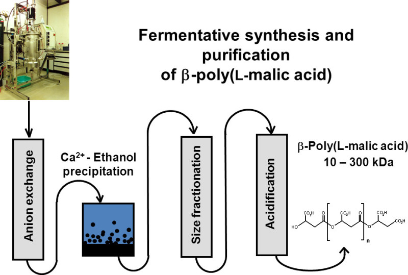 Figure 2. Production and purification of polymalic acid from cultured micro plasmodia of Physarum polycephalum (adapted from Lee et al.9). Polymalic acid (PMLA) is produced from plasmodia of Physarum polycephalum in a bioreactor and collected from the culture supernatant by binding on anion exchanger (DEAE). The eluent is ethanol-precipitated as calcium salt. Solutions of the calcium salt are Mw-fractionated over Sephadex-G25. PMLA containing fractions are converted from the Ca-salt into the acid over Amberlite IR-120H+. The free PMLA is finally lyophilized to yield dry white powder.
Chemical synthesis of the lead nano conjugate
P/mPEG 5000 (5%)/LOEt(40%)/AON(2.5%)/Herceptin(0.2%)/anti-MsTfRmAb(0.2%)
The schematic structures of the lead nano drug and of two precursor nano conjugates are shown in Figure 3. The lead contains all constituents, while the precursors were designed to lack either AONHER2 or the antibody Herceptin. Besides AON and mAbs, the compounds contained polyethylene glycol, mPEG5000, to minimize binding to plasma proteins, clearance through the reticulo-endothelial system (RES), and degradation by enzymatic cleavage. The antisense sequence 5’-CAT-GGT-GCT-CAC-TGC-GGC-TCC-GGC-3’ was complementary to Her2 mRNA and has been carefully tested in vitro on several HER2-expressing cell lines to obtain high specificity towards blocking HER2 synthesis and not showing off-target effects.
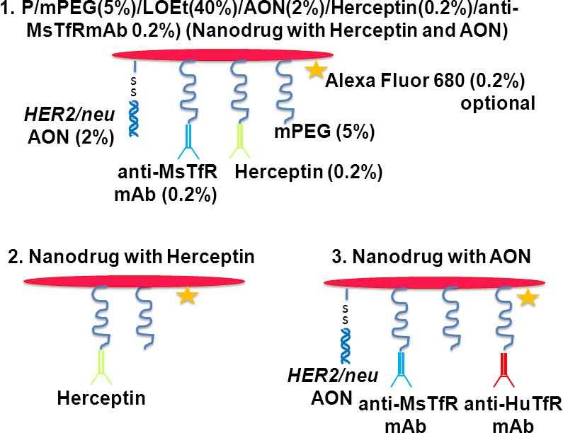 Figure 3. Nano conjugates to treat human HER2-positive breast cancer. Leading nano drug (top structure) contains two drugs (Herceptin, AONHER2). Versions 2 and 3 contain only one of the drugs. Uptake of 1 and 2 into human HER2-overexpressing tumor cells is managed by the binding of Herceptin. Nano conjugate 3, which does not contain Herceptin, received anti-HuTfRmAb for endosome uptake by the human cells. The conjugation with Alexa Fluor 680 was optional for imaging purposes. The red bar represents the nano conjugate platform PMLA/LOEt(40%). % indicates the fraction of malic acid residues consumed in binding of the indicated ligand (total amount of malic acid residues in the nanodrug = 100%).
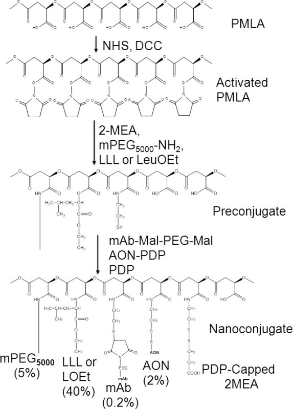 Figure 4. Chemical synthesis of nano conjugates P/LOEt/AONHER2/anti-MsTfR/Herceptin. From top to bottom: Synthesis of PMLA-N-hydroxy succinimidyl ester (PMLA-NHS) of pendant carboxylates (chemical activation). Replacement of NHS by amide formation with 2-mercapto-1-aminoethan (2-MEA), methyl-PEG5000-amine (mPEG5000-NH2), trileucine (LLL) or leucine ethylester (LOEt). This “preconjugate” attaches mAb-Mal-PEG-Mal by thioether formation and AON by formation of cleavable disulfide bonds. Leftover 2-MEA is capped forming amidoethyl-dithio-propionic acid. Only attachment of a single mAb is shown. Multiple attachments are managed by using mixtures of mAbs. Percentage % denotes fractions of carboxylates bound to various ligands (100% for free PMLA).
The nano conjugates were synthesized as outlined in Figure 4. The calculated molecular weight of the lead conjugate was 719,000. The overall yield of the synthesis was 45 ± 5% with regard to the malic acid content. On average, of the 862 malyl units (=100%) of the 100 kDa PMLA platform, 40% carried LOEt, 5% mPEG, 2% AON 2%, 0.2% Herceptin and 0.2% anti-MsTfR mAb. The amount for each antibody corresponded to an average of 1.7 molecules per molecule of PMLA platform. The % designed and the % verified by group analysis were the same within ±5%. An example is the case given in Table 1 for the calculation of the experimental content of malic acid, AON, mAb and mPEG5000 and in Table 2 shows the comparison with the content by design. The high purity according to the criteria under section 3 was achieved on the basis of high reaction yields and efficient separation by size exclusion (e.g. free mAb and mAb-nanoconjugate are separated by 1 min on sec-HPLC), and selective solubility in solvents. The activity of monoclonal antibodies was shown to be retained throughout nano drug synthesis, and it was confirmed that the two kinds of antibodies were assembled on the same polymer platform. The result of atypical ELISA experiment is shown in Figure 5. In general, the assays for quantitative group analysis for malic acid, AON, protein, PEG and for ELISA were robust, yielding reproducible results when conducted by different personnel and instrumentation not only in the case of the reported synthesis, but also when used for analysis of other synthesized nanoconjugates.
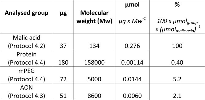 Table 1. Calculation of nanodrug composition with reference to malic acid content.
 Table 2. Comparison of experimental nano drug composition with designed composition.
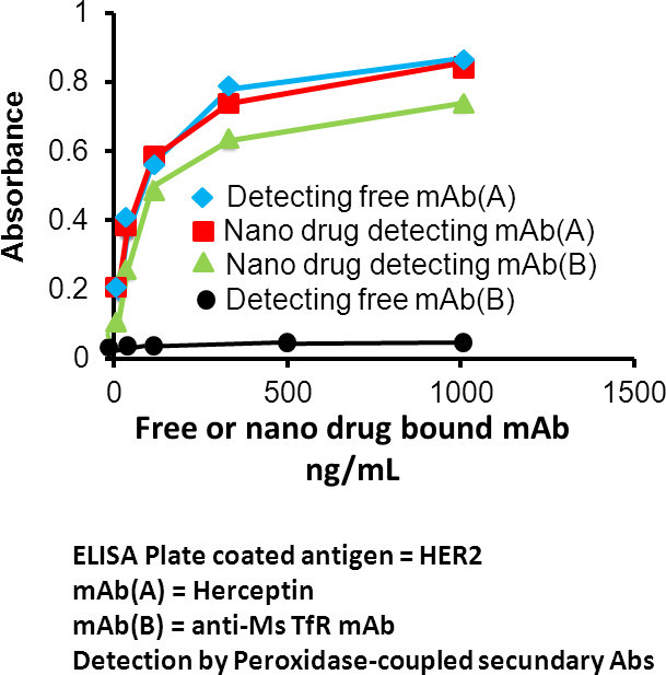 Figure 5. ELISA for measurement of antibody affinity after chemical synthesis, and for demonstration of multiple binding of different antibodies. The nano drug contains antibody mAb(A) and antibody mAb(B) against antigens A and B, respectively. ELISA plates are coated with antigen(A). Free mAb(A), free mAb(B), and nano drug are applied to ELISA plates. After washing, the antibodies are tested with secondary peroxidase coupled antibodies specific for either mAb(A) or mAb(B), while only mAb(A) is bound to antigen(A). It is seen that free and nano drug conjugated with mAb(A), and indirectly conjugated mAb(B) of the same nano drug molecule, but not free mAb(B), are retained on the plate. The concentration dependence shows comparable binding affinities for free and conjugated mAb(A), indicating that the chemical conjugation did not affect antibody binding activity. Moreover, the co-ligation of mAb(B) with mAb(A) on the same physical entity (nano platform) results in antibody mAb(B) detection. The experimental results shown here refer to anti-human TfRmAb (A) and anti-mouse TfRmAb (B). Similar results have been shown for other antibody couples tested, such as anti-MsTfR mAb and anti-HER2 mAb (Herceptin).
The physicochemical investigation indicated hydrodynamic diameter 22.1 ± 2.3 nm for P/mPEG(5%)/LOEt(40%)/AON(2%)/Herceptin(0.2%)/anti-MsTfRmAb(0.2%) (lead, the two-drug version), 20.1 ± 2.4 nm for P/mPEG(5%)/LOEt(40%)/AON(2%)/anti-MsTfRmAb(0.2%)/anti-HuTfRmAb (0.2%) (the AON drug version), and 15.1 ± 1.2 nm for P/mPEG(5%)/LOEt(40%)/Herceptin(0.2%) (the Herceptin drug version). Zeta-potentials at pH 7.5 were in this order -5.2 ± 0.4 mV, -5.7 ± 0.6 mV, and -4.1 ± 0.4 mV. It should be mentioned that the measured hydrodynamic diameter of nano conjugates did not follow additivity of the diameters measured for free components.
Delivery of nano drug through the tumor cell membrane and release into the cytoplasm after 0 hr and 3hr
The delivery of nano drug through the cell membrane by endosome uptake is shown in Figure 6 after 0 hr and 3 hr. Fluorescence labels are attached to the nano drug platform (Alexa Fluor 680, green) and to AON (Lissamine, red). Their superposition is shown in panels D&L and together with the fluorescence of labeled endosomes (blue) in panels G&O. Pearson’s correlation coefficients (R(r) were calculated from the images regarding co-localization of platform/endosomes, AON/endosomes, platform/AON for 0 hr and 3 hr. The 3 hr image indicated release from endosomes into the cytoplasm and dissociation of AON from the nano drug by glutathione-dependent disulfide cleavage in the cytoplasm.
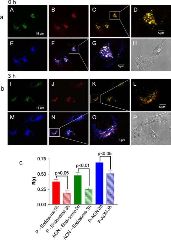 Figure 6. Confocal microscopy for nano conjugate uptake by target cells (adapted from Ding et al.11). Cells were incubated for 30 min at 37 °C with nano drug which had been double labeled with Alexa Fluor 680 (green) at the polymer platform and with Lissamine (red) attached to AON. In addition, endosomes were stained with FM1- 43 (blue). Localization is shown after 0 hr (A) and 3 hr (B) incubation. Panels A,a-b, & B,i-j show staining by the nano conjugate fluorophores alone, and their co-localization in A,c-d & B,k-l. Additionally, staining of endosomes is shown in panels A,e & B,m and the colocalization of nano conjugate and endosomesin panels A,f-g & B,n-o. Panels A,h & B,p show phase contrast for respective panels. (C) Pearson’s correlation coefficients R(r) for the colocalization of platform-endosomes, AON-endosomes, platform-AON calculated for 0 hr and 3 hr. Please click here to view a larger version of this figure.
Inhibition of human breast cancer in vitro and in vivo
In vitro growth inhibition of HER2 overexpressing cell line BT-474 in comparison with low expressing cell line MDA-MB-231 is shown in Figure 7. Degree of inhibition is 50% and 30%, respectively, by the lead nano conjugate P/mPEG/LOEt/AONHER2/Herceptin/anti-MsTfRmAb. Inhibition by the other compounds is less but higher for BT-474 than for MDA-MB-231 cells. BT-474 cell line was chosen for the treatment of xenogeneic breast cancer mouse.
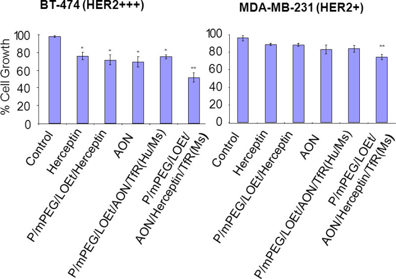 Figure 7. In vitro growth inhibition of HER2 overexpressing BT-474 breast cancer cells and low expressing MDA-MB-231 cells (adapted from Inoue et al.12). Comparison of growth inhibition for HER2 overexpressing BT-474 breast cancer cell line (left) and of HER2 low expressing breast cancer cell line MDA-MB-231 (right). TfR(s) denotes anti-MsTfRmAb and TfR(Hu/Ms) denotes anti-HuTfRmAb & anti-MsTfRmAb. The lead nano drug, P/mPEG/LOEt/AONHER2/Herceptin/TfR(Ms), and nano drugs devoid of either Herceptin or AONHER2 are compared with PBS, Herceptin and AONHER2. In the absence of Herceptin, anti-MsTfRmAb was conjugated to provide endosome uptake by the tumor cells. Concentrations were 40 μg/ml with regard to antibodies, 4 μM with regard to AONHER2, 4 μM endoporter (application of AON). Significance denoted by *, P<0.05: **, P<0.02: ***, P< 0.003 compared with PBS.
The results of in vivo treatment in Figure 8 are presented for mice bearing BT-474 Her2 overexpressing human breast cancer. Growth was inhibited >95% by the lead nanodrug containing Herceptin and HER2-specific AON in comparison to controls treated only with PBS (Figure 8). Inhibition was 60% or less with Herceptin alone, P/mPEG/LOEt/Herceptin or P/mPEG/LOEt/AON/anti-MsTfRmAb/anti-HuTfRmAb. Western blot analysis of tumor extracts showed inhibition of both HER2 synthesis and Akt phosphorylation, but no change of total Akt and housekeeping enzyme GAPDH. PARP was cleaved in correspondence with an increased level of apoptosis. Pictures of tumor after treatment, and tissue sections stained with H&E illustrating tumor regression are shown in Figure 9.
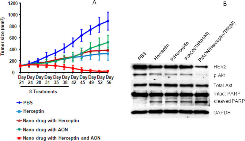 Figure 8. Tumor size and proteins during treatment with lead and precursor nano drugs (adapted from Inoue et al.12). A. Tumor size for the various treatments (day 21 to day 42, totally 8 injections). Bars indicate standard deviations from the mean. The lead conjugated with anti-Her2 AON and Herceptin was superior to either Herceptin alone or single drug containing nano conjugates. B. Effect of the various treatments on the expression of HER2, phosphorylated Akt, total Akt, PARB, cleaved PARB and GAPDH are shown by western blotting. Please click here to view a larger version of this figure.
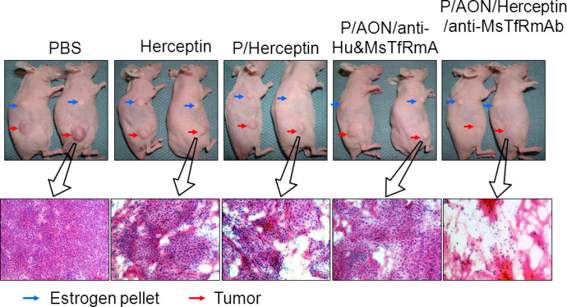 Figure 9. Treatment of HER2-positive BT-474 human breast cancer on nude mice. Tumor regression and necrosis/apoptosis in tissue sections (adapted from Ionue et al.12). Upper: Shrinkage of the tumor (red arrows). Indication of the estrogen pellet to support tumor growth (blue arrow). Lower: Hematoxilin & Eosine (H&E) staining of tumor sections. Proliferating tumor is shown by the dense packing of tumor cells. Necrotic tissue is seen where tumor cells have been eliminated. Please click here to view a larger version of this figure.
Discussion
The experimental route for the preparation of nano drugs from a biodegradable natural polymer is presented that can be used in the synthesis of personalized medicine. The description starts with the controlled production and purification of polymalic acid, which is a versatile platform for nano drug synthesis. Using reproducible techniques, the polymer is obtained with high molecular weight and in extreme purity suitable for pharmaceutical syntheses. The synthesis is described for a nano drug that is shown to efficiently treat HER2-positive breast cancer. The description can be translated into syntheses of most other nano drugs for the treatment of cancer. Targeting involves antibodies such as Herceptin that bind a tumor specific antigen such as HER2 protein or any other tumor marker that is efficiently internalized. The nano drug delivers a selection of antisense oligonucleotides and chemotherapeutics that will efficiently inhibit growth of the cancer under treatment. In the example, Herceptin binding to HER2 and specific antisense oligonucleotide annealing with HER2-coding mRNA resulted in sustained blocking of HER2-signaling and severe reduction of HER2-positive breast cancer. Based on the underlying principle of tumor targeting and inhibition of gene expression by antisense oligonucleotides we have to-date synthesized several other nano drugs and successfully inhibited preclinical human glioblastoma and triple-negative breast cancer11,14-18.
The synthetic work starts with the preparation of highly purified nano drug platform, which is polymalic acid from the culture supernatant of Physarum polycephalum (a species of the “slime mold” family). The preparation emphasizes high molecular weights of the polymer, in principle allowing attachment of numerous antibodies, peptides, oligonucleotides and other molecules functioning in active drug delivery and tumor growth inhibition. Following the controlled culturing and purification, reproducible quality of polymer in predictable yields has been produced. The polymer is stored under convenient conditions for any time.
The synthesis of PMLA nano conjugates beginning with the chemical activation of the polymer-pendant carboxylates is carried out in a few synthetic steps. In between steps, synthesis can be optionally placed on hold allowing preparation of arbitrary amounts of intermediates and thus can be used in up scaling. The progress of synthesis is followed by TLC and sec-HPLC, and both the composition and activity of the nano drug is controlled by group-specific quantitative chemical assays, ELISA, and a variety of physical measurements. Our experience is that these syntheses have been progressed smoothly and reproducibly with excellent yields and purity. By choosing the specificity of antibody and antisense oligonucleotides any variant of nano drug is favorably synthesized as needed in personalized medicine.
The modalities of the successful treatment of human HER2-positive breast cancer are valid representatives for preparation of mouse cancer models, application of nano drugs, imaging, and analysis of tumor growth. The results of in vitro viability tests are useful in selecting the cell line and the leading drug to be used in the animal experiment. In vivo Xenogen imaging validates that the nano drug is indeed delivered into the tumor. Results of western blotting reveals whether the level of certain proteins responded in the predicted fashion during cancer treatment. Measurement of tumor size informs about the success of the treatment, i.e. about inhibition, recession or regression. The described example reflects our experience with other polymalic acid-based nano drugs indicating a high degree of predictability, reproducibility and absence of notable toxicity. Recent results on toxicity and efficacy of multiple targeted polymalic acid conjugates for triple-negative breast cancer treatment are in strong support of this notion18. A key feature is that our nano drug is able to penetrate bio-barriers: the endothelial barrier by extravasation into the tumor interstitial, the tumor cell membrane by endosome uptake, and endosome membrane disruption by action of the leucine ethylester or trileucine groups. Extravasation and endosome uptake are reliably accomplished by the specific antibodies attached to the nano drug. Anti-mouse transferrin receptor antibody mediates efficient influx into the tumor by transcytosis, probably because the receptor is overexpressed on most tumor vasculature. A different antibody attached on the same nano conjugate molecule functions in specifically directing the nano drug into the recipient tumor cell. The presence of both antibodies, the one for transcytosis and the other one for endosome uptake, is essential for optimal functioning. Quantitative analysis of malic acid and antibody is highly recommended in order to control optimal composition of the nano drug. Finally it should be noted that the all-in-one covalent nano drug delivers drug in chemically attached form en route through the host vasculature into the recipient tumor cell. Chemical attachment renders most drugs inactive (prodrugs) until they are reconstituted as free drugs by cleavage from the nano conjugate platform at the targeted site. This is important because the reactivation modality provides a high degree of safety during delivery and a minimal chance to evoke harmful side effects. Versatility, efficacy and safety are indispensable attributes of good personalized medicine.
Disclosures
The authors Julia Y. Ljubimova, Keith L. Black, and Eggehard Holler are shareholders of Arrogene Technology Inc.
Acknowledgments
We greatly acknowledge financial support by NIH R01 CA123495, U01 CA151815, R01 CA136841, grants from the Department of Neurosurgery at Cedars-Cedars Medical Center and Arrogene Technology Inc.
References
- Koboldt DC, Fulton RS, McLellan MD. Comprehensive molecular portraits of human breast tumors. Cancer Genome Atlas Network. Nature. 2012;490:61–70. doi: 10.1038/nature11412. [DOI] [PMC free article] [PubMed] [Google Scholar]
- Kwong LN, Costello JC, et al. Oncogenic NRAS signaling differentially regulates survival and proliferation in melanoma. Nat. Med. 2012;18:1503–1510. doi: 10.1038/nm.2941. [DOI] [PMC free article] [PubMed] [Google Scholar]
- Gerlinger M, Rowan AJ, et al. Gerlinger M, Rowan AJ, editors. Intratumor heterogeneity and branched evolution revealed by multiregion sequencing. N. Engl. J. Med. 2012. pp. 883–892. [DOI] [PMC free article] [PubMed]
- Yap TA, Workman P. Exploiting the cancer genome: strategies for the discovery and clinical development of targeted molecular therapies. Annu. Rev. Pharmacol. Toxicol. 2012;52:549–573. doi: 10.1146/annurev-pharmtox-010611-134532. [DOI] [PubMed] [Google Scholar]
- Chatterjee S. Cancer Biomarkers: Knowing the Present and Predicting the Future. Future Oncol. 2005;1:37–50. doi: 10.1517/14796694.1.1.37. [DOI] [PubMed] [Google Scholar]
- Suh KS, Sarojini S, Youssif M, et al. Tissue Banking, Bioinformatics, and Electronic Medical Records: The Front-End Requirements for Personalized Medicine. J. Oncology. 2013. [DOI] [PMC free article] [PubMed]
- Ljubimova JY, Holler E. Biocompatible nanopolymers: the next generation of breast cancer treatment. Future Medicine, Nanomedicine. 2012;7:1–4. doi: 10.2217/nnm.12.115. [DOI] [PMC free article] [PubMed] [Google Scholar]
- Lee B-S, Vert M, Holler E. In: Water-soluble aiphatic polyesters: poly(malic acid)s. Biopolymers 3a. Steinbüchel A, editor. Weinheim (Bergstrasse): Wiley VCH; 2002. pp. 75–103. [Google Scholar]
- Lee B-S, Fujita M, et al. Polycefin, a new prototype of multifunctional nanoconjugate based on poly(beta-L-malic acid) for drug delivery. Bioconjugate Chem. 2006;17:317–326. doi: 10.1021/bc0502457. [DOI] [PMC free article] [PubMed] [Google Scholar]
- Nag A, Mitra G, et al. A colorimetric assay for estimation of polyethylene glycol and polyethylene glycolated protein using ammonium ferrothiocyanate. Analytical Biochem. 1996;237:224–231. doi: 10.1006/abio.1996.0233. [DOI] [PubMed] [Google Scholar]
- Ding H, Inoue S, et al. Inhibition of brain tumor growth by intravenous poly(beta-L-malic) acid nanobioconjugate with pH-dependent drug release. Proc. Natl. Acad. Sci. USA. 2010;107:18143–18148. doi: 10.1073/pnas.1003919107. [DOI] [PMC free article] [PubMed] [Google Scholar]
- Inoue S, Ding H, et al. Nanobioconjugate inhibition of HER2/neu signaling and synthesis provides efficient mouse breast cancer treatment. Cancer Research. 2011;71:1454–1464. doi: 10.1158/0008-5472.CAN-10-3093. [DOI] [PMC free article] [PubMed] [Google Scholar]
- Maeda H, Sawa T, Konno T. Mechanism of tumor-targeted delivery of macromolecular drugs, including the EPR effect in solid tumor and clinical overview of the prototype polymeric drug SMANS. J. Control. Release. 2001;74:47–61. doi: 10.1016/s0168-3659(01)00309-1. [DOI] [PubMed] [Google Scholar]
- Ljubimova JY, Fujita M, Ljubimov YJ, et al. Poly(malic acid) nanoconjugates containing various antibodies and oligonucleotides for multitargeting drug delivery. Nanomed. 2008;3:247–265. doi: 10.2217/17435889.3.2.247. [DOI] [PMC free article] [PubMed] [Google Scholar]
- Inoue S, Patil R, Portilla-Arias J, et al. Novel nanobioconjugate inhibiting EGFR expression in triple negative breast cancer. PLoS One. 2012;7:1–9. doi: 10.1371/journal.pone.0031070. [DOI] [PMC free article] [PubMed] [Google Scholar]
- Patil R, Portilla-Arias J, Ding H, et al. Cellular Delivery of Doxorubicin via pH-Controlled Hydrazone Linkage Using Multifunctional Nano Vehicle Based on Poly(β-L-malic Acid) Int. J. Mol. Sci. 2012;13:11681–11693. doi: 10.3390/ijms130911681. [DOI] [PMC free article] [PubMed] [Google Scholar]
- Patil R, Portilla-Arias J, Ding H, et al. Temozolomide delivery to tumor cells by a multifunctional nano vehicle based on poly(β-L-malic acid) Pharm. Res. 2010;27:2317–2329. doi: 10.1007/s11095-010-0091-0. [DOI] [PMC free article] [PubMed] [Google Scholar]
- Ljubimova JY, Portilla-Arias J, Patil R, et al. Toxicity and efficacy evaluation of multiple targeted polymalic acid conjugates for triple-negative breast cancer treatment. J. Drug Target. 2013;21:956–967. doi: 10.3109/1061186X.2013.837470. [DOI] [PMC free article] [PubMed] [Google Scholar]


