Abstract
Spiders of the Loxosceles species can cause dermonecrosis and acute kidney injury (AKI). Hemolysis, rhabdomyolysis and direct toxin-mediated renal damage have been postulated. There are very few reports of Loxoscelism from India. We report a case of AKI, hemolysis and a “gravitational” pattern of ulceration following the bite of the brown recluse spider (Loxosceles spp).
Keywords: Dermonecrosis, renal failure, spider bite
Introduction
Spider bite is endemic in parts of North America, Mexico, tropical belt of Africa and Europe and can cause serious systemic manifestations.[1,2,3,4,5] Loxosceles spiders belong to the family Loxoscelidae/Sicariidae. Of the 13 species of Loxosceles identified, at least 5 have been associated with necrotic arachnidism. Loxosceles reclusa, or the brown recluse spider, is the spider most commonly responsible for this injury.[6] Dermonecrotic arachnidism refers to the local skin and tissue injury as a result of spider-bite. Loxoscelism is the term used to describe the systemic clinical syndrome caused by envenomation from the brown spiders. Cutaneous manifestations occur in around 80% cases around the site of bite, predominantly the lower limbs.[3] The initial cutaneous manifestation is that of an erythematous halo with edema around the bite site. The erythematous margin around the site continues to enlarge peripherally, secondary to gravitational spread of the venom into the tissues. This gradually gives way to vesicles and finally a dark eschar or a necrotic ulcer.[7] Mild systemic effects such as fever, malaise, pruritus and exanthema are common, whereas intravascular hemolysis and coagulation, sometimes accompanied by thrombocytopenia and renal failure, occur in approximately 16% of those who receive the bite.[8] Rarely (<1% of the cases of suspected L. reclusa bites with a higher incidence in South American loxoscelism), recluse venom may cause hemolysis, disseminated intravascular coagulation, which can lead to serious injury and possibly death.[9] Although India is a home to a diverse array of arachnids, according to the latest updated list of spider species found in India,[6] Loxosceles rufescens is the only member of the Loxosceles genus described in India. Systemic envenomation (especially renal failure) from Loxosceles bite has been rarely described from India.[10]
Case Report
A 23-year-old male was bitten by a spider in the dorsal aspect of his right forearm. He developed a painful blister with red margins in the distal forearm, around the site of bite, which subsequently turned into a necrotic lesion. After a few hours, he developed nausea, vomiting and malaise. On the next day, he developed oliguria and brownish discoloration of urine. He had excruciating pain and erythematous swelling of the right hand and around the bite site. Initial blood urea was 133 mg/dl, creatinine 3.4 mg/dl, serum Na+ 131.6 meq/L and K+ 4.67 meq/L. Hemoglobin was 6.2 g/dl, total leucocyte count 12,000/cmm (N84L12E2M2B0) with normal platelet counts. He was referred to our hospital for worsening renal function and deteriorating skin lesions.
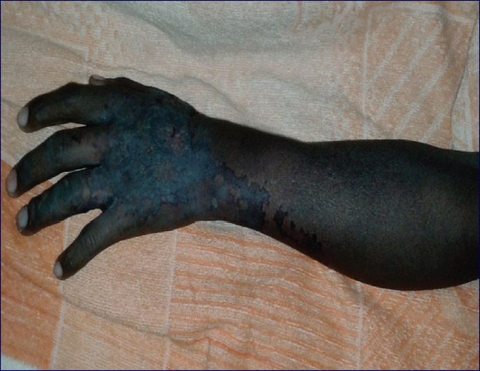
On arrival 5 days after the bite, he was conscious and alert. There was blackish discoloration of the right distal forearm and hands and a “gravitational” pattern of involvement from the bite site down into the hands [Figure 1]. Radial pulse was palpable and he had preserved sensation in the fingers of his right hand. He had a normal heart rate while blood pressure was 160/90 mm of Hg. Arterial Doppler of the right forearm revealed normal flow up to the radial arteries. Physical examination was normal otherwise. Though the spider was not brought, it was identified to be the brown recluse spider (Loxosceles spp) based on the description and on showing representative pictures.
Figure 1.

“Gravitational pattern” of dermonecrosis following the bite of brown recluse spider (Loxosceles spp), day 5
Investigations revealed hemoglobin of 4.3 gm/dl with normal leucocyte and platelet counts with a reticulocyte count of 9.1%. Urine showed 1 + proteinuria with no red blood cells or cast. He had advanced renal failure with normal electrolytes, serum urea 206 mg/dl and creatinine 6.6 mg/dl. Liver function tests were normal with mild unconjugated hyperbilirubinemia (total bilirubin was 2.4 mg/dl with unconjugated bilirubin of 1.9 mg/l). Serum creatine phosphokinase (CPK) was 90 U/l, lactate dehydrogenase (LDH) 588 U/l (normal range: <200 U/l) and serum haptoglobin was 18 mg/dl (normal). Serum and urine myoglobin were normal.
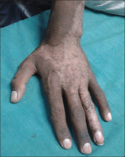
He received hemodialysis for 2 weeks before renal function eventually recovered. The hand ulcer also healed gradually with conservative management. He was discharged after 3 weeks with a daily urine output of 1.8 L, serum creatinine of 1.8 mg/dl, hemoglobin 9.2 gm/dl and LDH of 180 mg/dl. He was seen 4 weeks after discharge. Renal function had normalized (serum creatinine 0.9 mg/dl, normal urine microscopy) and ulcer had healed with desquamation of the involved skin [Figure 2].
Figure 2.

Healed skin lesion of the same patient at 6 weeks
Discussion
Acute intravascular hemolysis is a hallmark of loxoscelism.[11] In our case, intravascular hemolysis was evidenced by low hemoglobin level, elevated reticulocyte counts with elevated unconjugated bilirubin, high serum LDH and low serum haptoglobin levels. The likely etiology of renal failure in our case is hemolysis leading to acute tubular injury. There was no evidence of myonecrosis/rhabdomyolysis as evidenced by normal serum CPK and urine myoglobin levels. Although the spider was not brought by the patient for identification, the features were typical of loxoscelism, especially the “gravitational” pattern of dermonecrosis. Most of the case series of Loxoscelism have documented dermonecrosis with a few cases of hemolysis and rare cases of renal injury.[12] However, there have been very few reports from the Indian sub-continent.
Loxoscelism can manifest as necrotic, cutaneous or viscerocutaneous involvement.[2] No specific anti-venom is available for Loxoscelism. Treatment depends on the severity of the disease. Wound debridement, elevation, application of ice and immobilization of the affected area may help ameliorate the extent of cutaneous damage. Tetanus prophylaxis is warranted.[13] Dapsone has been recommended by some authorities to treat dermonecrosis on account of its leucocyte inhibiting properties. Antibiotics are recommended only for documented infection. Patients exhibiting signs of systemic toxicity should be admitted and evaluated for evidence of coagulopathy and renal failure. Urinalysis can provide early evidence of systemic involvement (e.g. hemoglobinuria, myoglobinuria). Hemodialysis support helps to tide over the renal injury.[14]
Spiders of the genus Loxosceles can cause complement-dependent intravascular hemolysis.[8] Glycophorins protect the erythrocytes against complement-mediated hemolysis. It has been found that sphingomyelinase activity of the Loxosceles toxin induces activation of an endogenous metalloproteinase, which then cleaves glycophorins thus rendering it susceptible to complement mediated lysis. Tambourgi et al., in their study observed the transfer of complement-dependent hemolysis to other cells, suggesting that the Loxosceles toxins can act on multiple cells.[8] This observation can explain the relatively significant extent of hemolysis observed in patients with inoculation of small amounts of the toxin (max 30 μg). Loxoscelism causes necrotic dermatologic injury through a unique enzyme; sphingomyelinase D. Loxosceles toxin has also been shown to have hyaluronidase, alkaline phosphatase and esterase activity. These cause degradation of the extracellular matrix and contribute to the spread of the toxin in tissue compartment. This may account for the ‘gravitational’ pattern of dermonecrosis. The dermatohistopathology of Loxosceles bites include dermal edema, thickening of blood vessel endothelium, leukocyte infiltration, intravascular coagulation, vasodilatation, destruction of blood vessel walls and hemorrhage.[15] Renal injury in loxoscelism has been attributed to pigmentary nephropathy due to hemoglobin or myoglobin, secondary to hemolysis or rhabdomyolysis.[16]
India, belonging to the tropical belt, is home to several insects. However, in the absence of effective reporting systems, many fatal or near-fatal envenomations go unreported. This is reflected by the paucity of case reports of Arachnid bites. Careful clinical and entomological studies should be done to look into this neglected disease entity. It should be borne in mind that a case presenting with acute dermal inflammation or ulceration in a “gravitational” pattern, along with features of hemolysis or rhabdomyolysis or, in rare instances, acute kidney injury could be due to Loxoscelism and it is a close mimicker of hemotoxic snake bite.
Footnotes
Source of Support: Nil
Conflict of Interest: None declared.
References
- 1.Gendron BP. Loxosceles reclusa envenomation. Am J Emerg Med. 1990;8:51–4. doi: 10.1016/0735-6757(90)90297-d. [DOI] [PubMed] [Google Scholar]
- 2.Futrell JM. Loxoscelism. Am J Med Sci. 1992;304:261–7. doi: 10.1097/00000441-199210000-00008. [DOI] [PubMed] [Google Scholar]
- 3.Schenone H, Saavedra T, Rojas A, Villarroel F. Loxoscelism in Chile. Epidemiologic, clinical and experimental studies. Rev Inst Med Trop Sao Paulo. 1989;31:403–15. doi: 10.1590/s0036-46651989000600007. [DOI] [PubMed] [Google Scholar]
- 4.Sezerino UM, Zannin M, Coelho LK, Gonçalves J, Júnior, Grando M, Mattosinho SG, et al. A clinical and epidemiological study of Loxosceles spider envenoming in Santa Catarina, Brazil. Trans R Soc Trop Med Hyg. 1998;92:546–8. doi: 10.1016/s0035-9203(98)90909-9. [DOI] [PubMed] [Google Scholar]
- 5.White J, Cardoso J, Fan H. Clinical toxinology of spider bites. In: Meier J, White J, editors. Handbook of Clinical Toxinology of Animal Venoms and Poisons. Boca Raton FL: CRC Press; 1995. p. 259. [Google Scholar]
- 6.Manju S, Molur S, Biswas B. Indian spiders (Arachnida: Araneae): updated checklist 2005. Zoos’ Print J. 2005;20:1999–2049. [Google Scholar]
- 7.Dyachenko P, Ziv M, Rozenman D. Epidemiological and clinical manifestations of patients hospitalized with brown recluse spider bite. J Eur Acad Dermatol Venereol. 2006;20:1121–5. doi: 10.1111/j.1468-3083.2006.01749.x. [DOI] [PubMed] [Google Scholar]
- 8.Tambourgi DV, Morgan BP, de Andrade RM, Magnoli FC, van Den Berg CW. Loxosceles intermedia spider envenomation induces activation of an endogenous metalloproteinase, resulting in cleavage of glycophorins from the erythrocyte surface and facilitating complement-mediated lysis. Blood. 2000;95:683–91. [PubMed] [Google Scholar]
- 9.Anderson PC. Missouri brown recluse spider: A review and update. Mo Med. 1998;95:318–22. [PubMed] [Google Scholar]
- 10.Golay V, Desai A, Hossain A, Roychowdhary A, Pandey R. Acute kidney injury with pigment nephropathy following spider bite: A rarely reported entity in India. Ren Fail. 2013;35:538–40. doi: 10.3109/0886022X.2013.768936. [DOI] [PubMed] [Google Scholar]
- 11.McDade J, Aygun B, Ware RE. Brown recluse spider (Loxosceles reclusa) envenomation leading to acute hemolytic anemia in six adolescents. J Pediatr. 2010;156:155–7. doi: 10.1016/j.jpeds.2009.07.021. [DOI] [PMC free article] [PubMed] [Google Scholar]
- 12.Bronstein AC, Spyker DA, Cantilena LR, Jr, Green JL, Rumack BH, Dart RC. 2010 Annual Report of the American Association of Poison Control Centers’ National Poison Data System (NPDS): 28th Annual Report. Clin Toxicol (Phila) 2011;49:910–41. doi: 10.3109/15563650.2011.635149. [DOI] [PubMed] [Google Scholar]
- 13.Swanson DL, Vetter RS. Loxoscelism. Clin Dermatol. 2006;24:213–21. doi: 10.1016/j.clindermatol.2005.11.006. [DOI] [PubMed] [Google Scholar]
- 14.Rees R, Campbell D, Rieger E, King LE. The diagnosis and treatment of brown recluse spider bites. Ann Emerg Med. 1987;16:945–9. doi: 10.1016/s0196-0644(87)80738-2. [DOI] [PubMed] [Google Scholar]
- 15.Pereira N, Kalapothakis E, Vasconcelos A, Chatzaki M, Campos L, Vieira F, et al. Histopathological characterization of experimentally induced cutaneous loxoscelism in rabbits inoculated with Loxosceles similis venom. J Venom Anim Toxins Incl Trop Dis. 2012;18:277–86. [Google Scholar]
- 16.Wright SW, Wrenn KD, Murray L, Seger D. Clinical presentation and outcome of brown recluse spider bite. Ann Emerg Med. 1997;30:28–32. doi: 10.1016/s0196-0644(97)70106-9. [DOI] [PubMed] [Google Scholar]


