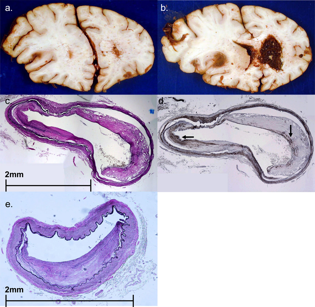
FIGURE 8.
Case #4. A,B. Coronal sections of fixed brain show a large left MCA territory subacute infarct, with associated edema and left to right shift of midline structures. There is also extensive intraventricular hemorrhage, especially in the right lateral ventricle. C,D. Cross sections of left MCA (C stained with EVG, D stained immunohistochemically using primary antibody to SMA) show relatively mild atheroma with a smooth muscle cell cap (arrows in D). At another level (panel E) the MCA shows significant atheroma, with estimated 55–60% stenosis of the lumen. None of the lumina shows a thrombo-embolus. (Panel E from a section stained with EVG).

