Abstract
Exposure to viruses and bacteria results in lung infections and places a significant burden on public health. The innate immune system is an early warning system that recognizes viruses and bacteria, which results in the rapid production of inflammatory mediators such as cytokines and chemokines and the pulmonary recruitment of leukocytes. When leukocytes emigrate from the systemic circulation through the extracellular matrix in response to lung infection they encounter proteoglycans, which consist of a core protein and their associated glycosaminoglycans. In this review, we discuss how proteoglycans serve to modify the pulmonary inflammatory response and leukocyte migration through a number of different mechanisms including: 1) The ability of soluble proteoglycans or fragments of glycosaminoglycans to activate Toll-like receptor signaling pathways; 2) The binding and sequestration of cytokines, chemokines, and growth factors by proteoglycans; 3) the ability of proteoglycans and hyaluronan to facilitate leukocyte adhesion and sequestration; and 4) The interactions between proteoglycans and matrix metalloproteinases that alter the function of these proteases. In conclusion, proteoglycans fine-tune tissue inflammation through a number of different mechanisms. Clarification of the mechanisms whereby proteoglycans modulate the pulmonary inflammatory response will most likely lead to new therapeutic approaches to inflammatory lung disease and lung infection.
Keywords: extracellular matrix, proteoglycan, glycosaminoglycans, lungs, infection, cytokine, chemokine, matrix metalloproteinases
INTRODUCTION
The primary function of the human lung is gas exchange, which requires the largest epithelial surface in the body to be exposed to up to 20, 000 liters of air each day. This makes the lungs the primary point of entry for a variety of human health hazards, including viruses, bacteria, particulate matter, pollutants, and noxious gases. Exposure to viruses and bacteria results in lung infections, which place a much higher burden on public health than better-recognized diseases such as HIV/AIDS, cancer, coronary heart disease, and strokes (Mizgerd, 2008). To protect the lung against microbial pathogens, an intricate system of host defenses has evolved that includes physical defenses and two interactive systems that recognize and remove pathogens from the alveolar space: the innate and the adaptive immune systems. The innate immune system includes the defenses that we are born with and is the body’s early warning system. Innate immunity includes physical defenses and pathogen recognition receptors such as Toll-like receptors (TLRs), which when activated through the recognition of bacteria and viruses results in the rapid production of inflammatory mediators, complement activation and the pulmonary recruitment of leukocytes (Akira, 2009). The adaptive immune system is immunity that is acquired following exposure to a pathogen. Adaptive immunity includes the production of cytokines, chemokines, and antibodies and the activation of B and T-cells in a response that is specific to the virus or bacteria causing the infection. Whereas the innate and adaptive immune systems are often treated as separate systems, the initial innate immune response influences adaptive immunity (Hoebe et al., 2004).
Inflammatory responses as a result of tissue infection require the emigration of leukocytes from the vasculature to the infected area as part of innate and adaptive immunity. Upon extravasation into the subendothelial compartment, leukocytes encounter the extracellular matrix (ECM) which functions as a scaffold for cell adhesion, retention and most probably cell activation (Vaday et al., 2001). It may be that specific components of the ECM interact with particular mediators of the immune response to provide the intrinsic signals needed to coordinate leukocyte adhesion to the ECM and their activation. For example, proteoglycans in the ECM can interact with chemokines, growth factors, proteases, and receptors on the surface of the immune cells to influence immune cell phenotype (Parish, 2006; Taylor and Gallo, 2006). Recent work shows that ECM molecules such as fragments of hyaluronan, soluble biglycan, and lumican are able to initiate the inflammatory response through interactions with CD14, CD44, and TLRs on myeloid cells (Jiang et al., 2005; Schaefer et al., 2005; Wu et al., 2007). Once bound, the leukocytes may in turn modify the ECM in such a way as to generate pro-inflammatory ECM fragments to further drive the inflammatory response (Schor et al., 2000; Vaday and Lider, 2000). Fragments of ECM affect multiple functional properties of inflammatory and immune cells (Adair-Kirk and Senior, 2008). Since different types of infection may demand extravasation of certain immune cell types, the ECM often undergoes compositional changes, which regulate the appropriate cellular responses. Such compositional changes may enrich for specific ECM molecules that actively participate in the recruitment and activation of specific immune cell types to either promote or inhibit the inflammatory cascade (Wight, 2008).
Once thought to be simply a structural element of the ECM, evidence now shows that proteoglycans play an important role in modifying the behavior of stromal and immune cells in inflamed lungs. Proteoglycans also control leukocyte emigration in tissues through interactions with cytokines, chemokines and adhesion molecules. The goal of this review is to provide an overview of proteoglycans in the normal lungs and discuss the mechanisms whereby proteoglycans control tissue inflammation and the innate immune response to lung infection.
PROTEOGLYCANS IN NORMAL LUNGS
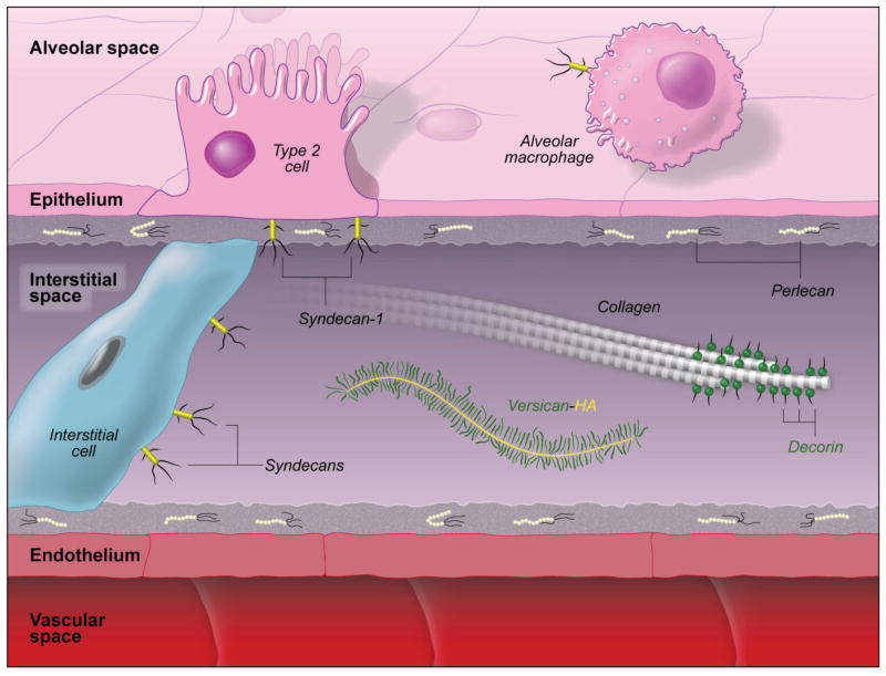
Proteoglycans are a family of charged molecules that contain a core protein and one or more covalently attached glycosaminoglycans. Proteoglycans are found in the extracellular matrix (ECM), plasma membrane of cells, and as intracellular structures in the lungs (Frevert and Wight, 2006) (Fig. 1). There are four classes of glycosaminoglycans, which includes: 1) hyaluronan, 2) chondroitin sulfate/dermatan sulfate, 3) heparan sulfate/heparin, and 4) keratan sulfate. All classes of glycosaminoglycan are found in normal lungs where the predominant glycosaminoglycan is heparan sulfate (40 to 60%) followed by chondroitin sulfate/dermatan sulfate (31%), hyaluronan (14%), and heparin (5%) (Sampson et al., 1984).
Figure 1.
Proteoglycans found in normal lungs. Perlecan, a HSPG, is found in the basal lamina of epithelial and endothelial cells. The CSPGs, versican and decorin, are found in the interstitial space of the lungs. Versican binds to the glycosaminoglycan, hyaluronan, to form high-molecular weight complexes. Decorin binds to collagen and helps stabilize the collagen-elastin network. Syndecans are membrane proteoglycans that interact with matrix proteins. This figure is not meant to represent the concentrations of the different proteoglycans in normal lungs and is meant to only show their approximate location in the alveolus. (This figure is from a chapter by Frevert, et al., (Frevert and Wight, 2006) with permission)
There are three families of ECM proteoglycans and these include the large aggregating chondroitin sulfate proteoglycans (lecticans), the small leucine-rich chondroitin sulfate proteoglycans (CSPGs), and the heparan sulfate proteoglycans (HSPGs) (Wight et al., 1991; Bosman and Stamenkovic, 2003; Kinsella et al., 2004). The lecticans, which are large aggregating CSPGs include aggrecan, neurocan, brevican, and versican. Versican, which is the predominant CSPG early in lung development decreases through gestation so that it is found at very low levels in adult lungs (Faggian et al., 2007). The small leucine rich CSPGs have relatively small core proteins (30–50 kDa) and examples in the lungs include decorin, biglycan, and lumican (Bianco et al., 1990; Dolhnikoff et al., 1998). The third family of proteoglycans in the ECM are the HSPGs, which includes perlecan, collagen XVIII, and agrin all of which are found in normal lungs (Murdoch et al., 1994; Groffen et al., 1998; Halfter et al., 1998).
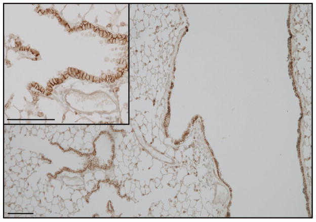
The transmembrane syndecans and the glycosylphosphoinositide-linked glypican are two HSPG families localized to cell surfaces (Bernfield et al., 1999). Syndecans are a family of four transmembrane HSPGs that interact with a number of proteins in the ECM. In adults, syndecans are expressed in specific cell and tissue patterns (Kim et al., 1994). In the lungs, syndecan-1 is expressed primarily on the basal lateral margins of airway epithelial cells and is localized to specific cells in the alveolar septa. Syndecan-2 is expressed on endothelial cells and fibroblasts and is expressed on human lung fibroblasts (David et al., 1990; Kim et al., 1994). Syndecan-3 is not found in normal lung tissue (Kim et al., 1994). Syndecan-4 is more ubiquitously expressed in many tissues including the lungs and is found on both stromal cells and leukocytes (Kim et al., 1994). Little is known about the expression of glypicans in normal lungs (Lories et al., 1992).
PROTEOGLYCANS IN INFECTED LUNGS
Changes in the composition of glycosaminoglycans and proteoglycans in the lungs have been reported in animal models and human lung disease. A consistent finding in animal models such as exposure to lipopolysaccharide and bleomycin is an increase in the synthesis of chondroitin sulfate and dermatan sulfate (Karlinsky, 1982; Blackwood et al., 1983). Gram-negative bacterial pneumonia results in the increased accumulation of chondroitin sulfate in the lungs of rabbits within 24 hours (Frevert and Sannes, 2006). Two patients with Mycobacterium tuberculosis pneumonia were reported to have an increased accumulation of the CSPG, versican, in granulomas associated with this lung infection (Bensadoun et al., 1997). Whereas very little is known about changes to CSPGs in response to lung infection, there are significant changes to versican, decorin, biglycan, and lumican in a number of acute and chronic lung diseases. There is an increased accumulation of versican and decorin in patients with pulmonary fibrosis (Bensadoun et al., 1996). In a rat model of pulmonary fibrosis the accumulation of versican, decorin, and biglycan were increased following exposure to bleomycin (Venkatesan et al., 2000). In humans with idiopathic pulmonary fibrosis, the versican-rich areas contain very little collagen whereas the decorin-rich areas have abundant collagen deposition (Bensadoun et al., 1996). Patients with ARDS,(Bensadoun et al., 1996) COPD, (Merrilees et al., 2008), and lymphangioleiomyomatosis (Merrilees et al., 2004) all have increased accumulation of versican in their lungs. In addition, the accumulation of versican, biglycan, and decorin is increased in remodeled airways of asthmatics (Bensadoun et al., 1996; Huang et al., 1999; de Medeiros Matsushita et al., 2005; Araujo et al., 2008) and an increased accumulation of versican has been observed in mice with asthma induced with IL-13 (Lowry et al., 2008). These studies suggest that significant changes to proteoglycans occur in response to lung injury and disease. Future work will need to determine what changes occur to the composition of the extracellular matrix during lung infection and how these changes will alter the inflammatory response.
PROTEOGLYCANS ROLE IN MODULATING THE INFLAMMATORY RESPONSE
Proteoglycans, once considered to be only structural components in tissue, are now recognized for the important role they play in controlling the inflammatory response (Gotte, 2003; Day and de la Motte, 2005; Mulloy and Rider, 2006; Parish, 2006; Taylor and Gallo, 2006; Coombe, 2008; Wight, 2008). Studies show that fragments of glycosaminoglycans and soluble proteoglycans initiate the inflammatory process through activation of Toll-like receptors (Taylor et al., 2004; Jiang et al., 2005; Schaefer et al., 2005; Wu et al., 2007). Proteoglycans in the ECM also interact and modify the function of chemokines, cytokines, adhesion molecules, and proteases to influence immune cell phenotype (Parish, 2006; Taylor and Gallo, 2006). Once activated, leukocytes may in turn modify the ECM in such a way as to generate pro-inflammatory ECM fragments to further drive the inflammatory response (Schor et al., 2000; Vaday and Lider, 2000). This growing evidence suggests four mechanisms whereby proteoglycans shape the inflammatory response and facilitate leukocyte migration into the airways of the lungs: 1) The direct activation of Toll-like receptor pathways; 2) The sequestration of cytokines, chemokines and growth factors in the ECM of the lungs; 3) The ability to facilitate and promote leukocyte adhesion and sequestration, and 4) Interactions between proteoglycans and matrix metalloproteinases. These four mechanisms suggest a key role for proteoglycans in the innate immune response to lung infection.
Fragments of Glycosaminoglycans and Soluble Proteoglycans Activate Toll-like Receptor Pathways
Toll-like receptors are proteins that recognize invariant structures on pathogens termed pathogen associated molecular patterns (Akira, 2009). Recognition of these invariant structures such as lipopolysaccharide (LPS) from gram-negative bacteria or single stranded RNA (e.g., PolyI:C) by TLRs results in the activation of the innate immune response. In addition to the recognition of pathogen associated molecular patterns, many studies have shown that TLRs recognize and respond to endogenous factors such as fragments of glycosaminoglycans and soluble proteoglycans. Fragments of hyaluronan and heparan sulfate cause increased pulmonary inflammation and injury, which occurs via activation of TLR2 and TLR4 (Johnson et al., 2004; Bai et al., 2005; Jiang et al., 2005; Noble and Jiang, 2006; Taylor and Gallo, 2006; Taylor et al., 2007; de la Motte et al., 2009). Whereas fragments of hyaluronan and heparan sulfate activate TLRs, intact soluble biglycan, upon release from the ECM or secretion from macrophages, activates TLR2 and TLR4 dependent signaling pathways resulting in increased inflammation (Schaefer et al., 2005; Schaefer and Iozzo, 2008). Similarly, the CSPG, versican, from Lewis lung carcinoma cells activates myeloid cells through a TLR2/TLR6 dependent process, which results in the increased expression of TNFα (Kim et al., 2009). Through a different mechanism, lumican activates the innate immune system by facilitating the presentation of LPS, a TLR4 agonist, to CD14 (Wu et al., 2007). This mechanism of action is similar to the presentation of LPS to CD14 by the LPS binding protein, which like lumican is a leucine-rich protein (Wright et al., 1990).
Therefore, proteoglycans and glycosaminoglycans initiate pulmonary inflammation through two distinct mechanisms. The first mechanism is the direct activation of TLRs by fragments of glycosaminoglycans and soluble proteoglycans, which act as endogenous “danger” signals following tissue injury. The second mechanism is the recognition and presentation of LPS to CD14 by the CSPG, lumican, which activates the innate immune system in response to bacterial pathogens. Whether soluble lumican is present in the airspaces of the lungs during lung infection will need to be determined.
Proteoglycans Bind and Sequester Cytokines, Chemokines and Growth Factors
There are numerous cytokines and chemokines that bind to glycosaminoglycans (Spillmann et al., 1998; Esko and Selleck, 2002; Parish, 2006; Taylor and Gallo, 2006). Sulfation of glycosaminoglycans provides a site to which cytokines and chemokines bind, which has several effects depending on the strength and location where proteins bind to glycosaminoglycans {Bishop, 2007 #7937). The number of inflammatory cytokines, chemokines, and growth factors that bind to proteoglycans suggest an important role for this interaction in modulating the inflammatory response in the lungs (Table 1).
Table 1.
Partial List of Cytokines, Chemokines, and Growth Factors that Bind to Glycosaminoglycans
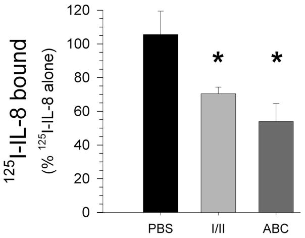
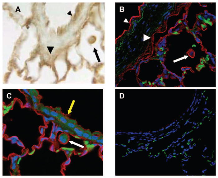
Chemokines are a family of chemotactic cytokines that bind to glycosaminoglycans. The binding of chemokines to glycosaminoglycan is a mechanism that was first suggested to promote leukocyte migration through the presentation of chemokines on the luminal surface of endothelial cell surfaces (Rot, 1992). Chemokines show considerable specificity in their interactions with glycosaminoglycans and several studies show that this interaction is critical for the recruitment of leukocytes into the peritoneum and lungs (Spillmann et al., 1998; Kuschert et al., 1999; Hirose et al., 2001; Li et al., 2002; Proudfoot et al., 2003). The binding of CXCL8 to heparin sulfate and chondroitin sulfate localizes this chemokine to specific sites in the lungs and facilitates the dimerization of CXCL8, a mechanism that increases both the amount and the retention of CXCL8 in lung tissue (Frevert et al., 2002; Frevert et al., 2003) (Figs. 2 and 3). Studies of chemokine-glycosaminoglycan interactions suggest that this is a mechanism that provides fine-tune control for chemokine function and the directional migration of leukocytes through inflamed tissue.
Figure 2.
The removal of the glycosaminoglycans heparan sulfate and chondroitin sulfate significantly decreases the low-affinity binding of 125I-CXCL8/IL-8 in lung tissue. Lung tissue was treated with heparinase I/II (I/II) and Chondroitinase ABC (ABC) prior to incubation of tissue with a trace amount of 125I-CXCL8/IL-8 [1 × 10⁻¹⁰ M] in combination with an excess of unlabeled CXCL8 [1 × 10⁻⁶ M]. The values are the mean + SEM (n = 4, *, p < 0.02). (This figure is from studies by Frevert, et al., (Frevert et al., 2003)with permission.)
Figure 3.
In situ tissue-binding assay showing the binding of rhCXCL8/IL-8 to alveolar macrophages (arrows), endothelial cells (small arrowhead) and perivascular lymphatics (large arrowheads) around a pulmonary artery (A and B) and to the cell surface of airway epithelial cells (yellow arrow) and alveolar macrophage (white arrow) (C). rhCXCL8 is stained brown (A) in the bright field image and red in the confocal images (B and C). The negative control is a serial tissue section incubated with PBS instead of rhCXCL8 (D). Nuclei are stained with methylene green in the bright field images (A) and ToPro-3 (Blue) in the confocal images (B, C, D). Tissue autofluorescence (green) was used to improve the image quality of the confocal images (B – D). (This figure is from studies by Frevert, et al., (Frevert et al., 2003) with permission.)
The importance of the interaction of cytokines with proteoglycans is emphasized by work in multiple organ systems. Interleukin-2 (IL-2), often considered a soluble cytokine, binds to the HSPG, perlecan, in the spleen. This interaction sequesters IL-2 to specific sites enriched in perlecan where the IL-2/perlecan complex controls T lymphocyte responses (Wrenshall and Platt, 1999; Wrenshall et al., 2003; Miller et al., 2007). Whether IL-2/perlecan complexes modulate T lymphocyte function in lungs has yet to be determined. The work of Bode et al. suggests that the binding of TNFα and IFNγ to HSPGs in the ECM or on cell surfaces sequesters these cytokines and decreases cytokine induced protein leakage in intestinal epithelial cells (Bode et al., 2008). The role that glycosaminoglycans play in controlling cytokine function in the lungs needs to be investigated.
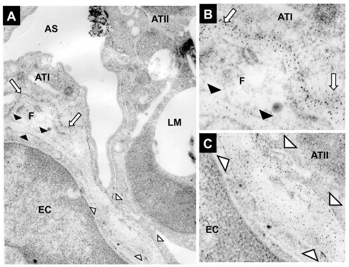
Studies show that considerable heterogeneity exists in the sulfation and the heparan sulfate epitopes in basement membrane underlying alveolar type I and type II cells in alveolus (Sannes, 1984; Smits et al., 2004). Increased sulfation of glycosaminoglycans under type I cells sequesters FGF2 to the ECM, which limits the ability of FGF-2 to bind to its receptor on the cell surface of alveolar type I cells (Fig. 4). In contrast, the under sulfated microdomains in the ECM beneath type II cells do not sequester FGF2, allowing this growth factor access to specific cell surface receptors, thus promoting activation of FGF signaling pathways in alveolar type II cells (Sannes et al., 1996; Sannes et al., 1998). These studies suggest that the highly sulfated ECM underlying Type I epithelial cells sequesters cytokines and chemokines to a greater degree than the ECM underlying Type II epithelial cells. A prominent path for migrating neutrophils in the alveolus is at the junction of Type I and Type II epithelial cells (Burns et al., 2003). Whether differences in the sulfation of ECM underlying Type I and Type II epithelial cells facilitates neutrophil migration at this location by modifying the biological activity of cytokines or the formation of chemokine gradients needs to be determined.
Figure 4.
An electron micrograph (EM) of normal lung tissue showing microdomains in the extracellular matrix which contain varying amounts of sulfation. Cytochemical visualization of sulfation in the extracellular matrix was performed using high iron diamine (HID), which binds to sulfates in lung tissue. The HID reaction product forms discrete 5–12 nm silver particles following appropriate intensification and is seen as discrete dark spots (Sannes, 1984). The basement membrane under a type I cell and an adjacent fibroblast (F) is highly sulfated (Arrows in 2A and 2B). The microdomains surrounding the pulmonary fibroblast show this cell to have microdomains of high and low sulfation. The extracellular matrix adjacent to the ATI cell is highly sulfated (white arrows in 2A and 2B) whereas the extracellular matrix adjacent to the endothelial cell (EC) is undersulfated (black arrowheads in 2A and 2B). In contrast to the ATI cell, this EM shows that the extracellular matrix underlying the type II cell (ATII) is undersulfated. White arrowheads in 2A and 2C define the extracellular matrix underlying the ATII cell and a pulmonary capillary EC (This figure is from studies by Frevert, et al., (Frevert and Sannes, 2006) with permission.).
Proteoglycans facilitate and Promote Leukocyte Adhesion and Sequestration
There is growing evidence that proteoglycans and glycosaminoglycans play a role in leukocyte adhesion and subsequent migration in tissue (Gotte, 2003; Parish, 2006). The anti-inflammatory effect of heparin is in part mediated through its ability to block CD11b/CD18, P- and L-selectin initiated adhesion to endothelial cells (Wang et al., 2002). CD44v3, a variant form of CD44 that contains a heparan sulfate side chain, is found on epithelial cells, and serves as a CD11b/CD18 counter-receptor during the transepithelial migration of neutrophils (Barbour et al., 2003; Zen et al., 2009). Lumican, which coats the surface of neutrophils as they cross endothelial cells, promotes neutrophil migration by binding to β2 Integrin (Lee et al., 2009).
The glycosaminoglycan, hyaluronan, plays an important role in leukocyte adhesion and sequestration through a CD44-dependent mechanism. The binding of hyaluronan by CD44 is responsible for the adhesion of monocytes to colon smooth muscle (de la Motte et al., 2003). Neutrophil CD44 and hyaluronan is required for the sequestration of neutrophils in inflamed liver sinusoids (McDonald et al., 2008). The work of Perigo and colleagues highlights the important role for versican in the hyaluronan-dependent binding of monocytes to the ECM of fibroblasts stimulated with PolyI:C (Potter-Perigo et al., 2009). Interactions between CD44 and high molecular weight hyaluronan in an intact extracellular matrix maintains immune tolerance through the persistence of induced CD4+CD25+ regulatory T cells (Bollyky et al., 2009). These studies suggests that in inflamed tissues, leukocytes will most likely interact with high-molecular weight complexes such as those consisting of hyaluronan, versican, and other matrix associated molecules (Day and de la Motte, 2005). Future studies will be needed to identify the role of high-molecular weight complexes in the extracellular matrix in influencing the phenotype of leukocytes and the pulmonary inflammatory response.
Proteoglycans Interact with Matrix Metalloproteinases
Matrix metalloproteinases (MMPs), a family of highly potent proteases, were first believed to be the enzymes responsible for turnover and degradation of the ECM. Whereas matrix degradation has been identified as a function of these proteinases, recent findings indicate that an important function of MMPs is to act on a number of molecules that regulate innate immunity (McCawley and Matrisian, 2001; Parks et al., 2004). The expression of MMPs, as well as their endogenous inhibitors, the tissue inhibitors of metalloproteinases (TIMPs), is altered following lung infection (Parks et al., 2004; Kassim et al., 2007; Page-McCaw et al., 2007). Growing evidence shows that interactions between proteoglycans and MMPs, which is mediated by glycosaminoglycans, alters MMP activity in the following ways: 1) glycosaminoglycans conceal proteolytic cleavage sites protecting chemokines, cytokines, and growth factors from proteolysis, 2) the proteolytic processing of proteoglycans results in bioactive fragments or shedding of syndecans from the cell surface creating chemotactic gradients, and 3) the binding of MMPs and TIMPs to glycosaminoglycans sequesters these proteins to specific sites in lung tissue leading to direct regulation of MMP activity.
Growing evidence shows that MMPs act on growth factors, cytokines, and chemokines, and that this proteolytic processing can either inhibit or potentiate the activity these inflammatory proteins in tissue (Parks et al., 2004). The binding of a protein to glycosaminoglycans conceals proteolytic cleavage sites, which protects heparin binding growth factors, cytokines, and chemokines, from proteolytic degradation (Gospodarowicz and Cheng, 1986; Rosengart et al., 1989; Webb et al., 1993; Sadir et al., 2004; Ellyard et al., 2007). Heparin and heparan sulfate limit the proteolytic cleavage of the carboxyl-terminal sequence of interferon-gamma which increases the activity of this cytokine by as much as 600% (Lortat-Jacob et al., 1996; Lortat-Jacob, 2006). Many chemokines, including CCL2/MCP1, CCL7/MCP3, CCL8/MCP2, CCL13/MCP4, CXCL5/LIX, and CXCL12/SDF1 are cleaved by MMPs, and this processing can either enhance or inhibit chemokine activity, depending on the specific chemokine and MMP involved (Van den Steen et al., 2000; McQuibban et al., 2001; McQuibban et al., 2002; Van Den Steen et al., 2003; Zhang et al., 2003; Tester et al., 2007). The binding of CCL11/eotaxin to heparin protects CCL11 from proteolysis by proteases, which potentiates the chemotactic activity of this chemokine in vivo (Ellyard et al., 2007). Therefore, an important function of proteoglycans during tissue inflammation is to sequester and protect proteolytic cleavage sites on cytokines and chemokines, preventing their proteolysis by proteases that are released in response to lung infection.
Post-translational processing of proteoglycans by proteinases, such as the MMPs, serves multiple functions. Degradation by MMPs serves to control the amount and localization of proteoglycans within the lung, but additionally, leads to the release or unmasking of cryptic fragments that can regulate cell behavior. The proteolytic cleavage of the HSPGs, perlecan and collagen XVIII, results in the anti-angiogenic fragments, endorepellin and endostatin (Zatterstrom et al., 2000; Mongiat et al., 2003). Interestingly, endostatin levels are increased in the plasma and bronchoalveolar lavage (BAL) of patients with acute lung injury and appear to correlate with the degree of neutrophilia and protein leak across the alveolar-capillary barrier (Perkins et al., 2009). This suggests that proteolytic processing of collagen XVIII may have a key pathophysiological role following lung injury or infection.
Versican, which can interact with infiltrating leukocytes, is expressed at low levels in the normal lung; however, the expression of versican is rapidly increased following lung injury (Venkatesan et al., 2000; Koslowski et al., 2001; Venkatesan et al., 2002; Faggian et al., 2007). Both MMPs and ADAMTSs (a disintegrin and metalloproteinase with thrombospondin motifs) are capable of degrading versican, thus, MMPs, and ADAMTSs may be required to control the accumulation of versican within the lung thereby regulating inflammatory cell influx and possibly activation (Sandy et al., 2001; de la Motte et al., 2003; Zheng et al., 2004; Porter et al., 2005; Kenagy et al., 2006).
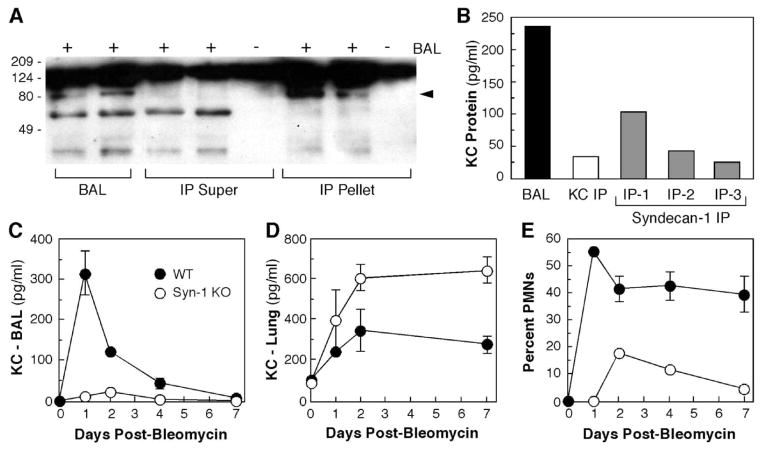
The shedding of syndecans from the surface of cells is a mechanism that controls pulmonary inflammation. Studies performed in a bleomycin-induced model of lung fibrosis show that shedding of syndecan-1/KC complexes by matrilysin (MMP7) directs neutrophil migration into the airspaces of the lungs (Li et al., 2002). This study demonstrates that syndecan-1, which is found on the cell surface of epithelial cells in the lungs is a substrate for MMP7 (Fig. 5). Li, et al. also show that syndecan-1 binds to the murine CXC-chemokine, KC, and that shedding of the syndecan-1/KC complex is a mechanism that controls neutrophil migration from the interstitium into the airspaces of the lungs (Fig. 6).
Figure 5.
Positive staining (brown) for syndecan-1 (syndecan-1) in normal mouse lungs shows a basal lateral distribution in airway epithelial cells and immunoreactivity in cells of the alveolar septa. Immunohistochemistry for syndecan-1 was performed with a rat anti-mouse syndecan-1 IgG (BD/Pharmingen, Franklin Lakes, NJ).
Figure 6.
Syndecan-1/KC Complexes control the pulmonary recruitment of neutrophils in mice treated with bleomycin. (A) KC was immunoprecipitated (IP) from BAL of WT mice 1 day post-bleomycin. BAL and post-IP supernatants and pellets were electrophoresed and blotted for syndecan-1 ectodomain. The arrowhead indicates the band specifically coprecipitated with KC. (B) KC and syndecan-1 were immunoprecipitated from BAL of WT mice 1 day post-bleomycin. Two additional syndecan-1 IPs were done on the post-IP supernatant. The level of KC protein in the BAL and in the post-IP supernatants was quantified by ELISA. (C–E) WT and syndecan-1 null mice (SYN1−/−) (n = 3) were instilled with 0.15 U bleomycin. KC levels in BAL (C) and lung homogenates (D) were determined by ELISA. (E) Neutrophils (PMNs) in BAL were counted and expressed as a percent of total leukocytes. Data are the mean ± SE.. (This figure is from studies by Li, et al., (Li et al., 2002) with permission.)
Recent work by Brule and co-workers demonstrated that CXCL12 binds to, and signals through, syndecan-4 on both macrophages and HeLa cells (Brule et al., 2006). The interaction between CXCL12 and syndecan-4 stimulates MMP9 expression and activation, which sheds syndecan-1 and -4 from the cell surface and disrupts this signaling pathway (Brule et al., 2006). Thus, cell-surface proteoglycans, like syndecan-4, are capable of acting as chemokine receptors following infection, and serve to regulate the expression and activation of potent enzymes, such as MMP9, which are capable of altering the inflammatory response in multiple ways (as has been previously discussed). syndecan-4, however, also serves as a key check point in this system as syndecan-4 shedding from the cell surface by MMP9 inhibits further enzyme release. Therefore, the proteolytic processing of proteoglycans by MMPs is capable of modulating pulmonary inflammation through several mechanisms including the development of bioactive fragments, controlling the accumulation of proteoglycans in tissue, and through the shedding of syndecans.
Proteoglycans may also prove to be very important in regulating activation and activity of MMPs (Ra and Parks, 2007). The direct binding of pro-MMP2 to chondroitin sulfate presents the catalytic domain of pro-MMP-2 to MT3-MMP. This interaction with chondroitin sulfate facilitates the generation of the active form of MMP-2 (Iida et al., 2007). As well, the binding of proteases to glycosaminoglycans can be inhibitory as shown by work of Sorensen et al. where heparanase promotes ADAM12 activity on the surface of cells by cleaving the inhibitory heparan sulfate (Sorensen et al., 2008). Furthermore, while TIMP1, 2, and 4 are considered to be soluble inhibitors; TIMP3 is associated with the matrix through interaction between its N-terminus and HSPG/CSPGs (Yu et al., 2000). Since TIMP3 is an efficient inhibitor of all of the MMPs and many of the ADAMs/ADAMTSs (Brew et al., 2000; Baker et al., 2002), localization to sulfated proteoglycans in the ECM, such as versican, places TIMP3 in an ideal location to regulate proteolytic processing of these proteins by enzymes such as MMP3 and 9, or by ADAM-TS4 or 5 (Hashimoto et al., 2001; Kashiwagi et al., 2001).
Thus, proteoglycans are able to bind to and sequester chemokines to protect them from proteolytic processing by metalloproteinases, which can both enhance and inhibit chemokine activity; however, shedding of these proteoglycan/chemokine complexes from the cell surface is also required for leukocyte extravasation into the alveolar space following lung injury. Furthermore, proteolytic processing of proteoglycans within the matrix by metalloproteinases can promote inflammation through the release of cryptic fragments, such as endostatin; however, degradation of the proteoglycans such as versican may also serve to abrogate inflammatory cell influx and therefore serve to restrict inflammation. Together, this data supports a model whereby interaction between proteoglycans and metalloproteinases or their inhibitors fine tunes the ability of proteoglycans to regulate leukocyte influx and this control is likely dependent on the mode of injury or infection.
VI) Conclusions
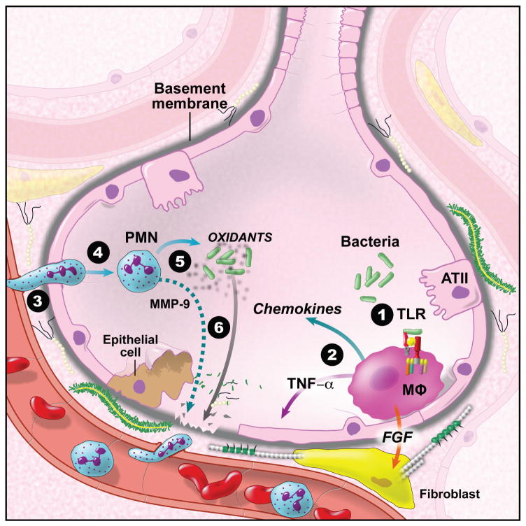
Inflammatory responses as a result of lung infection require the emigration of leukocytes from the vasculature to the infected area as part of the innate immune response (Fig. 7). Upon extravasation into the subendothelial compartment, leukocytes encounter the ECM and cell surface proteoglycans, which functions as a scaffold for leukocyte migration. A growing literature shows that proteoglycans and their associated glycosaminoglycans play an important role in modulating pulmonary inflammation through the activation of Toll-like receptor signaling pathways; the sequestration of cytokines, chemokines, and growth factors; and by facilitating leukocyte adhesion and sequestration. Moreover, interactions between proteoglycans and MMPs have the potential to play an important role in the regulation of the innate immune system. In conclusion, proteoglycans provide fine-tune control of tissue inflammation. Clarification of the mechanisms whereby proteoglycans modulate the pulmonary inflammatory response will most likely lead to new therapeutic approaches to inflammatory lung disease and lung infection.
Figure 7.
The recognition of bacteria and viruses in the lungs results in the activation of Toll-like receptor (TLR) signalling pathways, which leads to pulmonary inflammation and under ideal conditions the clearance of the pathogen. Proteoglycans and/or their glycosaminoglycans modify the inflammatory response in lungs through a number of different mechanisms. (1) The release of soluble proteoglycans such as biglycan or degradation of glycosaminoglycan such as hyaluronan and heparan sulfate can activate TLRs. (2) Cytokines, chemokines, and growth factors bind to glycosaminoglycans, which can either increase or decrease their biological activity. (3) Adhesion molecules including the selectins, integrins, and CD44 bind to proteoglycans and glycosaminoglycan, which suggests that these proteins play a critical role in leukocyte adhesion and migration. (4) Chemokine-glycosaminoglycan interactions provide fine-tune control of chemokine-gradient formation and leukocyte migration in tissue. (5) Activation of stromal and immune cells results in the release of MMP. Growing evidence shows that interactions between proteoglycans and MMPs play important roles in the regulation of the innate immune response. (6) Degradation of proteoglycans by MMPs and other proteases controls the amount and localization of proteoglycans in lungs. In addition, proteolytic cleavage of proteoglycans leads to the unmasking of cryptic fragments.
ABBREVIATIONS
- ADAMTS
A disintegrin and metalloproteinase with thrombospondin motifs
- CSPG
Chondroitin sulfate proteoglycan
- hr
Hour
- HSPG
Heparan sulfate proteoglycan
- LPS
Lipopolysaccharide
- MMPs
matrix metalloproteinases
- TIMP
Tissue inhibitors of metalloproteinases
- TLR
Toll-like receptors
LITERATURE CITED
- Adair-Kirk TL, Senior RM. Fragments of extracellular matrix as mediators of inflammation. Int J Biochem Cell Biol. 2008;40:1101–1110. doi: 10.1016/j.biocel.2007.12.005. [DOI] [PMC free article] [PubMed] [Google Scholar]
- Akira S. Innate immunity to pathogens: diversity in receptors for microbial recognition. Immunol Rev. 2009;227:5–8. doi: 10.1111/j.1600-065X.2008.00739.x. [DOI] [PubMed] [Google Scholar]
- Ali S, Palmer AC, Banerjee B, Fritchley SJ, Kirby JA. Examination of the Function of RANTES, MIP-1alpha, and MIP-1beta following Interaction with Heparin-like Glycosaminoglycans. J Biol Chem. 2000;275:11721–11727. doi: 10.1074/jbc.275.16.11721. [DOI] [PubMed] [Google Scholar]
- Allen BL, Filla MS, Rapraeger AC. Role of heparan sulfate as a tissue-specific regulator of FGF-4 and FGF receptor recognition. J Cell Biol. 2001;155:845–858. doi: 10.1083/jcb.200106075. [DOI] [PMC free article] [PubMed] [Google Scholar]
- Amara A, Lorthioir O, Valenzuela A, Magerus A, Thelen M, Montes M, Virelizier JL, Delepierre M, Baleux F, Lortat-Jacob H, Arenzana-Seisdedos F. Stromal cell-derived factor-1alpha associates with heparan sulfates through the first beta-strand of the chemokine. J Biol Chem. 1999;274:23916–23925. doi: 10.1074/jbc.274.34.23916. [DOI] [PubMed] [Google Scholar]
- Araujo BB, Dolhnikoff M, Silva LF, Elliot J, Lindeman JH, Ferreira DS, Mulder A, Gomes HA, Fernezlian SM, James A, Mauad T. Extracellular matrix components and regulators in the airway smooth muscle in asthma. Eur Respir J. 2008;32:61–69. doi: 10.1183/09031936.00147807. [DOI] [PubMed] [Google Scholar]
- Bai KJ, Spicer AP, Mascarenhas MM, Yu L, Ochoa CD, Garg HG, Quinn DA. The Role of Hyaluronan Synthase 3 in Ventilator-Induced Lung Injury. Am J Respir Crit Care Med. 2005;172:92–98. doi: 10.1164/rccm.200405-652OC. [DOI] [PMC free article] [PubMed] [Google Scholar]
- Baker AH, Edwards DR, Murphy G. Metalloproteinase inhibitors: biological actions and therapeutic opportunities. J Cell Sci. 2002;115:3719–3727. doi: 10.1242/jcs.00063. [DOI] [PubMed] [Google Scholar]
- Barbour AP, Reeder JA, Walsh MD, Fawcett J, Antalis TM, Gotley DC. Expression of the CD44v2-10 isoform confers a metastatic phenotype: importance of the heparan sulfate attachment site CD44v3. Cancer Res. 2003;63:887–892. [PubMed] [Google Scholar]
- Bensadoun ES, Burke AK, Hogg JC, Roberts CR. Proteoglycan deposition in pulmonary fibrosis. Am J Respir Crit Care Med. 1996;154:1819–1828. doi: 10.1164/ajrccm.154.6.8970376. [DOI] [PubMed] [Google Scholar]
- Bensadoun ES, Burke AK, Hogg JC, Roberts CR. Proteoglycans in granulomatous lung diseases. Eur Respir J. 1997;10:2731–2737. doi: 10.1183/09031936.97.10122731. [DOI] [PubMed] [Google Scholar]
- Bernfield M, Götte M, Park W, Reizes O, Fitzgerald M, Lincecum J, Zako M. Functions of cell surface heparan sulfate proteoglycans. Annu Rev Biochem. 1999;68:729–777. doi: 10.1146/annurev.biochem.68.1.729. [DOI] [PubMed] [Google Scholar]
- Bianco P, Fisher LW, Young MF, Termine JD, Robey PG. Expression and localization of the two small proteoglycans biglycan and decorin in developing human skeletal and non-skeletal tissues. J Histochem Cytochem. 1990;38:1549–1563. doi: 10.1177/38.11.2212616. [DOI] [PubMed] [Google Scholar]
- Blackwood RA, Cantor JO, Moret J, Mandl I, Turino GM. Glycosaminoglycan synthesis in endotoxin-induced lung injury. Proc Soc Exp Biol Med. 1983;174:343–349. doi: 10.3181/00379727-174-41746. [DOI] [PubMed] [Google Scholar]
- Bode L, Eklund EA, Murch S, Freeze HH. Heparan sulfate depletion amplifies TNF-alpha-induced protein leakage in an in vitro model of protein-losing enteropathy. Am J Physiol Gastrointest Liver Physiol. 2005;288:G1015–1023. doi: 10.1152/ajpgi.00461.2004. [DOI] [PubMed] [Google Scholar]
- Bode L, Salvestrini C, Park PW, Li JP, Esko JD, Yamaguchi Y, Murch S, Freeze HH. Heparan sulfate and syndecan-1 are essential in maintaining murine and human intestinal epithelial barrier function. J Clin Invest. 2008;118:229–238. doi: 10.1172/JCI32335. [DOI] [PMC free article] [PubMed] [Google Scholar]
- Bollyky PL, Falk BA, Wu RP, Buckner JH, Wight TN, Nepom GT. Intact extracellular matrix and the maintenance of immune tolerance: high molecular weight hyaluronan promotes persistence of induced CD4+CD25+ regulatory T cells. J Leukoc Biol. 2009;86:567–572. doi: 10.1189/jlb.0109001. [DOI] [PMC free article] [PubMed] [Google Scholar]
- Borghesi LA, Yamashita Y, Kincade PW. Heparan sulfate proteoglycans mediate interleukin-7-dependent B lymphopoiesis. Blood. 1999;93:140–148. [PubMed] [Google Scholar]
- Bosman FT, Stamenkovic I. Functional structure and composition of the extracellular matrix. J Pathol. 2003;200:423–428. doi: 10.1002/path.1437. [DOI] [PubMed] [Google Scholar]
- Brew K, Dinakarpandian D, Nagase H. Tissue inhibitors of metalloproteinases: evolution, structure and function. Biochim Biophys Acta. 2000;1477:267–283. doi: 10.1016/s0167-4838(99)00279-4. [DOI] [PubMed] [Google Scholar]
- Brule S, Charnaux N, Sutton A, Ledoux D, Chaigneau T, Saffar L, Gattegno L. The shedding of syndecan-4 and syndecan-1 from HeLa cells and human primary macrophages is accelerated by SDF-1/CXCL12 and mediated by the matrix metalloproteinase-9. Glycobiology. 2006;16:488–501. doi: 10.1093/glycob/cwj098. [DOI] [PubMed] [Google Scholar]
- Burns AR, Smith CW, Walker DC. Unique structural features that influence neutrophil emigration into the lung. Physiol Rev. 2003;83:309–336. doi: 10.1152/physrev.00023.2002. [DOI] [PubMed] [Google Scholar]
- Clarke D, Katoh O, Gibbs RV, Griffiths SD, Gordon MY. Interaction of interleukin 7 (IL-7) with glycosaminoglycans and its biological relevance. Cytokine. 1995;7:325–330. doi: 10.1006/cyto.1995.0041. [DOI] [PubMed] [Google Scholar]
- Coombe DR. Biological implications of glycosaminoglycan interactions with haemopoietic cytokines. Immunol Cell Biol. 2008 doi: 10.1038/icb.2008.49. [DOI] [PubMed] [Google Scholar]
- David G, Lories V, Decock B, Marynen P, Cassiman JJ, Van den Berghe H. Molecular cloning of a phosphatidylinositol-anchored membrane heparan sulfate proteoglycan from human lung fibroblasts. J Cell Biol. 1990;111:3165–3176. doi: 10.1083/jcb.111.6.3165. [DOI] [PMC free article] [PubMed] [Google Scholar]
- Day AJ, de la Motte CA. Hyaluronan cross-linking: a protective mechanism in inflammation? Trends Immunol. 2005;26:637–643. doi: 10.1016/j.it.2005.09.009. [DOI] [PubMed] [Google Scholar]
- de la Motte C, Nigro J, Vasanji A, Rho H, Kessler S, Bandyopadhyay S, Danese S, Fiocchi C, Stern R. Platelet-derived hyaluronidase 2 cleaves hyaluronan into fragments that trigger monocyte-mediated production of proinflammatory cytokines. Am J Pathol. 2009;174:2254–2264. doi: 10.2353/ajpath.2009.080831. [DOI] [PMC free article] [PubMed] [Google Scholar]
- de la Motte CA, Hascall VC, Drazba J, Bandyopadhyay SK, Strong SA. Mononuclear leukocytes bind to specific hyaluronan structures on colon mucosal smooth muscle cells treated with polyinosinic acid:polycytidylic acid: inter-alpha-trypsin inhibitor is crucial to structure and function. Am J Pathol. 2003;163:121–133. doi: 10.1016/s0002-9440(10)63636-x. [DOI] [PMC free article] [PubMed] [Google Scholar]
- de Medeiros Matsushita M, da Silva LF, dos Santos MA, Fernezlian S, Schrumpf JA, Roughley P, Hiemstra PS, Saldiva PH, Mauad T, Dolhnikoff M. Airway proteoglycans are differentially altered in fatal asthma. J Pathol. 2005;207:102–110. doi: 10.1002/path.1818. [DOI] [PubMed] [Google Scholar]
- Dolhnikoff M, Morin J, Roughley PJ, Ludwig MS. Expression of lumican in human lungs. Am J Respir Cell Mol Biol. 1998;19:582–587. doi: 10.1165/ajrcmb.19.4.2979. [DOI] [PubMed] [Google Scholar]
- Ellyard JI, Simson L, Bezos A, Johnston K, Freeman C, Parish CR. Eotaxin selectively binds heparin. An interaction that protects eotaxin from proteolysis and potentiates chemotactic activity in vivo. J Biol Chem. 2007;282:15238–15247. doi: 10.1074/jbc.M608046200. [DOI] [PubMed] [Google Scholar]
- Esko JD, Selleck SB. ORDER OUT OF CHAOS: Assembly of Ligand Binding Sites in Heparan Sulfate. Annu Rev Biochem. 2002;71:435–471. doi: 10.1146/annurev.biochem.71.110601.135458. [DOI] [PubMed] [Google Scholar]
- Faggian J, Fosang AJ, Zieba M, Wallace MJ, Hooper SB. Changes in versican and chondroitin sulfate proteoglycans during structural development of the lung. Am J Physiol Regul Integr Comp Physiol. 2007;293:R784–792. doi: 10.1152/ajpregu.00801.2006. [DOI] [PubMed] [Google Scholar]
- Fernandez-Botran R, Yan J, Justus DE. Binding of interferon gamma by glycosaminoglycans: a strategy for localization and/or inhibition of its activity. Cytokine. 1999;11:313–325. doi: 10.1006/cyto.1998.0438. [DOI] [PubMed] [Google Scholar]
- Frevert C, Wight TN. Matrix Proteoglycans. In: Laurent GJ, editor. The Encyclopedia of Respiratory Medicine. London, U.K: Elsevier; 2006. pp. 184–187. [Google Scholar]
- Frevert CW, Goodman RB, Kinsella MG, Kajikawa O, Ballman K, Clark-Lewis I, Proudfoot AE, Wells TN, Martin TR. Tissue-Specific Mechanisms Control the Retention of IL-8 in Lungs and Skin. J Immunol. 2002;168:3550–3556. doi: 10.4049/jimmunol.168.7.3550. [DOI] [PubMed] [Google Scholar]
- Frevert CW, Kinsella MG, Vathanaprida C, Goodman RB, Baskin DG, Proudfoot A, Wells TN, Wight TN, Martin TR. Binding of interleukin-8 to heparan sulfate and chondroitin sulfate in lung tissue. Am J Respir Cell Mol Biol. 2003;28:464–472. doi: 10.1165/rcmb.2002-0084OC. [DOI] [PubMed] [Google Scholar]
- Frevert CW, Sannes PL. Matrix proteoglycans as effector molecules for epithelial cell function. Eur Respir Rev. 2006;14:137–144. [Google Scholar]
- Gospodarowicz D, Cheng J. Heparin protects basic and acidic FGF from inactivation. J Cell Physiol. 1986;128:475–484. doi: 10.1002/jcp.1041280317. [DOI] [PubMed] [Google Scholar]
- Gotte M. Syndecans in inflammation. Faseb J. 2003;17:575–591. doi: 10.1096/fj.02-0739rev. [DOI] [PubMed] [Google Scholar]
- Groffen AJ, Buskens CA, van Kuppevelt TH, Veerkamp JH, Monnens LA, van den Heuvel LP. Primary structure and high expression of human agrin in basement membranes of adult lung and kidney. Eur J Biochem. 1998;254:123–128. doi: 10.1046/j.1432-1327.1998.2540123.x. [DOI] [PubMed] [Google Scholar]
- Halfter W, Dong S, Schurer B, Cole GJ. Collagen XVIII is a basement membrane heparan sulfate proteoglycan. J Biol Chem. 1998;273:25404–25412. doi: 10.1074/jbc.273.39.25404. [DOI] [PubMed] [Google Scholar]
- Hasan M, Najjam S, Gordon MY, Gibbs RV, Rider CC. IL-12 is a heparin-binding cytokine. J Immunol. 1999;162:1064–1070. [PubMed] [Google Scholar]
- Hashimoto G, Aoki T, Nakamura H, Tanzawa K, Okada Y. Inhibition of ADAMTS4 (aggrecanase-1) by tissue inhibitors of metalloproteinases (TIMP-1, 2, 3 and 4) FEBS Lett. 2001;494:192–195. doi: 10.1016/s0014-5793(01)02323-7. [DOI] [PubMed] [Google Scholar]
- Hirose J, Kawashima H, Yoshie O, Tashiro K, Miyasaka M. Versican interacts with chemokines and modulates cellular responses. J Biol Chem. 2001;276:5228–5234. doi: 10.1074/jbc.M007542200. [DOI] [PubMed] [Google Scholar]
- Hoebe K, Janssen E, Beutler B. The interface between innate and adaptive immunity. Nat Immunol. 2004;5:971–974. doi: 10.1038/ni1004-971. [DOI] [PubMed] [Google Scholar]
- Huang J, Olivenstein R, Taha R, Hamid Q, Ludwig M. Enhanced proteoglycan deposition in the airway wall of atopic asthmatics. Am J Respir Crit Care Med. 1999;160:725–729. doi: 10.1164/ajrccm.160.2.9809040. [DOI] [PubMed] [Google Scholar]
- Hurt-Camejo E, Rosengren B, Sartipy P, Elfsberg K, Camejo G, Svensson L. CD44, a cell surface chondroitin sulfate proteoglycan, mediates binding of interferon-gamma and some of its biological effects on human vascular smooth muscle cells. J Biol Chem. 1999;274:18957–18964. doi: 10.1074/jbc.274.27.18957. [DOI] [PubMed] [Google Scholar]
- Iida J, Wilhelmson KL, Ng J, Lee P, Morrison C, Tam E, Overall CM, McCarthy JB. Cell surface chondroitin sulfate glycosaminoglycan in melanoma: role in the activation of pro-MMP-2 (pro-gelatinase A) Biochem J. 2007;403:553–563. doi: 10.1042/BJ20061176. [DOI] [PMC free article] [PubMed] [Google Scholar]
- Jiang D, Liang J, Fan J, Yu S, Chen S, Luo Y, Prestwich GD, Mascarenhas MM, Garg HG, Quinn DA, Homer RJ, Goldstein DR, Bucala R, Lee PJ, Medzhitov R, Noble PW. Regulation of lung injury and repair by Toll-like receptors and hyaluronan. Nat Med. 2005 doi: 10.1038/nm1315. [DOI] [PubMed] [Google Scholar]
- Johnson GB, Brunn GJ, Platt JL. Cutting Edge: An Endogenous Pathway to Systemic Inflammatory Response Syndrome (SIRS)-Like Reactions through Toll-Like Receptor 4. J Immunol. 2004;172:20–24. doi: 10.4049/jimmunol.172.1.20. [DOI] [PubMed] [Google Scholar]
- Karlinsky JB. Glycosaminoglycans in emphysematous and fibrotic hamster lungs. Am Rev Respir Dis. 1982;125:85–88. doi: 10.1164/arrd.1982.125.1.85. [DOI] [PubMed] [Google Scholar]
- Kashiwagi M, Tortorella M, Nagase H, Brew K. TIMP-3 is a potent inhibitor of aggrecanase 1 (ADAM-TS4) and aggrecanase 2 (ADAM-TS5) J Biol Chem. 2001;276:12501–12504. doi: 10.1074/jbc.C000848200. [DOI] [PubMed] [Google Scholar]
- Kassim SY, Gharib SA, Mecham BH, Birkland TP, Parks WC, McGuire JK. Individual matrix metalloproteinases control distinct transcriptional responses in airway epithelial cells infected with Pseudomonas aeruginosa. Infect Immun. 2007;75:5640–5650. doi: 10.1128/IAI.00799-07. [DOI] [PMC free article] [PubMed] [Google Scholar]
- Kawashima H, Watanabe N, Hirose M, Sun X, Atarashi K, Kimura T, Shikata K, Matsuda M, Ogawa D, Heljasvaara R, Rehn M, Pihlajaniemi T, Miyasaka M. Collagen XVIII, a basement membrane heparan sulfate proteoglycan, interacts with L-selectin and monocyte chemoattractant protein-1. J Biol Chem. 2003:M212244200. doi: 10.1074/jbc.M212244200. [DOI] [PubMed] [Google Scholar]
- Keck RG, Berleau L, Harris R, Keyt BA. Disulfide structure of the heparin binding domain in vascular endothelial growth factor: characterization of posttranslational modifications in VEGF. Arch Biochem Biophys. 1997;344:103–113. doi: 10.1006/abbi.1997.0145. [DOI] [PubMed] [Google Scholar]
- Kenagy RD, Plaas AH, Wight TN. Versican degradation and vascular disease. Trends Cardiovasc Med. 2006;16:209–215. doi: 10.1016/j.tcm.2006.03.011. [DOI] [PMC free article] [PubMed] [Google Scholar]
- Kim CW, Goldberger OA, Gallo RL, Bernfield M. Members of the syndecan family of heparan sulfate proteoglycans are expressed in distinct cell-, tissue-, and development-specific patterns. Mol Biol Cell. 1994;5:797–805. doi: 10.1091/mbc.5.7.797. [DOI] [PMC free article] [PubMed] [Google Scholar]
- Kim S, Takahashi H, Lin WW, Descargues P, Grivennikov S, Kim Y, Luo JL, Karin M. Carcinoma-produced factors activate myeloid cells through TLR2 to stimulate metastasis. Nature. 2009;457:102–106. doi: 10.1038/nature07623. [DOI] [PMC free article] [PubMed] [Google Scholar]
- Kinsella MG, Bressler SL, Wight TN. The regulated synthesis of versican, decorin, and biglycan: extracellular matrix proteoglycans that influence cellular phenotype. Crit Rev Eukaryot Gene Expr. 2004;14:203–234. doi: 10.1615/critreveukaryotgeneexpr.v14.i3.40. [DOI] [PubMed] [Google Scholar]
- Koopmann W, Ediriwickrema C, Krangel MS. Structure and function of the glycosaminoglycan binding site of chemokine macrophage-inflammatory protein-1 beta. J Immunol. 1999;163:2120–2127. [PubMed] [Google Scholar]
- Koslowski R, Pfeil U, Fehrenbach H, Kasper M, Skutelsky E, Wenzel KW. Changes in xylosyltransferase activity and in proteoglycan deposition in bleomycin-induced lung injury in rat. Eur Respir J. 2001;18:347–356. doi: 10.1183/09031936.01.00085601. [DOI] [PubMed] [Google Scholar]
- Kuschert GS, Coulin F, Power CA, Proudfoot AE, Hubbard RE, Hoogewerf AJ, Wells TN. Glycosaminoglycans interact selectively with chemokines and modulate receptor binding and cellular responses. Biochemistry. 1999;38:12959–12968. doi: 10.1021/bi990711d. [DOI] [PubMed] [Google Scholar]
- Kuschert GS, Hoogewerf AJ, Proudfoot AE, Chung CW, Cooke RM, Hubbard RE, Wells TN, Sanderson PN. Identification of a glycosaminoglycan binding surface on human interleukin-8. Biochemistry. 1998;37:11193–11201. doi: 10.1021/bi972867o. [DOI] [PubMed] [Google Scholar]
- Lau EK, Paavola CD, Johnson Z, Gaudry J-P, Geretti E, Borlat F, Kungl AJ, Proudfoot AE, Handel TM. Identification of the glycosaminoglycan binding site of the CC chemokine, MCP-1; Implications for structure and function In vivo. J Biol Chem. 2004:M311224200. doi: 10.1074/jbc.M311224200. [DOI] [PubMed] [Google Scholar]
- Lee S, Bowrin K, Hamad AR, Chakravarti S. Extracellular Matrix Lumican Deposited on the Surface of Neutrophils Promotes Migration by Binding to {beta}2 Integrin. J Biol Chem. 2009;284:23662–23669. doi: 10.1074/jbc.M109.026229. [DOI] [PMC free article] [PubMed] [Google Scholar]
- Li Q, Park PW, Wilson CL, Parks WC. Matrilysin shedding of syndecan-1 regulates chemokine mobilization and transepithelial efflux of neutrophils in acute lung injury. Cell. 2002;111:635–646. doi: 10.1016/s0092-8674(02)01079-6. [DOI] [PubMed] [Google Scholar]
- Lipscombe RJ, Nakhoul AM, Sanderson CJ, Coombe DR. Interleukin-5 binds to heparin/heparan sulfate. A model for an interaction with extracellular matrix. J Leukoc Biol. 1998;63:342–350. doi: 10.1002/jlb.63.3.342. [DOI] [PubMed] [Google Scholar]
- Lories V, Cassiman JJ, Van den Berghe H, David G. Differential expression of cell surface heparan sulfate proteoglycans in human mammary epithelial cells and lung fibroblasts. J Biol Chem. 1992;267:1116–1122. [PubMed] [Google Scholar]
- Lortat-Jacob H. Interferon and heparan sulphate. Biochem Soc Trans. 2006;34:461–464. doi: 10.1042/BST0340461. [DOI] [PubMed] [Google Scholar]
- Lortat-Jacob H, Baltzer F, Grimaud JA. Heparin decreases the blood clearance of interferon-gamma and increases its activity by limiting the processing of its carboxyl-terminal sequence. J Biol Chem. 1996;271:16139–16143. doi: 10.1074/jbc.271.27.16139. [DOI] [PubMed] [Google Scholar]
- Lortat-Jacob H, Garrone P, Banchereau J, Grimaud JA. Human interleukin 4 is a glycosaminoglycan-binding protein. Cytokine. 1997;9:101–105. doi: 10.1006/cyto.1996.0142. [DOI] [PubMed] [Google Scholar]
- Lowry MH, McAllister BP, Jean JC, Brown LA, Hughey RP, Cruikshank WW, Amar S, Lucey EC, Braun K, Johnson P, Wight TN, Joyce-Brady M. Lung lining fluid glutathione attenuates IL-13-induced asthma. Am J Respir Cell Mol Biol. 2008;38:509–516. doi: 10.1165/rcmb.2007-0128OC. [DOI] [PMC free article] [PubMed] [Google Scholar]
- Luster AD, Greenberg S-M, Leder P. The IP-10 chemokine binds to a specific cell surface heparan sulfate site shared with platelet factor 4 and inhibits endothelial cell proliferation. J Exp Med. 1995;182:219–231. doi: 10.1084/jem.182.1.219. [DOI] [PMC free article] [PubMed] [Google Scholar]
- Lyon M, Rushton G, Gallagher JT. The interaction of the transforming growth factor-betas with heparin/heparan sulfate is isoform-specific. J Biol Chem. 1997;272:18000–18006. doi: 10.1074/jbc.272.29.18000. [DOI] [PubMed] [Google Scholar]
- McCawley LJ, Matrisian LM. Matrix metalloproteinases: they’re not just for matrix anymore! Curr Opin Cell Biol. 2001;13:534–540. doi: 10.1016/s0955-0674(00)00248-9. [DOI] [PubMed] [Google Scholar]
- McDonald B, McAvoy EF, Lam F, Gill V, de la Motte C, Savani RC, Kubes P. Interaction of CD44 and hyaluronan is the dominant mechanism for neutrophil sequestration in inflamed liver sinusoids. J Exp Med. 2008;205:915–927. doi: 10.1084/jem.20071765. [DOI] [PMC free article] [PubMed] [Google Scholar]
- McQuibban GA, Butler GS, Gong JH, Bendall L, Power C, Clark-Lewis I, Overall CM. Matrix Metalloproteinase Activity Inactivates the CXC Chemokine Stromal Cell-derived Factor-1. J Biol Chem. 2001;276:43503–43508. doi: 10.1074/jbc.M107736200. [DOI] [PubMed] [Google Scholar]
- McQuibban GA, Gong JH, Wong JP, Wallace JL, Clark-Lewis I, Overall CM. Matrix metalloproteinase processing of monocyte chemoattractant proteins generates CC chemokine receptor antagonists with anti-inflammatory properties in vivo. Blood. 2002;100:1160–1167. [PubMed] [Google Scholar]
- Merrilees MJ, Ching PS, Beaumont B, Hinek A, Wight TN, Black PN. Changes in elastin, elastin binding protein and versican in alveoli in chronic obstructive pulmonary disease. Respir Res. 2008;9:41. doi: 10.1186/1465-9921-9-41. [DOI] [PMC free article] [PubMed] [Google Scholar]
- Merrilees MJ, Hankin EJ, Black JL, Beaumont B. Matrix proteoglycans and remodelling of interstitial lung tissue in lymphangioleiomyomatosis. J Pathol. 2004;203:653–660. doi: 10.1002/path.1577. [DOI] [PubMed] [Google Scholar]
- Miller JD, Stevens ET, Smith DR, Wight TN, Wrenshall LE. Perlecan: a major IL-2-binding proteoglycan in murine spleen. Immunol Cell Biol. 2007 doi: 10.1038/sj.icb.7100128. [DOI] [PubMed] [Google Scholar]
- Mizgerd JP. Acute lower respiratory tract infection. N Engl J Med. 2008;358:716–727. doi: 10.1056/NEJMra074111. [DOI] [PMC free article] [PubMed] [Google Scholar]
- Mongiat M, Sweeney SM, San Antonio JD, Fu J, Iozzo RV. Endorepellin, a novel inhibitor of angiogenesis derived from the C terminus of perlecan. J Biol Chem. 2003;278:4238–4249. doi: 10.1074/jbc.M210445200. [DOI] [PubMed] [Google Scholar]
- Mulloy B, Rider CC. Cytokines and proteoglycans: an introductory overview. Biochem Soc Trans. 2006;34:409–413. doi: 10.1042/BST0340409. [DOI] [PubMed] [Google Scholar]
- Murdoch AD, Liu B, Schwarting R, Tuan RS, Iozzo RV. Widespread expression of perlecan proteoglycan in basement membranes and extracellular matrices of human tissues as detected by a novel monoclonal antibody against domain III and by in situ hybridization. J Histochem Cytochem. 1994;42:239–249. doi: 10.1177/42.2.7507142. [DOI] [PubMed] [Google Scholar]
- Noble PW, Jiang D. Matrix regulation of lung injury, inflammation, and repair: the role of innate immunity. Proc Am Thorac Soc. 2006;3:401–404. doi: 10.1513/pats.200604-097AW. [DOI] [PMC free article] [PubMed] [Google Scholar]
- Page-McCaw A, Ewald AJ, Werb Z. Matrix metalloproteinases and the regulation of tissue remodelling. Nat Rev Mol Cell Biol. 2007;8:221–233. doi: 10.1038/nrm2125. [DOI] [PMC free article] [PubMed] [Google Scholar]
- Parish CR. The role of heparan sulphate in inflammation. Nat Rev Immunol. 2006;6:633–643. doi: 10.1038/nri1918. [DOI] [PubMed] [Google Scholar]
- Parks WC, Wilson CL, Lopez-Boado YS. Matrix metalloproteinases as modulators of inflammation and innate immunity. Nat Rev Immunol. 2004;4:617–629. doi: 10.1038/nri1418. [DOI] [PubMed] [Google Scholar]
- Perkins GD, Nathani N, Richter AG, Park D, Shyamsundar M, Heljasvaara R, Pihlajaniemi T, Manji M, Tunnicliffe W, McAuley D, Gao F, Thickett DR. Type XVIII collagen degradation products in acute lung injury. Crit Care. 2009;13:R52. doi: 10.1186/cc7779. [DOI] [PMC free article] [PubMed] [Google Scholar]
- Petersen F, Bock L, Flad HD, Brandt E. A chondroitin sulfate proteoglycan on human neutrophils specifically binds platelet factor 4 and is involved in cell activation. J Immunol. 1998;161:4347–4355. [PubMed] [Google Scholar]
- Porter S, Clark IM, Kevorkian L, Edwards DR. The ADAMTS metalloproteinases. Biochem J. 2005;386:15–27. doi: 10.1042/BJ20040424. [DOI] [PMC free article] [PubMed] [Google Scholar]
- Potter-Perigo S, Johnson PY, Evanko SP, Chan CK, Braun KR, Wilkinson TS, Altman LC, Wight TN. Poly I:C Stimulates Versican Accumulation in the Extracellular Matrix Promoting Monocyte Adhesion. Am J Respir Cell Mol Biol. 2009 doi: 10.1165/rcmb.2009-0081OC. [DOI] [PMC free article] [PubMed] [Google Scholar]
- Proudfoot AEI, Handel TM, Johnson Z, Lau EK, LiWang P, Clark-Lewis I, Borlat F, Wells TNC, Kosco-Vilbois MH. Glycosaminoglycan binding and oligomerization are essential for the in vivo activity of certain chemokines. PNAS. 2003;100:1885–1890. doi: 10.1073/pnas.0334864100. [DOI] [PMC free article] [PubMed] [Google Scholar]
- Ra HJ, Parks WC. Control of matrix metalloproteinase catalytic activity. Matrix Biol. 2007;26:587–596. doi: 10.1016/j.matbio.2007.07.001. [DOI] [PMC free article] [PubMed] [Google Scholar]
- Ramsden L, Rider CC. Selective and differential binding of interleukin (IL)-1 alpha, IL-1 beta, IL-2 and IL-6 to glycosaminoglycans. Eur J Immunol. 1992;22:3027–3031. doi: 10.1002/eji.1830221139. [DOI] [PubMed] [Google Scholar]
- Rapraeger AC. Heparan sulfate-growth factor interactions. Methods Cell Biol. 2002;69:83–109. doi: 10.1016/s0091-679x(02)69009-0. [DOI] [PubMed] [Google Scholar]
- Rosengart TK, Kuperschmid JP, Maciag T, Clark RE. Pharmacokinetics and distribution of heparin-binding growth factor I (endothelial cell growth factor) in the rat. Circ Res. 1989;64:227–234. doi: 10.1161/01.res.64.2.227. [DOI] [PubMed] [Google Scholar]
- Rot A. Endothelial cell binding of NAP-1/IL-8: role in neutrophil emigration. Immunol Today. 1992;13:291–294. doi: 10.1016/0167-5699(92)90039-A. [DOI] [PubMed] [Google Scholar]
- Sadir R, Imberty A, Baleux F, Lortat-Jacob H. Heparan sulfate/heparin oligosaccharides protect stromal cell-derived factor-1 (SDF-1)/CXCL12 against proteolysis induced by CD26/dipeptidyl peptidase IV. J Biol Chem. 2004;279:43854–43860. doi: 10.1074/jbc.M405392200. [DOI] [PubMed] [Google Scholar]
- Salek-Ardakani S, Arrand JR, Shaw D, Mackett M. Heparin and heparan sulfate bind interleukin-10 and modulate its activity. Blood. 2000;96:1879–1888. [PubMed] [Google Scholar]
- Sampson PM, Boyd RB, Pietra GG, Fishman AP. Glycosaminoglycan biosynthesis in the isolated perfused rat lung. J Appl Physiol. 1984;57:1648–1654. doi: 10.1152/jappl.1984.57.6.1648. [DOI] [PubMed] [Google Scholar]
- Sandy JD, Westling J, Kenagy RD, Iruela-Arispe ML, Verscharen C, Rodriguez-Mazaneque JC, Zimmermann DR, Lemire JM, Fischer JW, Wight TN, Clowes AW. Versican V1 proteolysis in human aorta in vivo occurs at the Glu441-Ala442 bond, a site that is cleaved by recombinant ADAMTS-1 and ADAMTS-4. J Biol Chem. 2001;276:13372–13378. doi: 10.1074/jbc.M009737200. [DOI] [PubMed] [Google Scholar]
- Sannes PL. Differences in basement membrane-associated microdomains of type I and type II pneumocytes in the rat and rabbit lung. J Histochem Cytochem. 1984;32:827–833. doi: 10.1177/32.8.6747274. [DOI] [PubMed] [Google Scholar]
- Sannes PL, Khosla J, Cheng PW. Sulfation of extracellular matrices modifies responses of alveolar type II cells to fibroblast growth factors. Am J Physiol. 1996;271:L688–L697. doi: 10.1152/ajplung.1996.271.5.L688. [DOI] [PubMed] [Google Scholar]
- Sannes PL, Khosla J, Li CM, Pagan I. Sulfation of extracellular matrices modifies growth factor effects on type II cells on laminin substrata. Am J Physiol. 1998;275:L701–708. doi: 10.1152/ajplung.1998.275.4.L701. [DOI] [PubMed] [Google Scholar]
- Schaefer L, Babelova A, Kiss E, Hausser HJ, Baliova M, Krzyzankova M, Marsche G, Young MF, Mihalik D, Gotte M, Malle E, Schaefer RM, Grone HJ. The matrix component biglycan is proinflammatory and signals through Toll-like receptors 4 and 2 in macrophages. J Clin Invest. 2005;115:2223–2233. doi: 10.1172/JCI23755. [DOI] [PMC free article] [PubMed] [Google Scholar]
- Schaefer L, Iozzo RV. Biological functions of the small leucine-rich proteoglycans: from genetics to signal transduction. J Biol Chem. 2008;283:21305–21309. doi: 10.1074/jbc.R800020200. [DOI] [PMC free article] [PubMed] [Google Scholar]
- Schor H, Vaday GG, Lider O. Modulation of leukocyte behavior by an inflamed extracellular matrix. Dev Immunol. 2000;7:227–238. doi: 10.1155/2000/51902. [DOI] [PMC free article] [PubMed] [Google Scholar]
- Smits NC, Robbesom AA, Versteeg EM, Van De Westerlo EM, Dekhuijzen PN, Van Kuppevelt TH. Heterogeneity of heparan sulfates in human lung. Am J Respir Cell Mol Biol. 2004;30:166–173. doi: 10.1165/rcmb.2003-0198OC. [DOI] [PubMed] [Google Scholar]
- Sorensen HP, Vives RR, Manetopoulos C, Albrechtsen R, Lydolph MC, Jacobsen J, Couchman JR, Wewer UM. Heparan sulfate regulates ADAM12 through a molecular switch mechanism. J Biol Chem. 2008;283:31920–31932. doi: 10.1074/jbc.M804113200. [DOI] [PubMed] [Google Scholar]
- Spillmann D, Witt D, Lindahl U. Defining the Interleukin-8-binding Domain of Heparan Sulfate. J Biol Chem. 1998;273:15487–15493. doi: 10.1074/jbc.273.25.15487. [DOI] [PubMed] [Google Scholar]
- Taylor KR, Gallo RL. Glycosaminoglycans and their proteoglycans: host-associated molecular patterns for initiation and modulation of inflammation. Faseb J. 2006;20:9–22. doi: 10.1096/fj.05-4682rev. [DOI] [PubMed] [Google Scholar]
- Taylor KR, Trowbridge JM, Rudisill JA, Termeer CC, Simon JC, Gallo RL. Hyaluronan fragments stimulate endothelial recognition of injury through TLR4. J Biol Chem. 2004;279:17079–17084. doi: 10.1074/jbc.M310859200. [DOI] [PubMed] [Google Scholar]
- Taylor KR, Yamasaki K, Radek KA, Di Nardo A, Goodarzi H, Golenbock D, Beutler B, Gallo RL. Recognition of hyaluronan released in sterile injury involves a unique receptor complex dependent on Toll-like receptor 4, CD44, and MD-2. J Biol Chem. 2007;282:18265–18275. doi: 10.1074/jbc.M606352200. [DOI] [PubMed] [Google Scholar]
- Tester AM, Cox JH, Connor AR, Starr AE, Dean RA, Puente XS, Lopez-Otin C, Overall CM. LPS Responsiveness and Neutrophil Chemotaxis In Vivo Require PMN MMP-8 Activity. PLoS ONE. 2007;2:e312. doi: 10.1371/journal.pone.0000312. [DOI] [PMC free article] [PubMed] [Google Scholar]
- Vaday GG, Franitza S, Schor H, Hecht I, Brill A, Cahalon L, Hershkoviz R, Lider O. Combinatorial signals by inflammatory cytokines and chemokines mediate leukocyte interactions with extracellular matrix. J Leukoc Biol. 2001;69:885–892. [PubMed] [Google Scholar]
- Vaday GG, Lider O. Extracellular matrix moieties, cytokines, and enzymes: dynamic effects on immune cell behavior and inflammation. J Leukoc Biol. 2000;67:149–159. doi: 10.1002/jlb.67.2.149. [DOI] [PubMed] [Google Scholar]
- Van den Steen PE, Proost P, Wuyts A, Van Damme J, Opdenakker G. Neutrophil gelatinase B potentiates interleukin-8 tenfold by aminoterminal processing, whereas it degrades CTAP-III, PF-4, and GRO-alpha and leaves RANTES and MCP-2 intact. Blood. 2000;96:2673–2681. [PubMed] [Google Scholar]
- Van Den Steen PE, Wuyts A, Husson SJ, Proost P, Van Damme J, Opdenakker G. Gelatinase B/MMP-9 and neutrophil collagenase/MMP-8 process the chemokines human GCP-2/CXCL6, ENA-78/CXCL5 and mouse GCP-2/LIX and modulate their physiological activities. Eur J Biochem. 2003;270:3739–3749. doi: 10.1046/j.1432-1033.2003.03760.x. [DOI] [PubMed] [Google Scholar]
- Venkatesan N, Ebihara T, Roughley PJ, Ludwig MS. Alterations in large and small proteoglycans in bleomycin-induced pulmonary fibrosis in rats. Am J Respir Crit Care Med. 2000;161:2066–2073. doi: 10.1164/ajrccm.161.6.9909098. [DOI] [PubMed] [Google Scholar]
- Venkatesan N, Roughley PJ, Ludwig MS. Proteoglycan expression in bleomycin lung fibroblasts: role of transforming growth factor-beta(1) and interferon-gamma. Am J Physiol Lung Cell Mol Physiol. 2002;283:L806–814. doi: 10.1152/ajplung.00061.2002. [DOI] [PubMed] [Google Scholar]
- Wang L, Brown JR, Varki A, Esko JD. Heparin’s anti-inflammatory effects require glucosamine 6-O-sulfation and are mediated by blockade of L- and P-selectins. J Clin Invest. 2002;110:127–136. doi: 10.1172/JCI14996. [DOI] [PMC free article] [PubMed] [Google Scholar]
- Webb LM, Ehrengruber MU, Clark LI, Baggiolini M, Rot A. Binding to heparan sulfate or heparin enhances neutrophil responses to interleukin 8. Proc Natl Acad Sci USA. 1993;90:7158–7162. doi: 10.1073/pnas.90.15.7158. [DOI] [PMC free article] [PubMed] [Google Scholar]
- Wettreich A, Sebollela A, Carvalho MA, Azevedo SP, Borojevic R, Ferreira ST, Coelho-Sampaio T. Acidic pH modulates the interaction between human granulocyte-macrophage colony-stimulating factor and glycosaminoglycans. J Biol Chem. 1999;274:31468–31475. doi: 10.1074/jbc.274.44.31468. [DOI] [PubMed] [Google Scholar]
- Wight TN. Arterial remodeling in vascular disease: a key role for hyaluronan and versican. Front Biosci. 2008;13:4933–4937. doi: 10.2741/3052. [DOI] [PubMed] [Google Scholar]
- Wight TN, Heinegard DK, Hascall VC. Proteoglycans: Structure and Function. In: Hay E, editor. Cell Biology of the Extracellular Matrix. 2. New York: Plenum Press; 1991. pp. 45–77. [Google Scholar]
- Wrenshall LE, Platt JL. Regulation of T cell homeostasis by heparan sulfate-bound IL-2. J Immunol. 1999;163:3793–3800. [PubMed] [Google Scholar]
- Wrenshall LE, Platt JL, Stevens ET, Wight TN, Miller JD. Propagation and control of T cell responses by heparan sulfate-bound IL-2. J Immunol. 2003;170:5470–5474. doi: 10.4049/jimmunol.170.11.5470. [DOI] [PubMed] [Google Scholar]
- Wright SD, Ramos RA, Tobias PS, Ulevitch RJ, Mathison JC. CD14, a receptor for complexes of lipopolysaccharide (LPS) and LPS binding protein. Science. 1990;249:1431–1433. doi: 10.1126/science.1698311. [DOI] [PubMed] [Google Scholar]
- Wu F, Vij N, Roberts L, Lopez-Briones S, Joyce S, Chakravarti S. A novel role of the lumican core protein in bacterial lipopolysaccharide-induced innate immune response. J Biol Chem. 2007;282:26409–26417. doi: 10.1074/jbc.M702402200. [DOI] [PubMed] [Google Scholar]
- Yu WH, Yu S, Meng Q, Brew K, Woessner JF., Jr TIMP-3 binds to sulfated glycosaminoglycans of the extracellular matrix. J Biol Chem. 2000;275:31226–31232. doi: 10.1074/jbc.M000907200. [DOI] [PubMed] [Google Scholar]
- Zatterstrom UK, Felbor U, Fukai N, Olsen BR. Collagen XVIII/endostatin structure and functional role in angiogenesis. Cell Struct Funct. 2000;25:97–101. doi: 10.1247/csf.25.97. [DOI] [PubMed] [Google Scholar]
- Zen K, Liu DQ, Li LM, Chen CX, Guo YL, Ha B, Chen X, Zhang CY, Liu Y. The heparan sulfate proteoglycan form of epithelial CD44v3 serves as a CD11b/CD18 counter-receptor during polymorphonuclear leukocyte transepithelial migration. J Biol Chem. 2009;284:3768–3776. doi: 10.1074/jbc.M807805200. [DOI] [PMC free article] [PubMed] [Google Scholar]
- Zhang K, McQuibban GA, Silva C, Butler GS, Johnston JB, Holden J, Clark-Lewis I, Overall CM, Power C. HIV-induced metalloproteinase processing of the chemokine stromal cell derived factor-1 causes neurodegeneration. Nat Neurosci. 2003;6:1064–1071. doi: 10.1038/nn1127. [DOI] [PubMed] [Google Scholar]
- Zheng PS, Vais D, Lapierre D, Liang YY, Lee V, Yang BL, Yang BB. PG-M/versican binds to P-selectin glycoprotein ligand-1 and mediates leukocyte aggregation. J Cell Sci. 2004;117:5887–5895. doi: 10.1242/jcs.01516. [DOI] [PubMed] [Google Scholar]


