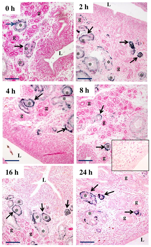
Figure 3.
Representative image of immunolocalization of PrPC protein in endometrium of E2-treated OVX ewes. Arrows indicate PrPC-positive (dark) staining; counterstaining (pink) was with nuclear fast red. Note a lack of positive staining in control (inset), where primary antibody was replaced with mouse IgG. L = lumen of uterus, g = uterine gland, * = blood vessel. Note that PrPC was expressed in arterioles and venules (primarily vascular smooth muscle and adventitia) at 0 to 24 h after E2, but PrPC was not present in glands or luminal epithelium. Size bar = 50 μm.

