Abstract
A broad range of anti-cancer agents, including glucocorticoids (GCs) and tyrosine kinase inhibitors (TKIs), kill cells by upregulating the pro-apoptotic BCL2 family member, BIM. A common germline deletion in the BIM gene was recently shown to favor the production of non-apoptotic BIM isoforms, and to predict inferior responses in TKI-treated chronic myeloid leukemia (CML) and EGFR-driven lung cancer patients. Given that both in vitro and in vivo GC resistance are predictive of adverse outcomes in acute lymphoblastic leukemia (ALL), we hypothesized that this polymorphism would mediate GC resistance, and serve as a biomarker of poor response in ALL. Accordingly, we used zinc finger nucleases to generate ALL cell lines with the BIM deletion, and confirmed the ability of the deletion to mediate GC resistance in vitro. In contrast to CML and lung cancer, the BIM deletion did not predict for poorer clinical outcome in a retrospective analysis of 411 pediatric ALL patients who were uniformly treated with GCs and chemotherapy. Underlying the lack of prognostic significance, we found that the chemotherapy agents used in our cohort (vincristine, L-asparaginase, and methotrexate) were each able to induce ALL cell death in a BIM-independent fashion, and resensitize BIM deletion-containing cells to GCs. Together, our work demonstrates how effective therapy can overcome intrinsic resistance in ALL patients, and suggests the potential of using combinations of drugs that work via divergent mechanisms of cell killing to surmount BIM deletion-mediated drug resistance in other cancers.
Introduction
Genome-wide profiling studies of acute lymphoblastic leukemia (ALL) have revealed it to be a highly heterogeneous disease [1]. In spite of this, the majority of ALL subtypes are treated with a remission-induction protocol that invariably consists of a glucocorticoid, vincristine and at least one other chemotherapy agent (L-asparaginase, an anthracycline, or both) [2]. Unfortunately, 15-20% of patients continue to relapse, and outcome remains poor for these individuals [3]. Consequently, there have been ongoing efforts to identify genetic factors that could account for this response heterogeneity and serve as prognostic markers for risk stratification or novel druggable targets in order to improve patient outcomes [4]–[6].
At the same time, recent reviews have underscored the notion that response heterogeneity can arise from not only somatic mutations but also germline polymorphisms [7], [8]. A number of examples of the latter have been described, including genetic variants that influence the pharmacokinetic and pharmacodynamic phenotype of the host, as well as those affecting the underlying biology of the leukemic cell and thereby cell intrinsic drug resistance/sensitivity [9]–[15]. Notably, however, studies correlating genetic variants with clinical phenotypes have been largely based on genetic epidemiology data and lack experimental validation at a mechanistic level. Such mechanistic studies have been hampered in part by the difficulty and cost of generating isogenic cell lines that either possess or lack a mutation of interest. More recently, a variety of methods that enable genome engineering to faithfully recapitulate mutations of interest have been developed and these will aid the functional validation of these variants in vitro [16].
Using such an approach, we recently validated the functional consequences of a germline deletion in the BIM gene in chronic myeloid leukemia (CML) [17]. Unlike in ALL, a single causative lesion, the 9;22 translocation, is known to be present in >95% of chronic myeloid leukemia (CML) cases [18]. Despite the targeted nature of tyrosine kinase inhibitors (TKIs), response heterogeneity is also a significant challenge in CML [19]. From a group of TKI-resistant CML patients, we identified a 2.9 kb intronic deletion in the BIM gene, and later verified it to be a polymorphism found in 12.3% of East Asians [17]. BIM encodes a potent pro-apoptotic BH3-only protein that is required for specific anti-cancer therapies to induce apoptotic cell death [20]–[25]. When we introduced the deletion into a CML cell line using zinc finger nuclease-based technology, the polymorphism was sufficient to cause intrinsic resistance to tyrosine kinase inhibitors. Mechanistically, we showed that the BIM deletion biases splicing toward BIM isoforms that lack the BH3 domain encoded in exon 4, resulting in the expression of BIM isoforms incapable of inducing apoptosis. Consistent with the in vitro data, both CML and EGFR-driven lung cancer patients carrying the polymorphism experienced inferior responses to treatment with tyrosine kinase inhibitors.
Since BIM is required for GC-induced apoptosis in lymphoid lineage cells, including ALL cells [26]–[32], and both in vitro and in vivo GC response has been shown to predict favorable treatment outcome in ALL [33]–[37], we wondered if the polymorphism could contribute to response heterogeneity in ALL patients. If this were the case, we expect that pharmacological restoration of BIM function using drugs such as BH3 mimetics would enable us to improve response in patients with the polymorphism [17], [25]. Furthermore, because multi-agent chemotherapy is essential to the long-term control of pediatric ALL, the clinical model of ALL could allow us to determine the interaction between a single germline variant and combination therapy.
Accordingly, we used zinc finger nucleases to generate de novo ALL cell lines with the BIM deletion polymorphism in both heterozygous and homozygous configurations. Using these lines, we found that the BIM deletion polymorphism was sufficient to confer GC resistance in vitro. However, analysis of a pediatric ALL cohort uniformly treated with GCs and chemotherapy [38] revealed that patients with the BIM deletion did not experience inferior response rates nor poorer clinical outcomes. Mechanistically, we determined that GC resistance conferred by the BIM polymorphism could be overcome with the addition of chemotherapeutic agents used in standard ALL protocols, and which likely act via a BIM-independent mechanism to cause cell death. Together, our data demonstrate that, whilst the BIM deletion is sufficient to confer resistance to GCs, the negative impact of polymorphic variants on single agent therapy can be overcome with multi-agent chemotherapy that kill cancer cells via divergent mechanisms. These results highlight the challenge of identifying genetic markers predictive of clinical outcome in populations treated with multi-agent therapy, the utility of genome editing technologies in the study of polymorphic variants, as well as the importance of using drug combinations that kill cancer cells via non-overlapping mechanisms.
Methods
Cell lines and culture conditions
CCRF-CEM was purchased from the American Type Culture Collection (Manassas, VA, USA). Cells were maintained in RPMI-1640 media (Nacalai Tesque, Japan) supplemented with 20% FBS, penicillin/streptomycin and L-glutamine (all from Thermo Scientific, Rockford, IL, USA). Dexamethasone (Rotexmedica, Germany), methotrexate (ABIC Ltd, Israel), vincristine (Korea United Pharm Inc, Korea) and L-asparaginase (Kyowa Hakko Kirin, Japan) were used at the dosages and times indicated in the figure legends. All experiments using cell lines were performed at least 3 times.
Creation of genome-edited lines
The zinc finger nucleases (ZFNs) targeting the BIM gene were custom-made (Sigma-Aldrich, St Louis, MO, USA) and the repair template was generated as described in a previous paper [17]. Plasmids encoding the repair template and ZFNs were transfected into CCRF-CEM cells using the Neon system (Invitrogen, Carlsbad, CA, USA). Clones were isolated by dilution cloning and screened for presence of the deletion using the following primers: Forward (5′-GGCCTTCAACCACTATCTCAGTGCAATGG-3′) and Reverse (5′- GGTTTCAGAGACAGAGCTGGGACTCC-3′). qPCR to determine exon 3 to exon 4 ratio was performed as previously described [17].
MTS assays
Cells were seeded at a density of 4×10⁴ per well in a 96-well plate and incubated with the indicated drugs. In each experiment, every treatment condition was repeated in triplicate wells. After 48 h, CellTiter AQueous One Solution Cell Proliferation reagent (Promega, Fitchburg, WI, USA) was added to each well and incubated for 2 h before an absorbance reading at 490 nm was taken.
Immunoblotting
Cells were washed once in PBS and lysed in RIPA lysis buffer (Millipore, Billerica, MA, USA) containing proteinase inhibitor (Roche, Indianapolis, IN, USA). Protein concentrations were assayed using the Quick Start Bradford protein assay kit (Bio-Rad) and bovine serum albumin as a standard. The following antibodies were used at these concentrations: phospho-glucocorticoid receptor (S211) (Cell Signaling Technology, Danvers, MA, USA #4161, 1∶1 000), glucocorticoid receptor (BD Transduction Laboratories, San Jose, CA, USA, 1∶2 000), β-actin (Sigma-Aldrich, 1∶10 000), PARP (Cell Signaling Technology #9542, 1∶2 000), caspase 3 (Cell Signaling Technology #9663, 1∶500) and BIM (Cell Signaling Technology #2819, 1∶1 000). HRP-conjugated secondary antibodies against mouse or rabbit IgG (Santa Cruz Biotechnology, Santa Cruz, CA, USA) were used at 1∶10 000. Western Lightning ECL reagent (PerkinElmer, Waltham, MA, USA) was used to visualize the protein bands. Any adjustments to contrast and intensity were applied uniformly to the images.
Patient recruitment
411 patients with newly-diagnosed ALL from the Malaysia-Singapore (Ma-Spore) acute lymphoblastic leukemia (ALL) 2003 study [38] were included on the basis of DNA availability. Written informed consent was obtained from the parents or the legal guardians of the patients. The study was approved by the National Healthcare Group Domain Specific Review Board (NHG DSRB). Since the BIM deletion is germline in nature, and will be present in both normal and leukemic samples, we were able to employ both remission (n = 362) and diagnostic (n = 49) samples for genotyping for this study. Patient risk stratification, details of the treatment protocol, minimal residual disease monitoring and molecular subgrouping were described previously [38].
Statistical analyses
Statistical analysis was performed using SPSS software (version 16.0 for Windows; IBM Corporation, Armonk, NY, USA). Comparisons between groups were examined by Fisher's exact test for categorical variables. A statistically significant difference was defined as a P value of <0.05. Survival curves were evaluated using Kaplan–Meier analysis. Event-free survival (EFS) was defined as the time from diagnosis to first recurrence of the disease, including induction failure, or death. Induction failure was defined as failure to achieve complete remission and considered as an event at one day after date of diagnosis. Overall survival (OS) was defined as the time from diagnosis to death. Patients who were alive and had no progression of disease or relapse were censored at the time of their last follow-up.
Results
De novo generation of ALL cell lines bearing the BIM deletion polymorphism
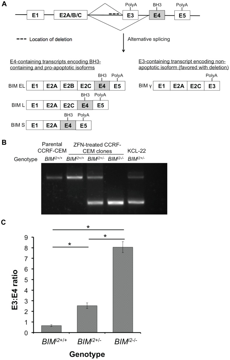
To determine if the BIM deletion polymorphism is sufficient to confer GC resistance to ALL cells, we used zinc finger nucleases to derive de novo ALL cell lines bearing the deletion. Because human cell lines vary in their amenability to transfection and genome editing by zinc fingers (personal communication, TK Ko and unpublished observations), we tested the ability of our approach to edit 3 different GC-sensitive ALL cell lines (CCRF-CEM, RS4;11, and PALL-2) [39]–[41] that did not have the polymorphism. Of these lines, we were only able to successfully generate clones containing the deletion in CCRF-CEM cells. The structure of the BIM gene and the location of the deletion in intron 2 are illustrated in Figure 1A. Using PCR primers that flank the deleted region, we identified subclones that either did not have the deletion (denoted BIMi2+/+) or were heterozygous (denoted BIMi2+/−) or homozygous (denoted BIMi2−/−) for the deletion polymorphism (Figure 1B). The deleted region contains splicing elements that either promote the production of functional, exon 4-containing isoforms or suppress the production of non-apoptotic, exon 3-containing isoforms [42]. Consequently, when deleted, an increase in exon 3 to exon 4-containing transcripts is expected. To confirm that the deletion produced the expected changes in the splicing of BIM, we measured the ratio of exon 3- to exon 4-containing transcripts in clones of each genotype by exon-specific RT-PCR, and found that it was increased in a polymorphism-dosage-dependent manner (Figure 1C).
Figure 1. Generation of isogenic CCRF-CEM cell lines with the BIM deletion polymorphism.
(A) Structure of the BIM gene and major splice isoforms. The BIM deletion polymorphism lies within intron 2 and upstream of exon 3, as indicated by the dashed line. Exon 4 contains the crucial BH3 domain required for apoptosis. Exon 3 (E3) and 4 (E4) are spliced in a mutually exclusive fashion, leading to the generation of either E4-containing isoforms with the BH3 domain (BIM EL, BIM L and BIM S) or E3-containing isoforms without the BH3 domain (BIM γ). When present, the deletion biases splicing towards E3-containing non-apoptotic isoforms. (B) Agarose gel of the products from a PCR reaction to detect the polymorphism in zinc finger nuclease (ZFN)-treated CCRF-CEM subclones, with the lower band indicating the presence of the deletion. Parental CCRF-CEM and KCL-22 cells (a CML cell line known to be heterozygous for the BIM deletion polymorphism) were included as controls. (C) The ratio of exon 3 to exon 4-containing transcripts (E3:E4) in CCRF-CEM BIMi2+/+, BIMi2+/− and BIMi2−/− clones as measured by qPCR. Error bars indicate mean ± SEM (n = 3). A student's t-test was performed for pairwise comparisons of E3:E4 ratio between genotypes. * indicates a significant difference with P<0.05.
The BIM deletion polymorphism is sufficient to confer GC resistance in ALL cells
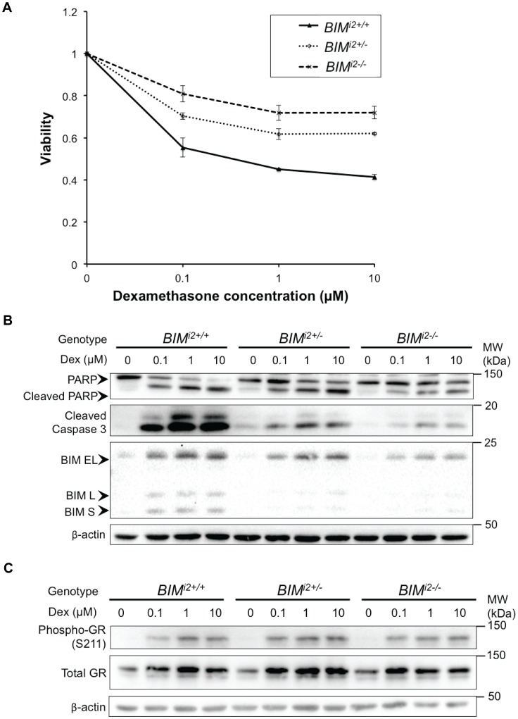
To determine if the deletion conferred resistance to GCs, we compared the effect of treating clones of each genotype with a range of dexamethasone concentrations. First, we quantified cell viability using the MTS assay and found that across the range tested, the deletion-containing clones exhibited increased cell viability in a polymorphism dosage-dependent manner (Figure 2A). Following this, we assessed the extent of apoptosis using the induction of cleaved poly ADP-ribose polymerase (PARP), as well as the level of cleaved caspase 3, using immunoblots performed on lysates of cells treated as in Figure 2A. We also probed for BIM using an antibody that only detects the pro-apoptotic E4-containing isoforms (BIM EL, L, and S). As a marker of glucocorticoid receptor (GR) activation, we probed for phospho-GR (S211). As predicted, when compared to wildtype clones, upregulation of E4-containing BIM isoforms was impaired in a polymorphism dosage-dependent manner. Furthermore, apoptosis was attenuated in clones with the deletion, as evidenced by an increase in cleaved PARP, as well as cleaved caspase 3 (Figure 2B). Importantly, this occurred in spite of equivalent GR phosphorylation and auto-induction upon GC treatment across the genotypes (Figure 2C). These results indicate that GC resistance in the deletion-containing clones takes place downstream of the GR, and is consistent with our hypothesis that GC resistance results from impaired expression of BH3-containing BIM isoforms. Taken together, our data demonstrate that the presence of the BIM deletion polymorphism is sufficient to confer GC resistance in ALL cells.
Figure 2. The BIM deletion confers dexamethasone resistance in CCRF-CEM cells.
(A) Cell viability following exposure of CCRF-CEM subclones to increasing concentrations of dexamethasone. Viability was measured by MTS assay at 48 h. Error bars indicate mean ± SEM (n = 3) of 3 independent replicates. (B) Western blot of cell lysates from CCRF-CEM BIMi2+/+, BIMi2+/− and BIMi2−/− clones following treatment with increasing doses of dexamethasone for 48 h. The induction of cleaved PARP and cleaved caspase 3 were used as readouts for apoptosis. An antibody that recognizes pro-apoptotic exon-4 containing BIM isoforms (BIM EL, L and S) was used to show the extent of BIM upregulation following GC exposure. β-actin was used as a loading control. (C) Western blot showing phosphorylation of the glucocorticoid receptor (Phospho-GR S211) in CCRF-CEM BIMi2+/+, BIMi2+/− and BIMi2−/− clones upon treatment with dexamethasone.
Because prior work has shown that in vitro GC responses per se is an important prognostic factor in childhood ALL [33], [34], we predicted that patients with the BIM deletion polymorphism would have inferior outcomes compared to those without.
The BIM deletion polymorphism does not predict inferior responses in pediatric ALL
To test our prediction that the BIM deletion polymorphism confers a poorer clinical outcome in pediatric ALL, we conducted a retrospective analysis correlating treatment outcome with the presence of the polymorphism in a group of uniformly-treated pediatric patients from the Malaysia-Singapore (Ma-Spore) ALL 2003 multicenter study. The Ma-Spore ALL 2003 protocol was based on a modified Berlin-Frankfurt-Münster regimen, where all patients received intrathecal methotrexate together with seven days of oral prednisolone at the point of diagnosis. Patients subsequently completed the rest of their induction regimen based on a common backbone of vincristine, L-asparaginase, and methotrexate, followed by risk-adapted consolidation and maintenance therapy as directed by their MRD status at day 33. Importantly, the design of this study allowed us to determine if the BIM deletion predicts for inferior clinical outcomes at three distinct assessment points: initial GC response (defined as absolute blast count ≥1000/µl at day 8), day 33 MRD following multi-agent induction chemotherapy, as well as overall survival (OS) and event-free survival (EFS) after consolidation and maintenance therapy.
Sufficient DNA from 411 individuals (out of a total of 556) from the Ma-Spore study was available for analysis for the BIM deletion polymorphism. Importantly, there was no difference in treatment outcome between this subgroup of 411 patients compared to the 556 patients in the full study (5-year EFS 82.0% vs 80.6%). Using this sample set, we determined the incidence of the BIM deletion to be 12.2%, which is consistent with the ethnic make up of the Ma-Spore cohort, as well as the incidence of the polymorphism in the normal population (Table 1, [17]). We also found that the BIM deletion polymorphism did not segregate according to any patient demographic except for Chinese ethnicity, which is as expected, or adverse prognostic indicators such as genetic subtype (Table 1).
Table 1. Biological and clinical features of patients from the Ma-Spore ALL 2003 trial genotyped for the BIM polymorphism.
| Characteristics | P value | ||||
| Wildtype (n = 361) | BIM polymorphism present (n = 50) | ||||
| No. % | No. % | ||||
| Age at diagnosis | 1.000 | ||||
| <1 or >10 | 71 | 19.7 | 10 | 20 | |
| 1–10 | 290 | 80.3 | 40 | 80 | |
| Sex | 0.094 | ||||
| Male | 213 | 59 | 23 | 46 | |
| Female | 148 | 41 | 27 | 54 | |
| Molecular subtype∧ | 0.127 | ||||
| ETV6-RUNX1 | 68 | 19 | 7 | 14.3 | |
| TCF-PBX1 | 17 | 4.7 | 7 | 14.3 | |
| BCR-ABL1 | 16 | 4.5 | 1 | 2 | |
| MLL rearrangements | 9 | 2.5 | 3 | 6.1 | |
| Hyperdiploidy | 65 | 18.2 | 12 | 24.5 | |
| Hypodiploidy | 4 | 1.1 | 0 | 0 | |
| T-ALL | 30 | 8.4 | 3 | 6.1 | |
| Others | 149 | 41.6 | 16 | 32.7 | |
| NCI Risk | 0.642 | ||||
| High | 138 | 38.2 | 17 | 34 | |
| Low | 223 | 61.8 | 33 | 66 | |
| Day 8 Prednisolone Response∧ | 0.804 | ||||
| Good | 320 | 88.9 | 44 | 91.7 | |
| Poor | 40 | 11.1 | 4 | 8.3 | |
| Day 33 PCR MRD∧ | 0.970 | ||||
| <0.01% | 146 | 43.6 | 19 | 46.3 | |
| 0.01–1% | 155 | 46.4 | 18 | 43.9 | |
| ≥1% | 34 | 10 | 4 | 9.8 | |
| PCR MRD Risk∧ | 0.966 | ||||
| Standard | 134 | 38.4 | 17 | 37 | |
| Intermediate | 194 | 55.6 | 27 | 58.7 | |
| High | 21 | 6 | 2 | 4.3 | |
| Ma-Spore Risk∧ | 0.463 | ||||
| Standard | 109 | 30.2 | 14 | 28 | |
| Intermediate | 177 | 49 | 29 | 58 | |
| High | 75 | 20.8 | 7 | 14 | |
| Ma-Spore Outcome | 0.608 | ||||
| CCR | 295 | 81.7 | 42 | 84 | |
| Induction Failure | 16 | 4.4 | 1 | 2 | |
| Relapse | 21 | 5.8 | 1 | 2 | |
| Death | 17 | 4.7 | 3 | 6 | |
| Abandonment | 12 | 3.3 | 3 | 6 | |
| Race | <0.001 | ||||
| Chinese | 156 | 43.2 | 35 | 70 | |
| Malay | 147 | 40.7 | 14 | 28 | |
| Indian & Others | 58 | 16.1 | 1 | 2 | |
Incidence of the BIM polymorphism is 50 out of 411 patients, or 12.2%. Abbreviations: Ma-Spore, Malaysia- Singapore; MRD, minimal residual disease; NCI, National Cancer Institute; PCR, polymerase chain reaction; CCR, continuous complete remission.
indicates that data was unavailable for some patients.
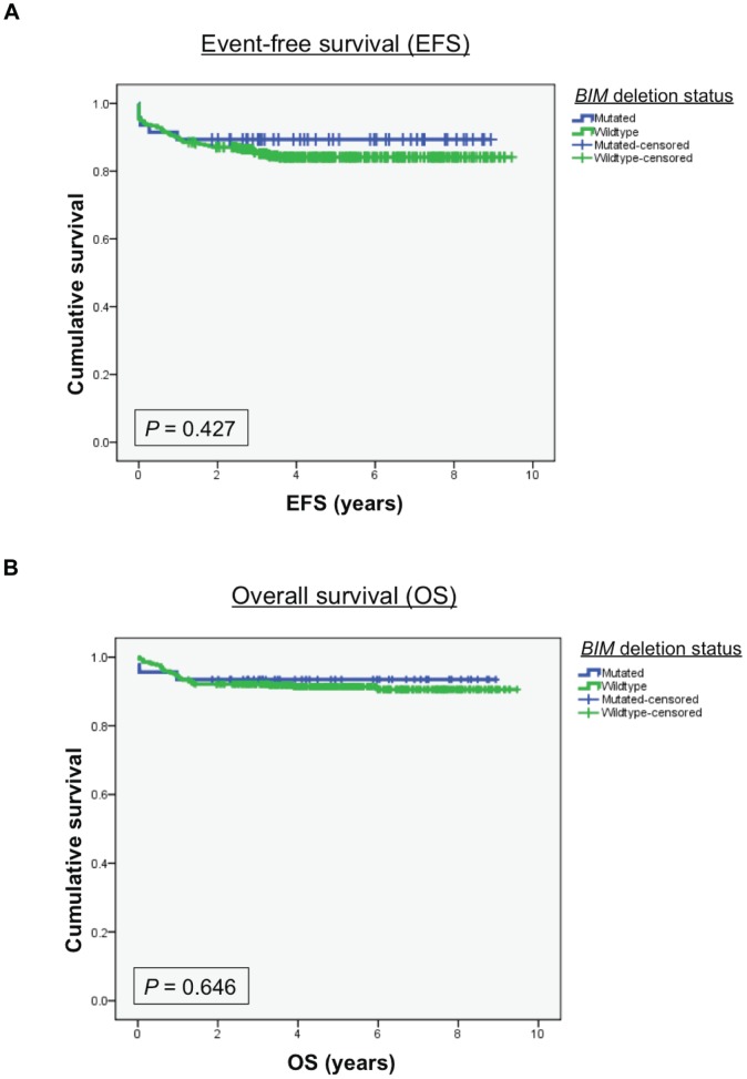
We next determined if the BIM deletion predicted for inferior outcomes at each of the three response assessment points described above. Here, and to our surprise, we found that there was no significant difference between patients with or without the deletion for GC response at day 8 (P = 0.804), MRD response at day 33 (P = 0.970), nor EFS (P = 0.427) or overall OS (P = 0.646) (Table 1, Figures 3A and 3B). Additionally, subgroup analysis by genetic subtype, race and risk category at diagnosis did not uncover any associations between the BIM deletion polymorphism and treatment outcome (data not shown). Together, these results demonstrate that the BIM deletion polymorphism does not predict for inferior outcomes following the administration of a modern GC-containing three-drug remission-induction regimen. Our clinical observations led us to propose that at least one or more of the chemotherapy agents employed during induction is able to overcome GC resistance conferred by the BIM deletion polymorphism.
Figure 3. Retrospective analysis of the Ma-Spore ALL 2003 cohort according to the presence or absence of the BIM deletion polymorphism.
Kaplan-Meier curves comparing event-free survival (A) or overall survival (B) in patients with or without the BIM deletion polymorphism are shown.
Chemotherapy overcomes GC resistance conferred by the BIM deletion polymorphism
To determine if any of the chemotherapy agents used in the induction regimen was able to overcome BIM deletion-mediated GC resistance, we treated the BIM deletion-containing clones with methotrexate, vincristine, and L-asparaginase individually, and in combination with dexamethasone. Cells were then assessed for activation of apoptotic cell death, BIM protein induction, and cell viability.
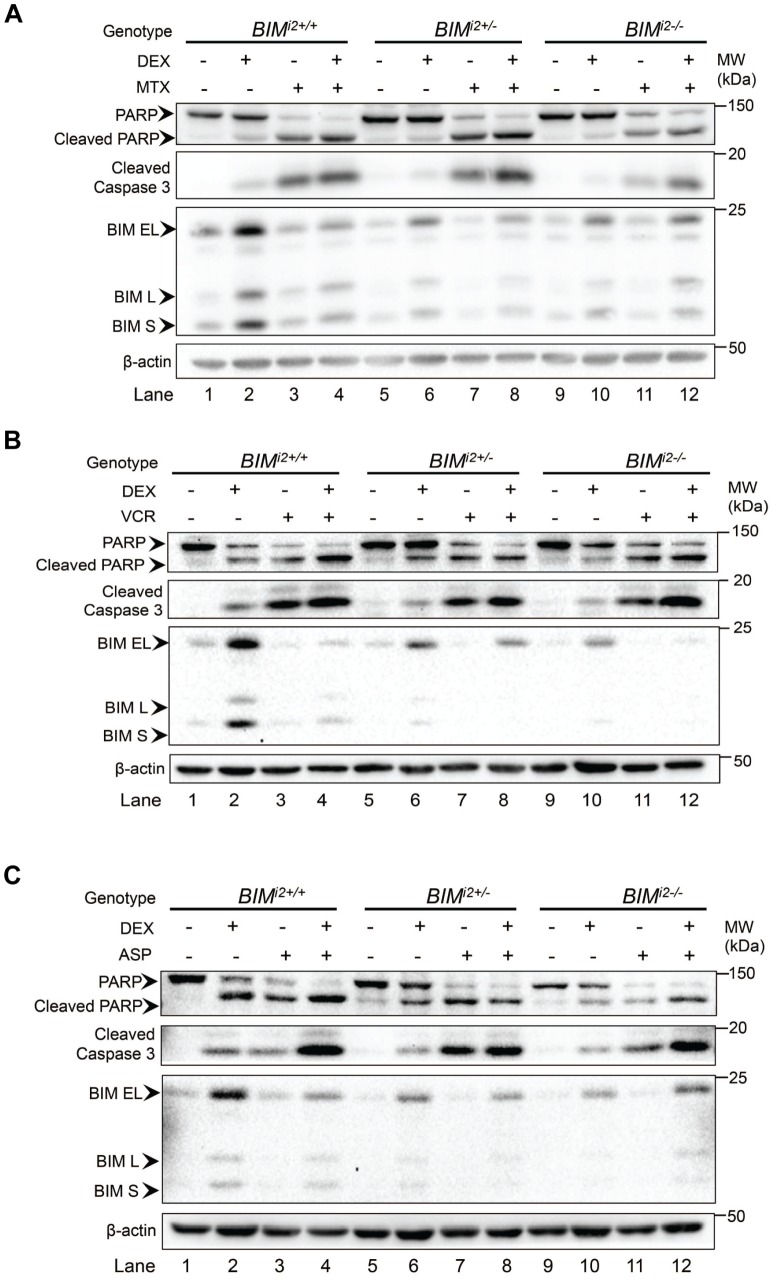
First, using immunoblot, we found that each of the three chemotherapy agents was individually able to induce equivalent levels of apoptotic cell death (as measured by the production of cleaved PARP and caspase 3) in the absence and presence of the BIM deletion (Figures 4A–C, compare lanes 3, 7 and 11). Importantly, we also observed that chemotherapy-induced apoptosis occurred without significant induction of any of the three BIM isoforms (BIM EL, L, and S) reported to be important for GC-induced apoptosis [31]. These results demonstrate that methotrexate, vincristine, and L-asparaginase are each able to induce ALL cell death in a BIM-independent manner, and that this occurred regardless of the BIM deletion status of the cell line.
Figure 4. Methotrexate, vincristine, and L-asparaginase activate apoptosis in a BIM-independent manner, and overcome BIM deletion-mediated GC resistance.
CCRF-CEM clones were treated with dexamethasone (DEX) (0.1 µM) with or without (A) methotrexate (MTX) (1 µM), (B) vincristine (VCR) (2 ng/ml), or (C) L-asparaginase (ASP) (0.5 IU/ml) for 48 h. Following incubation, cell lysates were obtained and analyzed for cleaved PARP and caspase 3, as well as BIM induction. β-actin was used as a loading control.
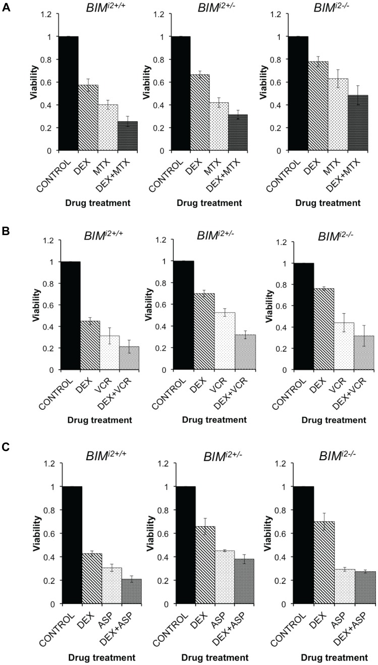
Next, we found that when dexamethasone was combined with methotrexate, vincristine or L-asparaginase, there was a consistent increase in the level of activated PARP and caspase 3 compared to GC alone (Figures 4A–C, lanes 4, 8 and 12). Similarly, when cell viability was assayed, the addition of methotrexate (Figure 5A), vincristine (Figure 5B), or L-asparaginase (Figure 5C) to dexamethasone augmented cell death in deletion-containing clones. Taken together, our in vitro data suggest that the ability of the BIM deletion polymorphism to confer GC resistance can be overcome by the co-administration of several of the cytotoxic components of the Ma-Spore regimen, including methotrexate, vincristine, and L-asparaginase.
Figure 5. The addition of methotrexate, vincristine or L-asparaginase resensitizes BIM deletion-containing CCRF-CEM clones to dexamethasone.
Cell viability was measured by MTS assay after (A) methotrexate (MTX) (1 µM), (B) vincristine (VCR) (2 ng/ml), or (C) L-asparaginase (ASP) (0.5 IU/ml) was used singly or in combination with dexamethasone (DEX) (0.1 µM) for 48 h. Values obtained for treated cells were normalized to the untreated control for the same genotype. Error bars indicate SEM (n = ) of 3 independent replicates.
Discussion
In the current work, we used a genome-editing approach to demonstrate that a common germline variant in the BIM gene is sufficient to confer GC resistance in ALL cell lines. Mechanistically, we confirm that cells harboring the deletion favor the splicing and expression of non-apoptotic isoforms of BIM, impairing the apoptotic response to GC exposure, and thereby promoting ALL cell survival. However, using a cohort of 411 uniformly-treated ALL patients, we also find that the deletion does not predict inferior responses to GC-containing multi-agent chemotherapy, and that this is associated with the ability of chemotherapy to induce BIM-independent cell death.
By generating CCRF-CEM subclones that were either wildtype, heterozygous or homozygous for the BIM polymorphism, we were able to demonstrate that the BIM deletion polymorphism is able to confer GC resistance in a T-ALL cell line. In these clones, the expected changes in splicing to favor the E3-containing, non-apoptotic splice variants were recapitulated in a polymorphism dose-dependent manner. We then showed that both upregulation of the E4-containing BIM isoforms and apoptosis upon GC treatment were impaired in the deletion-containing clones. Overall, our results are consistent with prior work demonstrating a critical role for BIM induction in GC-induced ALL cell death, particularly the EL, L, and S isoforms which harbor the E4- and BH3-containing isoforms capable of activating apoptosis [27], [31]. One limitation of our in vitro studies is the use of a single cell line, CCRF-CEM, which is a T-ALL line. Although we were unable to generate deletion-containing lines of other lineages, we expect that introduction of the deletion, which phenocopies a BIM knockdown of BIM, will likely confer glucocorticoid resistance in other ALL cell lines [27], [31], [32].
The inability of the BIM deletion to segregate poor versus good risk patients was somewhat surprising given previous reports describing the ability of in vitro as well as clinical GC responses to predict long-term outcomes in pediatric ALL [33]–[37]. Importantly, because we were able to generate isogenic cell lines with and without the BIM deletion, we could explore the mechanisms underlying our clinical observations. Here, we found that three other agents employed in the Ma-Spore ALL 2003 regimen, methotrexate, vincristine, and L-asparaginase, are each individually able to overcome BIM deletion-associated GC resistance. Our in vitro results also indicate that each of these drugs activate apoptotic cell death in a largely BIM-independent manner, and that this is likely to underlie their clinical efficacy in overcoming BIM deletion-mediated GC resistance. While the precise mechanisms by which methotrexate and L-asparaginase induce apoptosis remain ill-defined [43], it is interesting to note that the mechanism of vincristine-induced apoptosis has recently been described [44]. Here [44], and consistent with our observations, vincristine-induced apoptosis was shown to occur via the depletion of the pro-survival protein MCL1, a factor that has itself been shown to mediate GC resistance in ALL [45].
It is also important to highlight that in vitro resistance to GC, which has been shown to correlate with clinical responses [33], [34], may not necessarily readout for GC resistance per se. This is because such assays will also read out for more general mechanisms of resistance that would be expected to mediate cross-resistance among different drug classes, a conclusion that other studies have suggested [46], [47]. More recent work has also implicated other germline BIM variants in mediating drug resistance [48], [49]. Importantly, and reminiscent of our data, we note that it was the combination of a functional SNP in BIM with a SNP in the MCL1 promoter (associated with increased MCL1 expression) that best predicted OS in pediatric ALL [49], [50]. Together, our observations are consistent with a “BCL-2 rheostat” model where the cellular apoptotic threshold is set by the balance of pro-apoptotic BCL-2 family members such as BIM and anti-apoptotic members like MCL1 [27]. This model would predict that genetic variants affecting BCL2 family members may only be clinically important when two or more act in concert to alter the apoptotic threshold.
While there is increasing evidence that germline polymorphisms contribute to clinical heterogeneity in ALL [13]–[15], it is likely that only those variants capable of conferring alterations in biological behavior and/or multi-drug resistance will be associated with clinically meaningful endpoints. Thus, as we have demonstrated with the BIM deletion, polymorphisms that confer single-drug resistance in the setting of modern multi-agent ALL therapy are less likely to be of clinical importance. Indeed, variants that have been shown to predict poor response are enriched for genes expected to confer a multi-drug resistance phenotype, and include those that influence systemic drug clearance and intracellular drug concentrations [9], [12].
Finally, our observations also highlight the ability of at least three cytotoxic agents to induce apoptosis independently of BIM, and suggest that the success of modern day ALL regimens is due to the ability of individual agents to kill leukemia cells via targeting different components of the “BCL-2 rheostat”. Indeed, this general lesson may be applied to cancers where we have found that the BIM deletion does play a part in clinical drug resistance [17], and supports the use of judiciously chosen combination therapies to overcome BIM deletion-mediated drug resistance in these patients.
Acknowledgments
The authors thank the collaborators and patients in the Ma-Spore ALL 2003 study, as well as TK Ko for advice on genome editing and SK Kham for technical assistance.
Data Availability
The authors confirm that all data underlying the findings are fully available without restriction. All relevant data are within the paper and its Supporting Information files.
Funding Statement
STO was supported by the Leukemia & Lymphoma Society Translational Research Program Grant (R913-302-026-597) www.lls.org. AEY was supported by the Singapore National Medical Research Council (NMRC/0582/2001, NMRC/CSA/003/2008). www.nmrc.gov.sg, and by A*STAR (SCS-POU98, NMRC/CSI/004/2005). www.a-star.edu.sg, and by Children's Cancer Foundation, Singapore. www.ccf.org.sg, and by VIVA Foundation for Children with Cancer, Singapore. www.viva.sg. The funders had no role in study design, data collection and analysis, decision to publish, or preparation of the manuscript.
References
- 1. Mullighan CG (2012) The molecular genetic makeup of acute lymphoblastic leukemia. Hematology Am Soc Hematol Educ Program 2012: 389–396. [DOI] [PubMed] [Google Scholar]
- 2. Pui CH, Evans WE (2006) Treatment of acute lymphoblastic leukemia. N Engl J Med 354: 166–178. [DOI] [PubMed] [Google Scholar]
- 3. Locatelli F, Schrappe M, Bernardo ME, Rutella S (2012) How I treat relapsed childhood acute lymphoblastic leukemia. Blood 120: 2807–2816. [DOI] [PubMed] [Google Scholar]
- 4. Yeoh EJ, Ross ME, Shurtleff SA, Williams WK, Patel D, et al. (2002) Classification, subtype discovery, and prediction of outcome in pediatric acute lymphoblastic leukemia by gene expression profiling. Cancer Cell 1: 133–143. [DOI] [PubMed] [Google Scholar]
- 5. Martinelli G, Iacobucci I, Storlazzi CT, Vignetti M, Paoloni F, et al. (2009) IKZF1 (Ikaros) deletions in BCR-ABL1-positive acute lymphoblastic leukemia are associated with short disease-free survival and high rate of cumulative incidence of relapse: a GIMEMA AL WP report. J Clin Oncol 27: 5202–5207. [DOI] [PubMed] [Google Scholar]
- 6. Roberts KG, Morin RD, Zhang J, Hirst M, Zhao Y, et al. (2012) Genetic alterations activating kinase and cytokine receptor signaling in high-risk acute lymphoblastic leukemia. Cancer Cell 22: 153–166. [DOI] [PMC free article] [PubMed] [Google Scholar]
- 7. McLeod HL (2013) Cancer pharmacogenomics: early promise, but concerted effort needed. Science 339: 1563–1566. [DOI] [PMC free article] [PubMed] [Google Scholar]
- 8. Coate L, Cuffe S, Horgan A, Hung RJ, Christiani D, et al. (2010) Germline genetic variation, cancer outcome, and pharmacogenetics. J Clin Oncol 28: 4029–4037. [DOI] [PubMed] [Google Scholar]
- 9. Radtke S, Zolk O, Renner B, Paulides M, Zimmermann M, et al. (2013) Germline genetic variations in methotrexate candidate genes are associated with pharmacokinetics, toxicity, and outcome in childhood acute lymphoblastic leukemia. Blood 121: 5145–5153. [DOI] [PubMed] [Google Scholar]
- 10. Xu H, Cheng C, Devidas M, Pei D, Fan Y, et al. (2012) ARID5B genetic polymorphisms contribute to racial disparities in the incidence and treatment outcome of childhood acute lymphoblastic leukemia. J Clin Oncol 30: 751–757. [DOI] [PMC free article] [PubMed] [Google Scholar]
- 11. Trevino LR, Shimasaki N, Yang W, Panetta JC, Cheng C, et al. (2009) Germline genetic variation in an organic anion transporter polypeptide associated with methotrexate pharmacokinetics and clinical effects. J Clin Oncol 27: 5972–5978. [DOI] [PMC free article] [PubMed] [Google Scholar]
- 12. Rocha JC, Cheng C, Liu W, Kishi S, Das S, et al. (2005) Pharmacogenetics of outcome in children with acute lymphoblastic leukemia. Blood 105: 4752–4758. [DOI] [PMC free article] [PubMed] [Google Scholar]
- 13. Perez-Andreu V, Roberts KG, Harvey RC, Yang W, Cheng C, et al. (2013) Inherited GATA3 variants are associated with Ph-like childhood acute lymphoblastic leukemia and risk of relapse. Nat Genet 45: 1494–1498. [DOI] [PMC free article] [PubMed] [Google Scholar]
- 14. Yang JJ, Cheng C, Devidas M, Cao X, Campana D, et al. (2012) Genome-wide association study identifies germline polymorphisms associated with relapse of childhood acute lymphoblastic leukemia. Blood 120: 4197–4204. [DOI] [PMC free article] [PubMed] [Google Scholar]
- 15. Yang JJ, Cheng C, Yang W, Pei D, Cao X, et al. (2009) Genome-wide interrogation of germline genetic variation associated with treatment response in childhood acute lymphoblastic leukemia. JAMA 301: 393–403. [DOI] [PMC free article] [PubMed] [Google Scholar]
- 16. Gaj T, Gersbach CA, Barbas CF 3rd (2013) ZFN, TALEN, and CRISPR/Cas-based methods for genome engineering. Trends Biotechnol 31: 397–405. [DOI] [PMC free article] [PubMed] [Google Scholar]
- 17. Ng KP, Hillmer AM, Chuah CT, Juan WC, Ko TK, et al. (2012) A common BIM deletion polymorphism mediates intrinsic resistance and inferior responses to tyrosine kinase inhibitors in cancer. Nat Med 18: 521–528. [DOI] [PubMed] [Google Scholar]
- 18. Morel F, Ka C, Le Bris MJ, Herry A, Morice P, et al. (2003) Deletion of the 5′ABL region in Philadelphia chromosome positive chronic myeloid leukemia: frequency, origin and prognosis. Leuk Lymphoma 44: 1333–1338. [DOI] [PubMed] [Google Scholar]
- 19. Jabbour E, Cortes JE, Kantarjian HM (2009) Suboptimal response to or failure of imatinib treatment for chronic myeloid leukemia: what is the optimal strategy? Mayo Clin Proc 84: 161–169. [DOI] [PMC free article] [PubMed] [Google Scholar]
- 20. Aichberger KJ, Mayerhofer M, Krauth MT, Vales A, Kondo R, et al. (2005) Low-level expression of proapoptotic Bcl-2-interacting mediator in leukemic cells in patients with chronic myeloid leukemia: role of BCR/ABL, characterization of underlying signaling pathways, and reexpression by novel pharmacologic compounds. Cancer Res 65: 9436–9444. [DOI] [PubMed] [Google Scholar]
- 21. Bouillet P, Metcalf D, Huang DC, Tarlinton DM, Kay TW, et al. (1999) Proapoptotic Bcl-2 relative Bim required for certain apoptotic responses, leukocyte homeostasis, and to preclude autoimmunity. Science 286: 1735–1738. [DOI] [PubMed] [Google Scholar]
- 22. Cragg MS, Kuroda J, Puthalakath H, Huang DC, Strasser A (2007) Gefitinib-induced killing of NSCLC cell lines expressing mutant EGFR requires BIM and can be enhanced by BH3 mimetics. PLoS Med 4: 1681–1689 discussion 1690. [DOI] [PMC free article] [PubMed] [Google Scholar]
- 23. Gong Y, Somwar R, Politi K, Balak M, Chmielecki J, et al. (2007) Induction of BIM is essential for apoptosis triggered by EGFR kinase inhibitors in mutant EGFR-dependent lung adenocarcinomas. PLoS Med 4: e294. [DOI] [PMC free article] [PubMed] [Google Scholar]
- 24. Kuribara R, Honda H, Matsui H, Shinjyo T, Inukai T, et al. (2004) Roles of Bim in apoptosis of normal and Bcr-Abl-expressing hematopoietic progenitors. Mol Cell Biol 24: 6172–6183. [DOI] [PMC free article] [PubMed] [Google Scholar]
- 25. Kuroda J, Puthalakath H, Cragg MS, Kelly PN, Bouillet P, et al. (2006) Bim and Bad mediate imatinib-induced killing of Bcr/Abl+ leukemic cells, and resistance due to their loss is overcome by a BH3 mimetic. Proc Natl Acad Sci U S A 103: 14907–14912. [DOI] [PMC free article] [PubMed] [Google Scholar]
- 26. Erlacher M, Michalak EM, Kelly PN, Labi V, Niederegger H, et al. (2005) BH3-only proteins Puma and Bim are rate-limiting for gamma-radiation- and glucocorticoid-induced apoptosis of lymphoid cells in vivo. Blood 106: 4131–4138. [DOI] [PMC free article] [PubMed] [Google Scholar]
- 27. Ploner C, Rainer J, Niederegger H, Eduardoff M, Villunger A, et al. (2008) The BCL2 rheostat in glucocorticoid-induced apoptosis of acute lymphoblastic leukemia. Leukemia 22: 370–377. [DOI] [PMC free article] [PubMed] [Google Scholar]
- 28. Wang Z, Malone MH, He H, McColl KS, Distelhorst CW (2003) Microarray analysis uncovers the induction of the proapoptotic BH3-only protein Bim in multiple models of glucocorticoid-induced apoptosis. J Biol Chem 278: 23861–23867. [DOI] [PubMed] [Google Scholar]
- 29. Schmidt S, Rainer J, Riml S, Ploner C, Jesacher S, et al. (2006) Identification of glucocorticoid-response genes in children with acute lymphoblastic leukemia. Blood 107: 2061–2069. [DOI] [PubMed] [Google Scholar]
- 30. Bachmann PS, Piazza RG, Janes ME, Wong NC, Davies C, et al. (2010) Epigenetic silencing of BIM in glucocorticoid poor-responsive pediatric acute lymphoblastic leukemia, and its reversal by histone deacetylase inhibition. Blood 116: 3013–3022. [DOI] [PubMed] [Google Scholar]
- 31. Abrams MT, Robertson NM, Yoon K, Wickstrom E (2004) Inhibition of glucocorticoid-induced apoptosis by targeting the major splice variants of BIM mRNA with small interfering RNA and short hairpin RNA. J Biol Chem 279: 55809–55817. [DOI] [PubMed] [Google Scholar]
- 32. Jiang N, Koh GS, Lim JY, Kham SK, Ariffin H, et al. (2011) BIM is a prognostic biomarker for early prednisolone response in pediatric acute lymphoblastic leukemia. Exp Hematol 39: 321–329. [DOI] [PubMed] [Google Scholar]
- 33. Kaspers GJ, Pieters R, Van Zantwijk CH, Van Wering ER, Van Der Does-Van Den Berg A, et al. (1998) Prednisolone resistance in childhood acute lymphoblastic leukemia: vitro-vivo correlations and cross-resistance to other drugs. Blood 92: 259–266. [PubMed] [Google Scholar]
- 34. Den Boer ML, Harms DO, Pieters R, Kazemier KM, Gobel U, et al. (2003) Patient stratification based on prednisolone-vincristine-asparaginase resistance profiles in children with acute lymphoblastic leukemia. J Clin Oncol 21: 3262–3268. [DOI] [PubMed] [Google Scholar]
- 35. Lauten M, Moricke A, Beier R, Zimmermann M, Stanulla M, et al. (2012) Prediction of outcome by early bone marrow response in childhood acute lymphoblastic leukemia treated in the ALL-BFM 95 trial: differential effects in precursor B-cell and T-cell leukemia. Haematologica 97: 1048–1056. [DOI] [PMC free article] [PubMed] [Google Scholar]
- 36. Dordelmann M, Reiter A, Borkhardt A, Ludwig WD, Gotz N, et al. (1999) Prednisone response is the strongest predictor of treatment outcome in infant acute lymphoblastic leukemia. Blood 94: 1209–1217. [PubMed] [Google Scholar]
- 37. Schrappe M, Arico M, Harbott J, Biondi A, Zimmermann M, et al. (1998) Philadelphia chromosome-positive (Ph+) childhood acute lymphoblastic leukemia: good initial steroid response allows early prediction of a favorable treatment outcome. Blood 92: 2730–2741. [PubMed] [Google Scholar]
- 38. Yeoh AE, Ariffin H, Chai EL, Kwok CS, Chan YH, et al. (2012) Minimal residual disease-guided treatment deintensification for children with acute lymphoblastic leukemia: results from the Malaysia-Singapore acute lymphoblastic leukemia 2003 study. J Clin Oncol 30: 2384–2392. [DOI] [PubMed] [Google Scholar]
- 39. Foley GE, Lazarus H, Farber S, Uzman BG, Boone BA, et al. (1965) Continuous Culture of Human Lymphoblasts from Peripheral Blood of a Child with Acute Leukemia. Cancer 18: 522–529. [DOI] [PubMed] [Google Scholar]
- 40. Stong RC, Korsmeyer SJ, Parkin JL, Arthur DC, Kersey JH (1985) Human acute leukemia cell line with the t(4;11) chromosomal rearrangement exhibits B lineage and monocytic characteristics. Blood 65: 21–31. [PubMed] [Google Scholar]
- 41. Miyagi T, Ohyashiki J, Yamato K, Koeffler HP, Miyoshi I (1993) Phenotypic and molecular analysis of Ph1-chromosome-positive acute lymphoblastic leukemia cell lines. Int J Cancer 53: 457–462. [DOI] [PubMed] [Google Scholar]
- 42. Juan WC, Roca X, Ong ST (2014) Identification of cis-Acting Elements and Splicing Factors Involved in the Regulation of BIM Pre-mRNA Splicing. PLoS One 9: e95210. [DOI] [PMC free article] [PubMed] [Google Scholar]
- 43.DeVita VT Jr, Lawrence TS, Rosenberg SA (2008) DeVita, Hellman, and Rosenberg's Cancer: Principles & Practice of Oncology. Philadelphia, PA, USA: Wolters Kluwer/Lippincott Williams & Wilkins. pp 427, 451, 490.
- 44. Wertz IE, Kusam S, Lam C, Okamoto T, Sandoval W, et al. (2011) Sensitivity to antitubulin chemotherapeutics is regulated by MCL1 and FBW7. Nature 471: 110–114. [DOI] [PubMed] [Google Scholar]
- 45. Wei G, Twomey D, Lamb J, Schlis K, Agarwal J, et al. (2006) Gene expression-based chemical genomics identifies rapamycin as a modulator of MCL1 and glucocorticoid resistance. Cancer Cell 10: 331–342. [DOI] [PubMed] [Google Scholar]
- 46. Pieters R, Kaspers GJ, van Wering ER, Huismans DR, Loonen AH, et al. (1993) Cellular drug resistance profiles that might explain the prognostic value of immunophenotype and age in childhood acute lymphoblastic leukemia. Leukemia 7: 392–397. [PubMed] [Google Scholar]
- 47. Kaspers GJ, Pieters R, Van Zantwijk CH, Van Wering ER, Veerman AJ (1995) Clinical and cell biological features related to cellular drug resistance of childhood acute lymphoblastic leukemia cells. Leuk Lymphoma 19: 407–416. [DOI] [PubMed] [Google Scholar]
- 48. Augis V, Airiau K, Josselin M, Turcq B, Mahon FX, et al. (2013) A Single Nucleotide Polymorphism in cBIM Is Associated with a Slower Achievement of Major Molecular Response in Chronic Myeloid Leukaemia Treated with Imatinib. PLoS One 8: e78582. [DOI] [PMC free article] [PubMed] [Google Scholar]
- 49. Gagne V, Rousseau J, Labuda M, Sharif-Askari B, Brukner I, et al. (2013) Bim polymorphisms: influence on function and response to treatment in children with acute lymphoblastic leukemia. Clin Cancer Res 19(18): 5240–5249. [DOI] [PMC free article] [PubMed] [Google Scholar]
- 50. Sanchez R, St-Cyr J, Lalonde ME, Healy J, Richer C, et al. (2013) Impact of promoter polymorphisms in key regulators of the intrinsic apoptosis pathway in childhood acute lymphoblastic leukemia outcome. Haematologica. doi: 10.3324/haematol.2013.085340 [DOI] [PMC free article] [PubMed] [Google Scholar]
Associated Data
This section collects any data citations, data availability statements, or supplementary materials included in this article.
Data Availability Statement
The authors confirm that all data underlying the findings are fully available without restriction. All relevant data are within the paper and its Supporting Information files.


