Abstract
Metabarcoding is a powerful tool for exploring microbial diversity in the environment, but its accurate interpretation is impeded by diverse technical (e.g. PCR and sequencing errors) and biological biases (e.g. intra-individual polymorphism) that remain poorly understood. To help interpret environmental metabarcoding datasets, we investigated the intracellular diversity of the V4 and V9 regions of the 18S rRNA gene from Acantharia and Nassellaria (radiolarians) using 454 pyrosequencing. Individual cells of radiolarians were isolated, and PCRs were performed with generalist primers to amplify the V4 and V9 regions. Different denoising procedures were employed to filter the pyrosequenced raw amplicons (Acacia, AmpliconNoise, Linkage method). For each of the six isolated cells, an average of 541 V4 and 562 V9 amplicons assigned to radiolarians were obtained, from which one numerically dominant sequence and several minor variants were found. At the 97% identity, a diversity metrics commonly used in environmental surveys, up to 5 distinct OTUs were detected in a single cell. However, most amplicons grouped within a single OTU whereas other OTUs contained very few amplicons. Different analytical methods provided evidence that most minor variants forming different OTUs correspond to PCR and sequencing artifacts. Duplicate PCR and sequencing from the same DNA extract of a single cell had only 9 to 16% of unique amplicons in common, and alignment visualization of V4 and V9 amplicons showed that most minor variants contained substitutions in highly-conserved regions. We conclude that intracellular variability of the 18S rRNA in radiolarians is very limited despite its multi-copy nature and the existence of multiple nuclei in these protists. Our study recommends some technical guidelines to conservatively discard artificial amplicons from metabarcoding datasets, and thus properly assess the diversity and richness of protists in the environment.
Introduction
High-throughput sequencing of phylogenetic markers (metabarcoding) is becoming the gold standard approach for exploring microbial diversity in the environment [1], [2], [3]. The presence of the 18S rRNA across all eukaryotes, its extensive occurrence in public reference databases and the availability of generalist primers make this gene the best universal marker available to date for eukaryotes [4], [5]. Metabarcoding of microbial eukaryotes typically targets the short variable regions V4 and V9 of the 18S rRNA gene [2], [3]. From the reads generated (amplicons), definition of operational taxonomic units (OTUs) is classically used not only to identify taxonomic entities and describe community structure (e.g. diversity and richness), but also to assess the extent of the so-called “rare biosphere” [6], [7]. Different identity thresholds, ranging between 95% and 99%, have been used to delineate OTUs in various environmental surveys [8], [9], [10].
However, when using the 18S rRNA marker, heterogeneous evolutionary rates between taxa, intracellular polymorphism, rDNA copy number variation and presence of pseudogenes are potentially important, yet poorly understood, shortcomings for properly evaluating community composition [11], [12], [13]. For instance, intra-individual polymorphism of the 18S rRNA has been reported in different eukaryotes like benthic Foraminifera [14]. Pseudogenes, defined as non-functional gene copies [15], have been also found in different eukaryotic taxa, including metazoans and protists [16], [17]. Moreover, the ribosomal array can be composed of “alien” copies resulting from lateral transfer of one sequence from unrelated species. Recently, such lateral transfer of rRNA gene, though considered unique to prokaryotes [18], has been reported for the first time in eukaryotes (i.e. ciliates) [19]. Thus, considering the sequencing depth of the next generation technologies, the different copies, pseudogenes and other variants of the 18S rRNA of each organism, all can be potentially detected in metabarcoding surveys, and consequently lead to inflated diversity metrics by increasing the number of predicted OTUs. In this context, prior to studying specific taxa from metabarcoding of communities, it appears to be necessary to explore the genetic variation of the targeted barcode in single species or even in individual cells. Such calibration is paramount to carefully interpret the flow of sequences obtained from complex natural communities.
In addition to these biological concerns, PCR artifacts and sequencing errors that scale with the sequencing effort are known to artificially inflate diversity estimates [20], [21]. Discriminating between natural amplicons and technical artifacts is definitively a challenge that has to be addressed for accurate interpretation of large datasets in molecular ecology.
In this study, we investigated the intracellular diversity of the ribosomal barcodes V4 and V9 in eukaryotes using 454 pyrosequencing. We focused on two radiolarian taxa, Acantharia and Nassellaria, which are heterotrophic marine protists, from which no genomic data is available to date. Their large cells (100–500 µm in diameter) are supported by a mineral skeleton and can contain several nuclei [22]. Acantharia and Nassellaria are important components of planktonic communities due to their abundance, predation, contribution to the vertical flux of organic matter, and indirectly as primary producers through symbiosis with microalgae [23], [24], [25], [26]. These uncultivated planktonic organisms have also a widespread distribution in marine environments since numerous environmental 18S rRNA sequences have been found from diverse habitats, including coastal [27], deep [28], [29], polar [30], [31], and anoxic waters [32], [33], [34]. The ecological and biogeochemical significance of Acantharia and Nassellaria make them key players in pelagic ecosystems, and stress the need to define a proper analytical procedure to explore their molecular diversity in the environment.
Materials and Methods
Single-cell collection, PCR amplification and pyrosequencing of the V4 and V9 regions
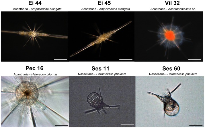
Radiolarian cells were collected in the Gulf of Eilat, Red Sea (the acantharians Ei 44 and Ei 45: Amphilonche elongata), Mediterranean Sea (the acantharians Vil 32 and Pec 16: Staurolithium sp. and Heteracon biformis, respectively) and Sesoko Island, NW Pacific Ocean (the nassellarians Ses 11 and Ses 60: Peromelissa phalacra) (see Figure 1; geographic coordinates of the locations are given in Materials S1). Individual cells were sampled from surface waters with a plankton net, micropipette isolated under a binocular microscope, and cleaned by several successive transfers into 0.22 µm-filtered seawater. No specific permits were required for the field sites, as the locations are not privately-owned or protected in any way (international oceanic waters), and the studied organisms did not involve endangered or protected species. Cells labeled Ei 44 and Ei 45 (A. elongata), and Ses 11 and Ses 60 (P. phalacra), belonging to the same acantharian and nassellarian morphospecies, respectively, are considered hereafter as biological replicates. The four acantharian morphospecies belong to the highly divergent phylogenetic clades C (Pec 16, H. biformis), D (Vil 32, Staurolithium sp.), and F (Ei 44 and Ei 45, A. elongata) [35].
Figure 1. Light microscopy pictures of individual acantharian (n = 4) and nassellarian (n = 2) cells of 100–300 µm in size, isolated in the Red Sea - Gulf of Eilat (the acantharians Ei 44 and Ei 45, Amphilonche elongata; scale bars = 50 µm), Mediterranean Sea (the acantharians Vil 32 - Heteracon biformis and Pec 16 - Staurolithium sp.; scale bars = 50 and 20 µm, respectively) and Pacific Ocean - Sesoko Island (the nassellarians Ses 11 and Ses 60, Peromelissa phalacra; scale bars = 30 µm).
DNA from each single cell was extracted as described in [35]. The V4 (ca. 380 bp) and V9 (ca. 130 bp) regions were PCR-amplified with eukaryote-specific primers that are regularly used in environmental protist surveys [3 and 2, respectively]. Because direct PCRs with 25 cycles yielded visible bands on agarose gel, Whole Genome Amplification (WGA) or nested PCR were not required to ensure the amplification of the V4 and V9 regions. Each sample was amplified in triplicate to increase the yield of amplicons, which were subsequently pooled and purified using the NucleoSpin Extract II kit (Macherey-Nagel, Hoerdt, France). To obtain a similar number of amplicons for each sample, purified PCR products were quantified with the Quant-iT™ PicoGreen dsDNA kit (Invitrogen) and then mixed in equal concentrations. Finally, amplicons were sequenced with the 454 GS-FLX Titanium pyrosequencing technology [36] (see Materials S1 for methodological details). Prior to PCR amplification and sequencing, the DNA extracts from two cells (Ei 44 and Pec 16) were split into two separate sub-samples, considered hereafter as technical replicates (Ei 44-1–Ei 44-2 and Pec 16-1–Pec 16-2).
Filtering and taxonomic assignation of 454 pyrosequencing amplicons
A three-step filtering method was adopted to eliminate ambiguous amplicons: 1) denoising was performed with Acacia v1.52.b0 and AmpliconNoise v1.29 as described in Materials S1 [37], [38]; 2) amplicons not containing the exact distal primer sequence were removed; 3) chimeras were eliminated using UCHIME with default parameters after Acacia denoising [39]. Finally, primer sequences were trimmed off and amplicons were assigned to their closest hit in the Protist Ribosomal Reference database (PR2, version August 13, 2012 [4]) using ggsearch [40]. Amplicons corresponding to symbiotic microalgae (e.g. Phaeocystis) or contaminants (e.g. fungi, metazoans or other distant radiolarians) were not included in subsequent analyses. The filtered amplicons assigned to Acantharia or Nassellaria were aligned with Muscle, implemented in Seaview v.4.2.6 [41], and clustered at different identity thresholds, from 80% to 99%, using usearch ([42]; v.6.0.203_i86linux32). The V4 and V9 alignments of each individual cell did not exhibit ambiguous sections since amplicons are short and highly similar. The secondary structure of the V4 and V9 amplicons was predicted using the RNAfold server available on the Vienna RNA web servers (http://rna.tbi.univie.ac.at).
Another analytical approach, called the linkage method, was applied to infer dominant patterns and eliminate random noise [43]. Details of the methodological procedures are given in Materials S1. The raw V4 and V9 sequences have been deposited in the Short Read Archive under the accession number PRJEB4199.
Results and Discussion
From each individual cell of Acantharia and Nassellaria, an average of 4,000 V4 and 2,380 V9 raw amplicons were obtained after pyrosequencing (Tables 1 and S1). Prior to assignation, two denoising algorithms, Acacia and AmpliconNoise, were used to filtered these amplicons. The total number of amplicons assigned to Acantharia or Nassellaria was highly variable between samples and denoising programs (Table 1). Using Acacia, from 4 to 957 V4 and 61 to 1,037 V9 amplicons were obtained. In general, AmpliconNoise retrieved more amplicons than Acacia, ranging from 386 to 2,594 for V4 and 30 to 1,080 for V9. This variability was also observed between technical replicates: in the sample Ei 44-2 we found six times more V4 amplicons than in Ei 44-1, despite comparable numbers of raw amplicons (7,197 and 6,942, respectively). Note that the acantharian Pec 16-2 and the nassellarian Ses 60 had no valid V4 amplicons after filtering in both denoising programs, probably because of the low initial number of raw amplicons obtained from these samples (1,081 and 1,088, respectively). Some of these raw amplicons were partial (distal primers were missing), and the majority were assigned to fungi, stramenopiles or metazoans that could correspond to preys or contaminants.
Table 1. Number of raw and filtered 454-pyrosequenced amplicons using the denoising programs Acacia [37] and AmpliconNoise [38].
| Radiolarian amplicons filtered with Acacia | Radiolarian amplicons filtered with AmpliconNoise | Number of OTUs at different identity cut-off levels (Acacia OTUs | AmpliconNoise OTUs) | Linkage method | |||||||||||||||||||
| Samples | Region | Raw amplicons | Total amplicons | Unique amplicons | Total amplicons | Unique amplicons | 99% | 98% | 97% | 96% | 95% | 94% | 93% | 92% | 91% | 90% | 89% | 88% | 87% | 86% | 85% | Amplicon patterns |
| Ei 44_1 | V4 | 6942 | 190 | 18 (15) | 386 | 4 (2) | 8|4 | 4|4 | 3|4 | 3|4 | 2|3 | 2|3 | 1|2 | 1|2 | 1|2 | 1|2 | 1|2 | 1|2 | 1|2 | 1|1 | 1|1 | 7 |
| Ei 44_2 | V4 | 7197 | 957 | 52 (37) | 2594 | 3 | 19|3 | 10|3 | 5|3 | 2|3 | 2|1 | 1|1 | 1|1 | 1|1 | 1|1 | 1|1 | 1|1 | 1|1 | 1|1 | 1|1 | 1|1 | 21 |
| Ei 45 | V4 | 5142 | 907 | 76 (52) | 1984 | 3 | 19|2 | 5|2 | 2|2 | 2|1 | 1|1 | 1|1 | 1|1 | 1|1 | 1|1 | 1|1 | 1|1 | 1|1 | 1|1 | 1|1 | 1|1 | 23 |
| Pec 16_1 | V4 | 2444 | 13 | 8 (5) | 0 | 0 | 3|0 | 2|0 | 1|0 | 1|0 | 1|0 | 1|0 | 1|0 | 1|0 | 1|0 | 1|0 | 1|0 | 1|0 | 1|0 | 1|0 | 1|0 | 3 |
| Pec 16_2 | V4 | 1081 | 0 | 0 | 0 | 0 | 0|0 | 0|0 | 0|0 | 0|0 | 0|0 | 0|0 | 0|0 | 0|0 | 0|0 | 0|0 | 0|0 | 0|0 | 0|0 | 0|0 | 0|0 | 0 |
| Ses 11 | V4 | 5460 | 4 | 1 | 0 | 0 | 1|0 | 1|0 | 1|0 | 1|0 | 1|0 | 1|0 | 1|0 | 1|0 | 1|0 | 1|0 | 1|0 | 1|0 | 1|0 | 1|0 | 1|0 | 1 |
| Ses 60 | V4 | 1088 | 0 | 0 | 0 | 0 | 0|0 | 0|0 | 0|0 | 0|0 | 0|0 | 0|0 | 0|0 | 0|0 | 0|0 | 0|0 | 0|0 | 0|0 | 0|0 | 0|0 | 0|0 | 0 |
| Vil 32 | V4 | 2906 | 399 | 18 (15) | 1226 | 1 | 1|1 | 1|1 | 1|1 | 1|1 | 1|1 | 1|1 | 1|1 | 1|1 | 1|1 | 1|1 | 1|1 | 1|1 | 1|1 | 1|1 | 1|1 | 1 |
| Ei 44_1 | V9 | 2746 | 656 | 18 (14) | 30 | 3 (1) | 10|3 | 8|3 | 5|3 | 3|3 | 3|3 | 3|3 | 2|2 | 2|2 | 2|2 | 2|2 | 1|1 | 1|1 | 1|1 | 1|1 | 1|1 | 7 |
| Ei 44_2 | V9 | 3899 | 1001 | 11 (5) | 587 | 2 | 8|2 | 7|2 | 4|2 | 3|2 | 3|2 | 3|2 | 3|2 | 3|2 | 3|2 | 3|2 | 2|1 | 2|1 | 2|1 | 2|1 | 2|1 | 7 |
| Ei 45 | V9 | 1331 | 577 | 6 (3) | 369 | 3 (1) | 5|3 | 4|3 | 3|3 | 3|3 | 3|3 | 3|3 | 2|2 | 2|2 | 2|2 | 2|2 | 1|1 | 1|1 | 1|1 | 1|1 | 1|1 | 2 |
| Pec 16_1 | V9 | 1538 | 832 | 4 (3) | 887 | 1 | 1|1 | 1|1 | 1|1 | 1|1 | 1|1 | 1|1 | 1|1 | 1|1 | 1|1 | 1|1 | 1|1 | 1|1 | 1|1 | 1|1 | 1|1 | 1 |
| Pec 16_2 | V9 | 1330 | 785 | 7(6) | 808 | 1 | 2|1 | 1|1 | 1|1 | 1|1 | 1|1 | 1|1 | 1|1 | 1|1 | 1|1 | 1|1 | 1|1 | 1|1 | 1|1 | 1|1 | 1|1 | 1 |
| Ses 11 | V9 | 3913 | 61 | 1 | 64 | 1 | 1|1 | 1|1 | 1|1 | 1|1 | 1|1 | 1|1 | 1|1 | 1|1 | 1|1 | 1|1 | 1|1 | 1|1 | 1|1 | 1|1 | 1|1 | 1 |
| Ses 60 | V9 | 2488 | 108 | 3(2) | 110 | 1 | 2|1 | 2|1 | 1|1 | 1|1 | 1|1 | 1|1 | 1|1 | 1|1 | 1|1 | 1|1 | 1|1 | 1|1 | 1|1 | 1|1 | 1|1 | 1 |
| Vil 32 | V9 | 1793 | 1037 | 1 | 1080 | 1 | 1|1 | 1|1 | 1|1 | 1|1 | 1|1 | 1|1 | 1|1 | 1|1 | 1|1 | 1|1 | 1|1 | 1|1 | 1|1 | 1|1 | 1|1 | 1 |
The number of singletons is indicated in brackets in the “Unique amplicons” column. OTUs were then calculated from each individual cells for the V4 (top) and V9 (bottom) regions of the ribosomal 18S rRNA gene. The numbers separated by a “|” symbol indicate the number of OTUs obtained after Acacia and AmpliconNoise denoising, respectively.
From a single cell, after denoising with Acacia and merging strictly identical amplicons, the number of unique amplicons was as high as 76 for the V4 and 18 for the V9. On average, 70% of these unique amplicons are singletons (amplicons occurring only once in the dataset). With AmpliconNoise, the number of unique amplicons was much lower (up to 4 V4 and 3 V9 amplicons from a single cell). AmpliconNoise appeared to be more stringent than Acacia since most singletons were discarded. However, this denoising algorithm did not retrieved V4 amplicons in Ses 11, Ses 60, Pec 16-1 and Pec 16-2. Among the radiolarians sampled in this study, Acantharia had more amplicons (total and unique) than Nassellaria, presumably because of the presence of multiple nuclei in acantharian cells. For the technical replicates Ei 44-1 and Ei 44-2, 18 and 52 unique V4 amplicons were found with Acacia, respectively, while the corresponding biological replicate Ei 45 had 76 unique V4 amplicons. Different number of unique amplicons between replicates were also observed with AmpliconNoise for the same cells, but to a lesser extent. These inconsistencies between replicates show that, in similar conditions, the PCR and sequencing steps can yield significantly different results in terms of amplicon number and diversity from the same morphospecies and even from the same DNA extract.
OTU-based approaches are classically used by microbial ecologists to assess species diversity and richness in the environment. Therefore, we investigated whether the various unique amplicons from single cells could form distinct OTUs. At the 97% identity level, a clustering threshold traditionally used in microbial diversity studies [8], [9], a single radiolarian cell can contain up to 5 V4 and 5 V9 radiolarian OTUs with Acacia, and up to 4 V4 and 3 V9 OTUs with the more stringent AmpliconNoise (Table 1). The highest numbers of OTUs were observed in acantharian cells (Ei 44 and Ei 45), presumably because of their higher number of unique amplicons. Notably, up to 3 V4 and 3 V9 OTUs were still found at 94% identity in the acantharian cells Ei 44 and Ei 45, both belonging to the species Amphilonche elongata (clade F). Furthermore, for both V4 and V9 regions, the number of OTUs was different between the biological and technical replicates, showing again the consequences of the fluctuating PCR and sequencing outcomes.
Overall, the distinct amplicon sequences and OTUs obtained from a single cell may indicate the existence of natural intracellular variability of the 18S rRNA in these radiolarians, more particularly in Acantharia. However, there was generally one numerically dominant amplicon sequence, and other amplicon sequences were in single or few copies (minor variants). Similarly, most amplicons from a single cell grouped in a single OTU, whereas other OTUs contained only few amplicons (Figure S1): between 88 and 100% of the total amplicons clustered in a single OTU at the 98% identity level. Thus, we argue that these acantharian and nassellarian morphospecies have one dominant 18S rRNA ribotype, but we cannot rule out at this stage the presence of distinct minor ribosomal variants.
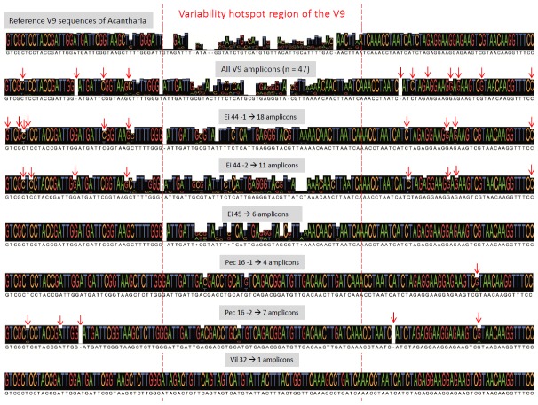
It is difficult to ascertain whether these minor variants represent natural intracellular variability, or whether they are artificially produced during the PCR and sequencing steps. This distinction is critical for carefully interpreting deep sequencing of environmental barcodes. Inspection of V4 and V9 alignments containing reference Sanger sequences and amplicons produced in this study (filtered with Acacia) revealed that most minor variants contained substitutions that seem to be randomly distributed and were not preferentially located in the variability hotspot region of reference sequences (Figure 2). For instance, Ei 44-2 had 11 unique V9 amplicons that represent 7 OTUs at 97%, but 9 of these amplicons contain nucleotide changes in regions that are conserved across all acantharian clades. Some of these substitutions might therefore represent PCR or sequencing errors that accumulate and can ultimately lead to the delineation of distinct OTUs when using high-level clustering thresholds. In addition, these substitutions can change the secondary structure of the V4 and V9 amplicon sequences found in individual cells (Figure S2). The secondary structure of the minor variants is generally different from the one of the dominant amplicon sequence, confirming that most substitutions are probably artificial.
Figure 2. V9 alignment comparison between reference sequences of all the clades of Acantharia obtained in [35] (upper sequence consensus), and the unique V9 amplicons generated in this study filtered with Acacia (n = 47) from each individual acantharian cells (Ei 44-1, Ei 44-2, Ei 45, Pec 16-1, Pec 16-2 and Vil 32).
The red dashed lines delimit the variability hotspot region of the V9 reference sequences, and the red arrows represent base substitutions (insertions or deletions) occurring outside the variability hotspot region in the pyrosequenced amplicons.
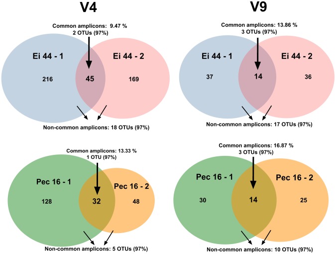
In a further attempt to differentiate natural and artificial amplicons, we examined the amplicons that were shared between technical replicates (Ei 44-1 and Ei 44-2; Pec 16-1 and Pec 16-2). We choose to work with the radiolarian amplicons containing both primers without any additional quality-based filtering in order to compare between a simple noise removal procedure based on replicates and denoising programs (Acacia and AmpliconNoise). From the same single-cell DNA extract, we found that only 9 to 13% for the V4 and 13 to 16% for the V9 of the total unique amplicons were common between replicates (Figure 3). The majority of common amplicons were the most abundant ones, corresponding to the dominant ribotypes found previously, although some common amplicons were also present in low copy numbers (as low as 2 copies). The common amplicons formed 2 or 3 OTUs at the 97% clustering threshold in the acantharians Ei 44 and Pec 16 for both V4 and V9 regions (Figure 3 and Table S2). By contrast, amplicons found in only one of the replicates typically occurred in low abundance, most of them being singletons, and exhibited mutations in highly-conserved regions (outside the variability hotspot region; Figure S3). Remarkably, despite their low copy numbers, the non-common amplicons can form up to 18 OTUs at the 97% identity level, which is on average 4.7 times more than OTUs with common amplicons (Figure 3, Table S2). This additional line of evidence demonstrates that many amplicons are artificially produced during the PCR and sequencing steps, and are divergent enough to lead to an overestimation of OTU richness.
Figure 3. Venn diagrams showing the V4 and V9 amplicons shared between single-celled technical replicates (PCR and sequencing from the same DNA extract), or only found in one of the replicates.
The number of OTUs formed with these common and non-common amplicons at 97% identity level is indicated.
In addition to the more complex and computationally-intensive denoising algorithms [37], [38], [44], sample replication and cross-validation (selecting amplicons shared by replicates) could be an efficient and biologically meaningful method to differentiate technical artifacts from real biological signal in metabarcoding surveys. The identification of common amplicons between replicates also has the advantage of circumventing the use of arbitrary criteria and thresholds for inclusion/rejection of amplicons (e.g. minimum copy abundance and identity cut-off values), therefore allowing comparison between different environmental metabarcoding datasets. Considering the continually decreasing cost of sequencing and increasing size of new datasets, sample replication and cross-validation should be considered as an additional denoising step to ensure accurate estimates of environmental microbial diversity (“Replicate or lie” as claimed in [45]), though it remains to be properly tested on complex microbial communities.
To better understand the intracellular diversity of V4 and V9 regions, another approach, called the linkage method, was applied to the same single-cell datasets [43]. By detecting SNP combination patterns in sliding windows along the sequence, this method found that many pyrosequenced amplicons contained numerous random errors (Table S3). These amplicons that had unique patterns with no redundancy in each cell were therefore excluded for subsequent analyses (more details in File S1). The linkage method detected from 1 to 23 and 1 to 7 amplicon patterns in each acantharian and nassellarian cells for the V4 and V9, respectively (Table 1, Table S3). The results confirm that intra-individual polymorphism of the 18S rRNA is low or even absent in the acantharian and nassellarian cells. Between the technical replicates Ei 44-1 and Ei 44-2, three pairs of identical V4 and V9 sequences were recognized. For V4, one pair was numerically less abundant and more divergent compared to the other two pairs. These two pairs with one indel were also detected in a different cell of the same species (Ei 45, Amphilonche elongata), indicating that at least two different ribotypes of the 18S rRNA are present in this acantharian species. Pec 16-1 and Pec 16-2 shared a single and identical V9 amplicon, but the absence of V4 amplicons in Pec 16-2 prevented us from concluding that this acantharia has a single 18S rRNA ribotype. Similarly, the nassellarian species Ses 11 and Ses 60 had one unique V9 amplicon with a 1-substitution difference, but the number of amplicons obtained is not sufficient to define the ribotype number in these cells.
Conclusion and Perspectives
Because of their ubiquitous distribution in marine environments and recurrent molecular detection from distinct environmental surveys, the radiolarian taxa Acantharia and Nassellaria will undoubtedly represent a significant part of sequence data in forthcoming environmental metabarcoding studies. The main goal of our study was to assess the intracellular variability of two genetic barcodes in these radiolarians by deep single-cell sequencing, and improve our ability to interpret metabarcoding datasets. Although several amplicon sequences and OTUs were found in a single cell, we assert that intra-individual polymorphism (defined as the divergence and relative abundance of the distinct copies) is limited in Acantharia and Nassellaria as cells contained a dominant ribotype with low-abundant variants. The ribosomal array seems therefore to evolve in concert despite its multi-copy and heterogeneous nature, and more particularly the presence of multiple nuclei in Acantharia. Based on the combination of alignment visualization, technical replicates, denoising algorithms, secondary structure prediction and a pattern-based method, we provided good evidence that many of the minor variants are artificially produced during the PCR and sequencing steps. More particularly, as also highlighted in other studies [46], we showed that amplicon sequencing approaches are not reproducible and require replicates for rigorous interpretation. Consequently, we recommend a conservative approach to discard artifacts from metabarcoding datasets: 1) two or three technical replicates (parallel PCR and sequencing steps from the same environmental sample), 2) a denoising procedure including cross-validation of amplicons between replicates, and 3) if working on specific taxon like Acantharia, alignment visualization with reference sequences to further remove ambiguous amplicons. The remaining amplicons that correspond to the dominant ribotypes of each cell should better reflect the natural diversity and richness of radiolarians in the environment. This conservative approach is critical to properly infer and compare diversity estimates of a particular taxon or a whole community across different samples, and so irrespective of the high-throughput sequencing technique. In addition, low error-rate polymerases, low cycle numbers and ideally PCR-free methods should be favored to alleviate technical biases in future metabarcoding studies.
Moreover, our approach allowed improving the 18S rRNA reference database of radiolarians by adding information about the number of ribotypes found in each species. For instance, we detected two ribotypes in the acantharian species Amphilonche elongata (Ei 44 and Ei 45). Although one ribotype is numerically more abundant than the other, both can be detected in environmental metabarcoding datasets. Similar approach should be adopted on more eukaryotic taxa to fine-tune the assignation of environmental barcodes and avoid inflating diversity estimates in metabarcoding surveys.
An obvious next step for radiolarians is to assess the rRNA copy number in single cells in order to estimate the abundance of these protists from metabarcoding datasets. For instance, a recent study using qPCR assays estimated the rRNA copy number per cell in benthic Foraminifera (i.e. 10,000–30,000) [47]. This allowed the establishment of normalization factors that were used to correctly determine abundance of species by removing “excess” of amplicons. Similar normalization has been also applied in bacteria based on the known copy number in reference genomes [48]. Alternatively, a single-copy gene could be selected as a barcode to assess the abundance of radiolarians in the environment, but the lack of genomic data remains the main barrier to select and validate such barcode among radiolarians.
Extending the approach conducted here on radiolarian single cells to other microbial taxa will help to define taxonomically meaningful and relevant genetic entities, but also to contribute to a better understanding of the potential and limitations of the 18S rRNA gene marker for environmental metabarcoding studies. A careful representation of the diversity and relative abundances of microbial organisms is critical for the establishment of biodiversity monitoring projects and the assessment of the impact of anthropogenic changes.
Supporting Information
Number and size of the V4 and V9 OTUs found in different individual cells of Radiolaria, based on amplicons filtered with the denoising program Acacia. Each OTU is represented by a single color, and its number of amplicons is indicated in the bar.
(PDF)
Predicted secondary structures of the V4 amplicons found in the sample Ei 44_1. The numbers indicate the abundance of the given amplicon.
(PDF)
V9 alignment comparison between reference sequences of all the clades of Acantharia (upper sequence consensus) and the common and non-common pyrosequenced amplicons obtained from technical replicates without Acacia denoising (Ei 44-1/Ei 44-2 and Pec 16-1 and Pec 16-2). Compared to non-common amplicons, common amplicons tend to have fewer substitutions in highly-conserved regions.
(PDF)
Number of amplicons at the different consecutive filtering steps: 1- denoising with AmpliconNoise or Acacia, 2- selection of amplicons with the exact distal primer sequence and 3- detection of chimeras with UCHIME after Acacia denoising. (T) and (U) indicate the number of total and unique amplicons, respectively.
(PDF)
Number of common and non-common radiolarian amplicons (without Acacia and AmpliconNoise denoising) between single-celled technical replicates (PCR and sequencing on the same DNA extract). OTU reconstruction was performed with these amplicons at different identity levels.
(PDF)
Number of amplicons detected by the linkage method (See File S1). The number of unique and redundant amplicons are indicated in the “Unique amplicon (Linkage)” and “Redundant amplicon (>1)” columns, respectively. The number of identical sequences between technical replicates or cells is given in the right part of the table (“Number of overlapped amplicons”).
(PDF)
(HTML)
(HTML)
Acknowledgments
We thank the marine stations which hosted us for field sampling: LOV Villefranche-sur-Mer, the Interuniversity Institute for Marine Sciences in Eilat (EU FP7 ASSEMBLE program funded this sampling mission), and the Sesoko Marine Station in Okinawa. We are also grateful to the Genoscope for sequencing, and C. Berney and John Dolan for valuable comments on the manuscript.
Funding Statement
This work was supported by the PhD fellowship DIPHOPE from the Region Bretagne (J.D.), the BioMarKs project (2008-6530, ERA-net Biodiversa, EU), and a JST-CNRS exchange program (F.N). F.M. was supported by the Deutsche Forschungsgemeinschaft (grant #DU1319/1-1). This study was also supported by the project OCEANOMICS, that has received funding from the French government, managed by the Agence Nationale de la Recherche, under the grant agreement “Investissement d′Avenir” ANR-11-BTBR-0008. The funders had no role in study design, data collection and analysis, decision to publish, or preparation of the manuscript.
References
- 1. Sogin ML, Morrison HG, Huber JA, Welch DM, Huse SM, et al. (2006) Microbial diversity in the deep sea and the underexplored ‘rare biosphere’. Proc Natl Acad Sci USA 103: 12115–12120. [DOI] [PMC free article] [PubMed] [Google Scholar]
- 2. Amaral-Zettler LA, McCliment EA, Ducklow HW, Huse SM (2009) A method for studying protistan diversity using massively parallel sequencing of V9 hypervariable regions of small-subunit ribosomal RNA genes. PLoS ONE 4(7): e6372 10.1371/journal.pone.0006372 [DOI] [PMC free article] [PubMed] [Google Scholar]
- 3. Stoeck T, Bass D, Nebel M, Christen R, Jones MD, et al. (2010) Multiple marker parallel tag environmental DNA sequencing reveals a highly complex eukaryotic community in marine anoxic water. Mol Ecol 19: 21–31. [DOI] [PubMed] [Google Scholar]
- 4. Guillou L, Bachar D, Audic S, Bass D, Berney C, et al. (2012) The Protist Ribosomal Reference database (PR2): a catalog of unicellular eukaryote Small Sub-Unit rRNA sequences with curated taxonomy. Nucl Acids Res 41: 1–8 10.1093/nar/gks1160 [DOI] [PMC free article] [PubMed] [Google Scholar]
- 5. Pawlowski J, Audic S, Adl S, Bass D, Belbahri L, et al. (2012) CBOL Protist Working Group: Barcoding eukaryotic richness beyond the animal, plant, and fungal kingdoms. PLoS Biol 10(11): e1001419 10.1371/journal.pbio.1001419 [DOI] [PMC free article] [PubMed] [Google Scholar]
- 6. Pedrós-Alió C (2007) Dipping into the rare biosphere. Science 315: 192–193. [DOI] [PubMed] [Google Scholar]
- 7. Nebel M, Pfabel C, Stock A, Dunthorn M, Stoeck T (2010) Delimiting operational taxonomic units for assessing ciliate environmental diversity using small-subunit rRNA gene sequences. Environ Microbiol Rep 3: 154–158. [DOI] [PubMed] [Google Scholar]
- 8. Countway PD, Gast RJ, Dennett MR, Savai P, Rose JM, et al. (2007) Distinct protistan assemblages characterize the euphotic zone and deep sea (2500 m) of the western North Atlantic (Sargasso Sea and Gulf Stream). Environ Microbiol 9: 1219–1232. [DOI] [PubMed] [Google Scholar]
- 9. Stoeck T, Kasper J, Bunge J, Leslin C, Ilyin V, et al. (2007) Protistan diversity in the arctic: a case of paleoclimate shaping modern biodiversity? PLoS ONE 2: e728. [DOI] [PMC free article] [PubMed] [Google Scholar]
- 10. Caron DA, Countway PD, Savai PS, Gast RJ, Schnetzer A, et al. (2009) Defining DNA-based operational taxonomic units for microbial-eukaryote ecology. Appl Environ Microbiol 75: 5797–5808. [DOI] [PMC free article] [PubMed] [Google Scholar]
- 11. Zhu F, Massana R, Not F, Marie D, Vaulot D (2005) Mapping of picoeucaryotes in marine ecosystems with quantitative PCR of the 18S rRNA gene. FEMS Microbiol Ecol 52: 79–82. [DOI] [PubMed] [Google Scholar]
- 12. Medinger R, Nolte V, Pandey RV, Jost S, Ottenwälder B, et al. (2010) Diversity in a hidden world: potential and limitation of next-generation sequencing for surveys of molecular diversity of eukaryotic microorganisms. Mol Ecol 19: 32–40. [DOI] [PMC free article] [PubMed] [Google Scholar]
- 13. Gong J, Dong J, Liu X, Massana R (2013) Extremely high copy numbers and polymorphisms of the rDNA operon estimated from single cell analysis of Oligotrich and Peritrich ciliates. Protist 164: 369–379. [DOI] [PubMed] [Google Scholar]
- 14. Pillet L, Fontaine D, Pawlowski J (2012) Intra-genomic ribosomal RNA polymorphism and morphological variation in Elphidium macellum suggests inter-specific hybridization in Foraminifera. PLoS ONE 7(2): e32373. [DOI] [PMC free article] [PubMed] [Google Scholar]
- 15. Mighell AJ, Smith NR, Robinson PA, Markham AF (2000) Vertebrate pseudogenes. FEMS letters 468: 109–114. [DOI] [PubMed] [Google Scholar]
- 16. Márquez LM, Miller DJ, MacKenzie JB, van Oppen MJH (2003) Pseudogenes contribute to the extreme diversity of nuclear ribosomal DNA in the hard coral Acropora . Mol Biol Evol 20 7: 1077–1086. [DOI] [PubMed] [Google Scholar]
- 17. Santos SR, Kinzie RA 3rd, Sakai K, Coffroth MA (2003) Molecular characterization of nuclear small subunit (18S)-rDNA pseudogenes in a symbiotic dinoflagellate (Symbiodinium, Dinophyta). J Eukaryot Microbiol 50 6: 417–421. [DOI] [PubMed] [Google Scholar]
- 18. van Berkum P, Terefework Z, Paulin L, Suomalainen S, Lindström K, Eardly BD (2003) Discordant phylogenies within the rrn loci of Rhizobia. J Bacteriol 185: 2988–2998. [DOI] [PMC free article] [PubMed] [Google Scholar]
- 19. Yabuki A, Toyofuku T, Takishita K (2014) Lateral transfer of eukaryotic ribosomal RNA genes: an emerging concern for molecular ecology of microbial eukaryotes. ISME J 1–4. [DOI] [PMC free article] [PubMed] [Google Scholar]
- 20. Kunin V, Engelbrektson A, Ochman H, Hugenholtz P (2010) Wrinkles in the rare biosphere: pyrosequencing errors lead to artificial inflation of diversity estimates. Environ Microbiol 12: 118–123. [DOI] [PubMed] [Google Scholar]
- 21. Lee CK, Herbold CW, Polson SW, Wommack KE, Williamson SJ, et al. (2012) Groundtruthing next-gen sequencing for microbial ecology-biases and errors in community structure estimates from PCR amplicon pyrosequencing. PLoS ONE 7 9: e44224 10.1371/journal.pone.0044224 [DOI] [PMC free article] [PubMed] [Google Scholar]
- 22. Suzuki N, Aita Y (2011) Radiolaria: achievements and unresolved issues: taxonomy and cytology. Plankt & Benth Res 6: 69–91. [Google Scholar]
- 23. Caron DA, Michaels AF, Swanberg NR, Howes FA (1995) Primary productivity by symbiont-bearing planktonic sarcodines (Acantharia, Radiolaria, Foraminifera) in surface waters near Bermuda. J Plankton Res 17: 103–129. [Google Scholar]
- 24. Swanberg NR, Caron DA (1991) Patterns of sarcodine feeding in epipelagic oceanic plankton. J Plankton Res 13: 287–322. [Google Scholar]
- 25. Lampitt RS, Salter I, John D (2009) Radiolaria: major exporters of organic carbon to the deep ocean. Global Biogeochem Cycles 23 GB1010. [Google Scholar]
- 26. Decelle J, Probert I, Bittner L, Desdevises Y, Colin S, et al. (2012) An original mode of symbiosis in open ocean plankton. Proc Natl Acad Sci USA 109: 18000–18005. [DOI] [PMC free article] [PubMed] [Google Scholar]
- 27. Marie D, Shi XL, Rigaut-Jalabert F, Vaulot D (2010) Use of flow cytometric sorting to better assess the diversity of small photosynthetic eukaryotes in the English Channel. FEMS Microbiol Ecol 72: 165–178. [DOI] [PubMed] [Google Scholar]
- 28. Not F, Gausling R, Azam F, Heidelberg JF, Worden AZ (2007) Vertical distribution of picoeukaryotic diversity in the Sargasso Sea. Environ Microbiol 9: 1233–1252. [DOI] [PubMed] [Google Scholar]
- 29. Quaiser A, Zivanovic Y, Moreira D, López-García P (2010) Comparative metagenomics of bathypelagic plankton and bottom sediment from the Sea of Marmara. ISME J 5: 285–304. [DOI] [PMC free article] [PubMed] [Google Scholar]
- 30. López-García P, Rodríguez-Valera F, Pedrós-Alió C, Moreira D (2001) Unexpected diversity of small eukaryotes in deep-sea Antarctic plankton. Nature 409: 603–607. [DOI] [PubMed] [Google Scholar]
- 31. Lovejoy C, Massana R, Pedrós-Alió C (2006) Diversity and distribution of marine microbial eukaryotes in the Arctic Ocean and adjacent seas. Appl Environ Microbiol 72: 3085–3095. [DOI] [PMC free article] [PubMed] [Google Scholar]
- 32. Alexander E, Stock A, Breiner HW, Behnke A, Bunge J, et al. (2009) Microbial eukaryotes in the hypersaline anoxic L'Atalante deep-sea basin. Environ Microbiol 11: 360–381. [DOI] [PubMed] [Google Scholar]
- 33. Stoeck T, Taylor GT, Epstein SS (2003) Novel eukaryotes from the permanently anoxic Cariaco Basin (Caribbean Sea). Appl Environ Microbiol 69: 5656–5663. [DOI] [PMC free article] [PubMed] [Google Scholar]
- 34. Orsi W, Edgcomb V, Jeon S, Leslin C, Bunge J, et al. (2011) Protistan microbial observatory in the Cariaco Basin, Caribbean. II. Habitat specialization. ISME J 5: 1357–1373. [DOI] [PMC free article] [PubMed] [Google Scholar]
- 35. Decelle J, Suzuki N, Mahé F, de Vargas C, Not F (2012) Molecular phylogeny and morphological evolution of the acantharia (Radiolaria). Protist 163: 435–450. [DOI] [PubMed] [Google Scholar]
- 36. Mardis ER (2008) Next-generation DNA sequencing methods. Annu Rev Genomics Hum Genet 9: 387–402. [DOI] [PubMed] [Google Scholar]
- 37. Bragg L, Stone G, Imelfort M, Hugenholtz P, Tyson GW (2012) Fast, accurate error-correction of amplicon pyrosequences using Acacia. Nature Methods 9: 425–426. [DOI] [PubMed] [Google Scholar]
- 38. Quince C, Lanzen A, Davenport RJ, Turnbaugh PJ (2011) Removing noise from pyrosequenced amplicons. BMC Bioinformatics 12: 38. [DOI] [PMC free article] [PubMed] [Google Scholar]
- 39. Edgar RC, Haas BJ, Clemente JC, Quince C, Knight R (2011) UCHIME improves sensitivity and speed of chimera detection. Bioinformatics 27: 2194–2200. [DOI] [PMC free article] [PubMed] [Google Scholar]
- 40. Mackey AJ, Haystead TA, Pearson WR (2002) Getting more from less: algorithms for rapid protein identification with multiple short peptide sequences. Mol Cell Proteomics 1: 139–147. [DOI] [PubMed] [Google Scholar]
- 41. Gouy M, Guindon S, Gascuel O (2010) SeaView version 4: a multiplatform graphical user interface for sequence alignment and phylogenetic tree building. Mol Biol Evol 27: 221–224. [DOI] [PubMed] [Google Scholar]
- 42. Edgar RC (2010) Search and clustering orders of magnitude faster than BLAST. Bioinformatics 26: 2460–2461. [DOI] [PubMed] [Google Scholar]
- 43. Sasaki E, Sugino RP, Innan H (2013) The linkage method: a novel approach for SNP detection and haplotype reconstruction from a single diploid individual using next-generation sequence data. Mol Biol Evol 30: 2187–2196. [DOI] [PubMed] [Google Scholar]
- 44. Gaspar JM, Thomas WK (2013) Assessing the consequences of denoising marker-based metagenomic data. PLoS ONE 8 3: e60458 10.1371/journal.pone.0060458 [DOI] [PMC free article] [PubMed] [Google Scholar]
- 45. Prosser JI (2010) Replicate or lie. Environ Microbiol 12: 1806–1810. [DOI] [PubMed] [Google Scholar]
- 46. Zhou J, Wu L, Deng Y, Zhi X, Jiang YH, et al. (2011) Reproducibility and quantitation of amplicon sequencing-based detection. ISME J 5: 1303–1313. [DOI] [PMC free article] [PubMed] [Google Scholar]
- 47. Weber AA-T, Pawlowski J (2013) Can abundance of protists be inferred from sequence data: a case study of Foraminifera. PLoS ONE 8 2: e56739 10.1371/journal.pone.0056739 [DOI] [PMC free article] [PubMed] [Google Scholar]
- 48. Kembel SW, Wu M, Eisen JA, Green JL (2012) Incorporating 16S gene copy number information improves estimates of microbial diversity and abundance. PLoS Comput Biol 8 10: e1002743. [DOI] [PMC free article] [PubMed] [Google Scholar]
Associated Data
This section collects any data citations, data availability statements, or supplementary materials included in this article.
Supplementary Materials
Number and size of the V4 and V9 OTUs found in different individual cells of Radiolaria, based on amplicons filtered with the denoising program Acacia. Each OTU is represented by a single color, and its number of amplicons is indicated in the bar.
(PDF)
Predicted secondary structures of the V4 amplicons found in the sample Ei 44_1. The numbers indicate the abundance of the given amplicon.
(PDF)
V9 alignment comparison between reference sequences of all the clades of Acantharia (upper sequence consensus) and the common and non-common pyrosequenced amplicons obtained from technical replicates without Acacia denoising (Ei 44-1/Ei 44-2 and Pec 16-1 and Pec 16-2). Compared to non-common amplicons, common amplicons tend to have fewer substitutions in highly-conserved regions.
(PDF)
Number of amplicons at the different consecutive filtering steps: 1- denoising with AmpliconNoise or Acacia, 2- selection of amplicons with the exact distal primer sequence and 3- detection of chimeras with UCHIME after Acacia denoising. (T) and (U) indicate the number of total and unique amplicons, respectively.
(PDF)
Number of common and non-common radiolarian amplicons (without Acacia and AmpliconNoise denoising) between single-celled technical replicates (PCR and sequencing on the same DNA extract). OTU reconstruction was performed with these amplicons at different identity levels.
(PDF)
Number of amplicons detected by the linkage method (See File S1). The number of unique and redundant amplicons are indicated in the “Unique amplicon (Linkage)” and “Redundant amplicon (>1)” columns, respectively. The number of identical sequences between technical replicates or cells is given in the right part of the table (“Number of overlapped amplicons”).
(PDF)
(HTML)
(HTML)


