Abstract
A major Bacillus anthracis spore coat protein of 13.4 kDa, designated Cotα, was found only in the Bacillus cereus group. A stable ca. 30-kDa dimer of this protein was also present in spore coat extracts. Cotα, which is encoded by a monocistronic gene, was first detected late in sporulation, consistent with a σK-regulated gene. On the basis of immunogold labeling, the protein is in the outer spore coat and absent from the exosporium. In addition, disruption of the gene encoding Cotα resulted in spores lacking a dark-staining outer spore coat in thin-section electron micrographs. The mutant spores were stable upon heating or storage, germinated at the same rate as the wild type, and were resistant to lysozyme. They were, however, more sensitive than the wild type to phenol, chloroform, and hypochlorite but more resistant to diethylpyrocarbonate. In all cases, resistance or sensitivity to these reagents was restored by introducing a clone of the cotα gene into the mutant. Since Cotα is an abundant outer spore coat protein of the B. cereus group with a prominent role in spore resistance and sensitivity, it is a promising target for the inactivation of B. anthracis spores.
A major concern regarding Bacillus anthracis as a biological weapon centers on the spore because of its resistance to heat, radiation, etc., and its ability to survive in the dormant state for a prolonged period (15). Killing dispersed spores effectively has proven difficult and at present requires the use of agents, such as chlorine dioxide gas (7), which have very broad toxicity. It would be beneficial, therefore, to be able to selectively inactivate B. anthracis spores. This could be achieved by finding components unique to these spores which are critical for resistance and targeting them.
One such spore structure is the multilayered spore coat first analyzed in Bacillus cereus (2). In the best-studied species, Bacillus subtilis, these coats contain a complex mixture of at least 24 proteins which can be resolved into two or three morphological layers (2, 9, 11, 13). Coat assembly also requires regulatory proteins and key morphogenetic factors (9, 11). Many of the B. subtilis homologues encoding coat structural and regulatory proteins are present in the genome of B. anthracis, with the notable absence of some genes encoding proteins likely to be in the outer coat of B. subtilis spores (10).
In addition to a protective function, the coat has some role in germination (5, 6), perhaps by providing access for germinants to the germination proteins located in the inner forespore membrane. A lytic enzyme required for digestion of the spore cortex during germination may also be located in the coat (8, 19). In most cases, the precise function of various spore coat proteins has been difficult to determine, since gene disruptions had no measurable phenotypic effects, i.e., no alteration in spore sensitivity to lysozyme or solvents and generally no detectable changes in germination properties (9, 11). There are a few exceptions, especially due to disruptions of genes encoding the major morphogenetic factors. The absence of detectable phenotypic changes could be due to redundant functions of the various proteins or the lack of suitable phenotypic tests. In either case, the reason for the complexity of this structure, especially if its function was primarily protective, is not understood.
In the B. cereus group (which includes B. anthracis), there is a prevalent spore coat protein of ca. 13 kDa (2, 4), and preliminary data had indicated that it may be a major component of the spore outer coat and could thus contribute to spore resistance. We have cloned the gene encoding this protein and have found that its disruption resulted in spores which lacked an intact outer coat and were altered in their sensitivity or resistance to various chemical agents. Given the abundance and outer coat location of this protein, it is a promising target for the selective inactivation of B. anthracis spores.
MATERIALS AND METHODS
Growth and sporulation.
The B. anthracis Weymouth strain designated UM44 (from T. Koehler, University of Texas Medical Center, Houston) was grown in NSM (24) or on NSM plates for 24 to 48 h until sporulation was complete. Spores were purified as previously described (3, 24). For extraction, spores were suspended in 1 ml of deionized water to an A580 value of 1.2. Following centrifugation in a Microfuge for 6 min, the spores were suspended in 15 μl of 6 M urea-50 mM dithiothreitol-1% sodium dodecyl sulfate (SDS), pH 10.0 (UDS), and incubated at 37°C for 20 min. After centrifugation in an Eppendorf Microfuge for 6 min, the extraction was repeated and the two supernatants were pooled. Five to fifteen microliters was used for electrophoresis in SDS-15% polyacrylamide gels.
Cloning of the cotα gene.
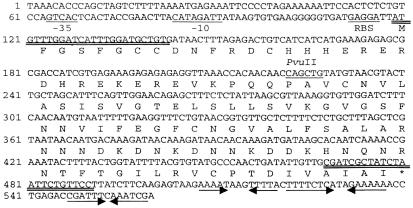
There was a major stained band of ca. 14 kDa (see Fig. 1), which was transferred to a polyvinylidene difluoride membrane for sequencing in the Purdue Center for Macromolecular Structure. This sequence, MFGSFGXXDNFRDXH (where X is unknown), was used to identify an open reading frame in the B. anthracis genome, designated cotα (as shown in Fig. 3). On the basis of the gene sequence, oligonucleotides (5′ATGTTTGGATCATTTGGATGCTG and 5′GGAAGAGAAATCGATAGCGATCGC) were used for amplification and cloning in the vector pCR T7/CT-TOPO (Invitrogen) containing a carboxy-terminal six-His tag. This clone was sequenced (GenBank accession no. AY489558; protein identification no. AAR33045) to confirm the sequence found in the B. anthracis genome. The protein expressed in Escherichia coli BL21(DE3)/pLysS was found to migrate in SDS-polyacrylamide gel electrophoresis (PAGE) at ca. 18 kDa, as expected, and to react with Cotα antibody (unpublished results). The Cotα-six-His conjugate was purified on an Ni2+-agarose column (Invitrogen). The protein that was eluted with 0.3 M imidazole was dialyzed extensively against deionized water, and suspensions of 200 μg (extensively aggregated due to disulfide bonds) were used for the preparation of rabbit antibody as previously described (23).
FIG. 1.
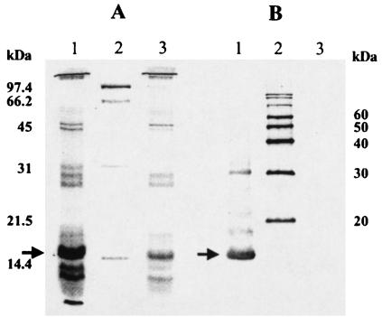
SDS-PAGE stained profile of spore coat proteins (A) and an immunoblot with Cotα antibody (B). Lane 1, strain UM44; lane 2, molecular mass markers; lane 3, strain 3 (Δcotα). The arrows indicate the Cotα protein bands of 14 to 15 kDa. A similar band in lane A3 and those of smaller sizes in lanes A1 and A3 are discussed in the text.
FIG. 3.
Sequence of the B. anthracis cotα gene and the deduced amino acid sequence. Also included are a potential ribosome binding site (RBS) and a possible σK promoter (−10 and −35, underlined). The oligonucleotides used for PCR amplification of the gene are double underlined. Inverted arrows indicate palindromes as potential terminators. The PvuII site was used for insertion of the Kmr cassette.
For expression in bacilli, the cotα gene, containing 277 bp of the upstream region, was amplified using the oligonucleotide 5′CATACTGTACGATCCATCGTACC plus the downstream oligonucleotide listed above and cloned into pCR T7/CT-TOPO. A XhoI/BamHI fragment was then subcloned into the shuttle vector pHT315 (1). This clone was electroporated into mutant 3 (discussed below) with selection for resistance to erythromycin (7 μg ml−1).
For gene replacement, the cotα gene region was recloned into pCRT7/CT-TOPO using oligonuceotides complementary to regions 1 kb up- and downstream of the coding sequence, as follows: 5′TAGGTGATTTAGGAGCTCATAAAAAC and 5′TGTAACAAGTGGTCGACATCAC. A kanamycin resistance cassette (aph3) of ca. 2.1 kb (18) was inserted into the PvuII site within the cotα gene, and then an XhoI/HindIII fragment of 4.2 kb was subcloned into the shuttle vector pUTE29 (12). This clone was linearized with BstEII, treated with shrimp alkaline phosphatase for 1 h at 37°C, heated at 65°C for 20 min, and then used for electroporation of B. anthracis UM44 with screening for resistance to kanamycin (50 μg ml−1).
For electroporation, cells were grown in a sidearm flask in 30 ml of brain heart infusion medium supplemented with 0.5 M sorbitol (22) to a Klett value (660-nm filter) of 150. In this medium, cells were well defined in short chains. They were collected by centrifugation at 6,000 rpm for 8 min in a Sorvall SS-34 rotor. The pellet was suspended in 3 ml of 0.5 M sorbitol-0.5 M mannitol-10% glycerol (SMG), and 100 μg of proteinase K was added per ml (J. Hoch, personal communication). The suspensions were shaken slowly at 37°C for 20 min. This step enhanced the efficiency of transformation two- to threefold, presumably by removing the S-layer protein which coats vegetative cells. The suspensions were diluted to 10 ml with SMG, centrifuged as described above, and washed an additional time with 10 ml of SMG. Cell suspensions in about 200 μl of SMG were used for electroporation with 80 μl per cuvette plus 4 to 5 μg of linearized plasmid DNA (dialyzed against water for 20 to 30 min to remove salts). Electroporation in a Bio-Rad gene pulser was at 2.5 kV, 400 Ω, and 3.0 uF. The contents of each cuvette were diluted to 1 ml with brain heart infusion-sorbitol medium, and the tubes were shaken slowly at 37°C for 90 min. Fifty- to two hundred-microliter aliquots were plated on Luria-Bertani medium containing 50 μg of kanamycin ml−1. Colonies were screened for sensitivity to 5 μg of tetracycline ml−1 (indicative of the absence of the pUTE29 vector) and resistance to kanamycin. Those which were Tcs and Kmr were screened for gene replacement by PCR using the oligonucleotides designed for cloning of the cotα gene. Conditions using the Taq polymerase were as specified with the minicycler (MJ Research, Inc.). One mutant, designated mutant 3, was further analyzed.
Preparation of cell extracts and immunoblotting.
Cells of strain UM44 were grown in 30 ml of NSM in a sidearm flask on a rotary shaker at 37°C. Growth was monitored in a Klett colorimeter (660-nm filter), and 5-ml samples were removed, commencing 90 min after the end of growth and at 2-h intervals thereafter. Samples were examined in the phase-contrast microscope to determine the percent phase-white and phase-bright endospores. Cells were washed and lysed in UDS (50 μl) as previously described (21). Five microliters was used for electrophoresis in SDS-15% PAGE. Immunoblotting and treatment with rabbit anti-Cotα antibody were done as previously described (6, 24).
Characterization of spores.
Sensitivity to lysozyme (0.01 to 1.0 mg ml−1) was measured by the decrease in A600 nm at 37°C for suspensions in 0.05 M Tris plus 5 mM EDTA, pH 8.0. Sensitivity to chloroform was determined by plating dilutions on Luria-Bertani agar before and after mixing with 10% chloroform, vortexing at top speed for 1 min, and then sonicating for 10 s to break up clumps of spores. For treatment with diethylpyrocarbonate (DPC), spores were suspended to 3 × 10⁸ to 5 × 10⁸ ml−1 in 0.1 ml of 0.05 M sodium phosphate, pH 6.0. A sample was removed for viable counts, and then DPC was added to a 0.025 M concentration and the tubes were incubated at 27°C for 1 h. Sensitivities to 10% n-octanol, 5 and 10% phenol, 10% hydrogen peroxide, and 4% sodium hypochlorite (0.5% available chlorine) were measured as described elsewhere (16, 23). Spore germination in l-alanine plus inosine was also done as described elsewhere (5).
Microscopy.
Procedures for fixation, thin sectioning, and staining for electron microscopy were as described previously (20). Immunolocalization was carried out on thin sections mounted on Formvar plus carbon-coated 100-mesh nickel grids (20). Secondary antibody was goat anti-rabbit immunoglobulin G conjugated to 10-nm colloid gold (Ted Pella, Inc., Redding, Calif.). All samples were imaged using an FEI/Philips CM-10 Biotwin transmission electron microscope operated at 80 kV with images captured on Kodak S-136 film. Images were scanned at 600 dpi, adjusted in Adobe Photoshop 7, and printed using a Codonics NP-1660 thermal/dye sublimation printer.
RESULTS
Characterization of Cotα.
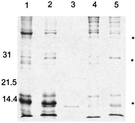
The SDS-PAGE profiles of soluble spore coat extracts of B. anthracis (Fig. 1) were very similar to those for B. cereus (3, 4) but differed substantially from those for B. subtilis (3, 4, 9). Among the former, there is a prevalent band of ca. 15 kDa, designated Cotα (Fig. 1), in contrast to multiple bands of <10 kDa in B. subtilis coat extracts (3). For B. anthracis, additional smaller bands are often present (Fig. 1A, lane 1), especially in spores stored for a prolonged period (Fig. 2). A heterogeneous amino acid sequence was found for these smaller bands, indicating that they were primarily degradation products. The properties of many of the other stained bands are under investigation (14).
FIG. 2.
SDS-PAGE profiles of spore coat extracts from stored spores of strain UM44 (lanes 1 and 2) and mutant 3 (lanes 4 and 5). Lane 3 contains standards, as indicated on the left. Equal quantities of spores were extracted immediately after purification (lanes 1 and 4) and after storage in deionized water at 4°C for 5 days (lanes 2 and 5). Two bands with decreased intensity after storage are marked by asterisks; the lowest asterisk refers to a region of increased staining intensity.
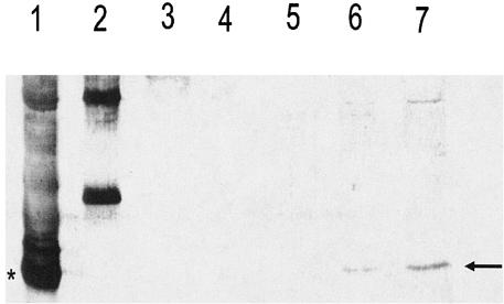
The major band of ca. 15 kDa was eluted and sequenced as described in Materials and Methods, and an open reading frame identified in the B. anthracis genome has been designated cotα (Fig. 3). It encodes a protein of 13.4 kDa which migrates slightly more slowly than expected in SDS-PAGE. This gene appeared to be monocistronic, since there were no open reading frames encoding more than 15 amino acids within 500 bp either up- or downstream. There was also a consensus σK promoter sequence, consistent with the appearance of Cotα antigen at about stage IV of sporulation in immunoblots of extracts from sporulating cells (Fig. 4). A Cotα-coding sequence was found in the B. cereus genome (87% amino acid identity), but no other orthologues or homologues were found in GenBank. Cotα thus appears to be novel to spores of the B. cereus group. The gene was cloned (see Materials and Methods), and a protein of ca. 18 kDa (larger due to the six-His tag and other residues in the pCR T7/CT-TOPO cloning vector) was found in SDS-polyacrylamide stained gels after induction of the E. coli clone with isopropyl-β-d-thiogalactopyranoside for 3 h. This band and the one of 13.4k Da from B. anthracis spores reacted with the Cotα antibody (Fig. 1B).
FIG. 4.
Time course of appearance of Cotα antigen in immunoblots of extracts of B. anthracis cells. Lane 1, extract of UM44 spores with the dark-staining Cotα band marked with an asterisk; lane 2, standards of 20 and 30 kDa; lanes 3 to 7, extracts of sporulating cells sampled at 2-h intervals commencing at 90 min after the end of growth through a time when >90% of the cells contained phase-bright endospores. The arrow on the right indicates the Cotα antigen. A Cotα dimer of ca. 30 kDa is present near the top of lanes 1 and 7.
Disruption of the cotα gene.
The cotα gene was disrupted with a 2.1-kb kanamycin resistance cassette (18) as described in Materials and Methods. A gene replacement in a strain designated 3 was confirmed by PCR (as was retention of plasmid pXO1, as demonstrated by PCR of two genes known to be in this plasmid, pag and atxA). The Cotα protein was absent from extracts of purified spores of this mutant (Fig. 1), as was a band of ca. 30 kDa. A partial sequence of the latter (15 residues) was identical to that of Cotα, so this band is a dimer which is stable in the presence of 100 mM β-mercaptoethanol. Since most of Cotα is solubilized by such a treatment, an alternative cross-linking may be present. There was a heterogeneous amino acid sequence of the stained band of ca. 14k Da present in extracts of mutant 3 (Fig. 1A, lane 3), indicative of degradation of coat proteins in older spore preparations (Fig. 2). There was, however, a prevalent sequence which is a novel protein of 14 kDa encoded in the B. anthracis genome. This protein is likely to be present in the wild type as part of the broad Cotα band (Fig. 1A).
Properties of mutant spores.
The spores of strain 3 were resistant to lysozyme, hydrogen peroxide, and n-octanol but sensitive to chloroform, phenol, and hypochlorite (Table 1). Interestingly, mutant 3 spores were more resistant to DPC. The mutant spores germinated with l-alanine and inosine at about the same rate as the wild type and were stable when stored at 4°C for prolonged periods. Wild-type resistance and sensitivity properties were restored when strain 3 was transformed with a plasmid containing the cotα gene (Table 1).
TABLE 1.
Sensitivity of wild-type and mutant spores to various reagentsa
| Strain | % Survival after treatment with:
|
|||
|---|---|---|---|---|
| Chloroform | Phenol | Hypochlorite | DPCc | |
| UM44 (wild type) | 98 | 99 | 98 | 20-30b |
| 3 (Δcotα) | 4-18b | 5-15b | 3-6b | 70-80b |
| 3/pHT315-cotα | 98 | 98 | 98 | 30-35b |
See Materials and Methods for assay procedures.
For the mutant and for spores treated with DPC, a range of values resulting from at least three independent experiments is presented. Other values are averages for three separate experiments (±2% for 1 standard deviation).
25 mM diethylpyrocarbonate.
Microscopy.
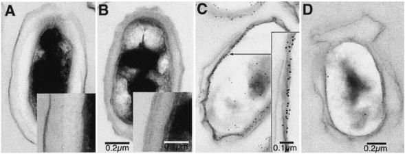
Thin-section electron micrographs (Fig. 5A and B) demonstrated the absence of a dark-staining outer spore coat layer in mutant 3. This coat layer reacted with immunogold-labeled Cotα antibody in wild-type spores but not in the mutant (Fig. 5C and D). There was no labeling of the loose-fitting exosporium, but there were some grains over the spore cytoplasm, especially in the wild type (Fig. 5C and D). There was no evidence for soluble Cotα antigen in extracts of spores treated to remove the coat and the cortex. The reason for this apparent cytoplasmic labeling is not known.
FIG. 5.
Thin-section electron micrographs of B. anthracis wild-type strain UM44 (A) and mutant strain 3 (B) showing the absence of the dark-staining outer spore coat in the mutant. Treatment of sections with immunogold-labeled secondary antibody are shown for the wild type (C) and mutant 3 (D). Grains in panel C are prevalent over the spore surface, with no labeling of the loose-fitting exosporium. There are also some grains over the spore core.
DISCUSSION
Cotα is a novel major outer spore coat layer protein unique to the B. cereus group. There are six Cys residues in this protein, primarily at the amino end (Fig. 3), which are likely to be cross-linked by inter- and perhaps intramolecular disulfide bonds. Such cross-linking is consistent with the requirement for a disulfide reducing agent to completely solubilize coat proteins from intact spores and the extensive aggregation of purified Cotα in the absence β-mercaptoethanol. There may be other cross-linking of this protein, since a 30-kDa band comprised of Cotα was not reduced to monomers by incubation with up to 0.2 M β-mercaptoethanol. One possibility is ɛ-(γ-glutamyl) lysine cross-links, as reported for B. subtilis (11).
The absence of Cotα had a marked effect on the appearance of the outer spore coat (Fig. 5A and B) as well as the resistance and sensitivity properties of the spores. There was no obvious pleiotropic effect on the spore coat profile due to the disruption of the cotα gene, so Cotα is unlikely to be involved in coat assembly. In addition, it is synthesized late in sporulation from a presumptive σK promoter.
As noted in the introduction, genes encoding what are believed to be outer spore coat proteins in B. subtilis are absent from the B. anthracis genome (10). In addition, greater than 95% of the coat protein can be solubilized from B. anthracis spores, in contrast to only about 70% from B. subtilis spores (3, 11). The remaining 30% “insoluble” B. subtilis coat protein appears to be extensively cross-linked and is likely to be part of the outer coat (11, 24). Deletion of several genes encoding presumptive outer spore coat proteins of B. subtilis had no phenotypic effect (24), in contrast to the altered sensitivity and resistance properties of B. anthracis spores resulting from disruption of the cotα gene. For whatever reason (ecology, presence of a well-defined exosporium, etc.), the outer coat organization in these two species differs substantially. It will be of interest to know if well-characterized morphogenetic factors in B. subtilis (such as CotE, GerE, etc.), which are present in the B. anthracis genome, function in the same way in both species. Because of the apparent relative simplicity of the outer coat in B. anthracis, the assembly process of this layer should be more amenable to analysis.
The absence of Cotα resulted in a significant decrease in spore resistance to at least three reagents (Table 1) at concentrations which did not affect wild-type spores. In contrast, spores of the mutant (and B. subtilis JH642 wild-type spores which lack Cotα) were more resistant to DPC, indicating that Cotα is the likely target. At pH 6.0, DPC preferentially targets His and Tyr residues (17), so spore inactivation may be due to the cluster of three His residues near the amino end of Cotα (Fig. 3). In all cases, resistance or sensitivity was restored by introducing a clone of the cotα gene.
As further evidence for the protective function of Cotα, a clone of this gene was found to restore resistance to lysozyme in B. cereus spores which had been selected for sensitivity to this enzyme (2) (unpublished results). These lysozyme-sensitive spores were altered in the appearance of the outer spore coat (2), which was apparently repaired by the B. anthracis Cotα protein.
Of practical interest is the presence of a major spore coat protein at or near the surface of the spore which is important for resistance. It should be possible to identify key residues in Cotα contributing to this property by site-directed mutagenesis and rescue of the resistance or sensitivity phenotype in mutant spores lacking Cotα. While the sequences of the Cotα proteins from B. anthracis and B. cereus (ATCC 1457 or strain T) are 87% identical, there are some nonconserved differences which may permit selective inactivation of the B. anthracis protein. Targeting Cotα could be very useful for decontaminating areas known to contain high concentrations of spores of this pathogen. We are now attempting to determine the residues in Cotα most critical to its functions in spore resistance and for its assembly into the outer coat.
Acknowledgments
This research was supported by NIH grant AI48505.
REFERENCES
- 1.Arantes, O., and D. Lereclus. 1991. Construction of cloning vectors for Bacillus thuringiensis. Gene 108:115-119. [DOI] [PubMed] [Google Scholar]
- 2.Aronson, A., and P. C. FitzJames. 1976. Structure and morphogenesis of the bacterial spore coat. Bacteriol. Rev. 40:360-402. [DOI] [PMC free article] [PubMed] [Google Scholar]
- 3.Aronson, A. I., and N. K Pandey. 1977. Comparative structural and functional aspects of spore coats, p. 54-61. In G. Chambliss and J. Vary (ed.), Spores VII. American Society for Microbiology, Washington, D.C.
- 4.Aronson, A. I., D. J. Tyrell, P. C. FitzJames, and L. A. Bulla, Jr. 1982. Relationship of the synthesis of spore coat protein and parasporal crystal protein in Bacillus thuringiensis. J. Bacteriol. 153:399-410. [DOI] [PMC free article] [PubMed] [Google Scholar]
- 5.Behravan, J., H. Chirakkal, A. Masson, and A. Moir. 2000. Mutations in the gerP locus of Bacillus subtilis and Bacillus cereus affect access of germinants to their targets in spores. J. Bacteriol. 182:1987-1994. [DOI] [PMC free article] [PubMed] [Google Scholar]
- 6.Bourne, N., P. C. FitzJames, and A. I. Aronson. 1991. Structural and germination defects of Bacillus subtilis spores with altered contents of a spore coat protein. J. Bacteriol. 173:6618-6625. [DOI] [PMC free article] [PubMed] [Google Scholar]
- 7.Boyce, N. 2001. Bleach, gas, or foam? U.S. News World Rep. 131:54. [PubMed] [Google Scholar]
- 8.Chen, Y., S. Fukuoka, and S. A Makino. 2000. Novel spore peptidoglycan hydrolase of Bacillus cereus: biochemical characterization and nucleotide sequence of the corresponding gene, sleL. J. Bacteriol. 182:1499-1506. [DOI] [PMC free article] [PubMed] [Google Scholar]
- 9.Driks, A. 2002. Proteins of the spore core and coat, p. 527-536. In A. L. Sonenshein, J. A. Hoch, and R. Losick (ed.), Bacillus subtilis and its closest relatives. ASM Press, Washington, D.C.
- 10.Driks, A. 2002. Maximum shields: the armor plating of the bacterial spore. Trends Microbiol. 10:251-255. [DOI] [PubMed] [Google Scholar]
- 11.Henriques, A. O., and C. P. Moran, Jr. 2000. Structure and assembly of the bacterial endospore coat. Methods 20:95-110. [DOI] [PubMed] [Google Scholar]
- 12.Koehler, T. M., Z. Dai, and M. K. Yarbray. 1994. Regulation of the Bacillus anthracis protective antigen gene: CO2 and a trans-acting element activate transcription from one of two promoters. J. Bacteriol. 176:586-595. [DOI] [PMC free article] [PubMed] [Google Scholar]
- 13.Kuwana, T. R., Y. Kasahara, M. Fujibayashi, H. Takamatsu, N. Ogasawara, and K. Watabe. 2002. Proteomics characterization of novel spore coat proteins of Bacillus subtilis. Microbiology 148:3971-3982. [DOI] [PubMed] [Google Scholar]
- 14.Lai, E.-M., N. D. Phadke, M. T. Kachman, R. Giorno, S. Vazquez, J. A. Vazquez, J. R. Maddock, and A. Driks. 2003. Proteomic analysis of the spore coats of Bacillus subtilis and Bacillus anthracis. J. Bacteriol. 185:1443-1454. [DOI] [PMC free article] [PubMed] [Google Scholar]
- 15.Manchee, R. J. 1981. Bacillus anthracis on Gruinard Island. Nature 294:254-255. [DOI] [PubMed] [Google Scholar]
- 16.McDonnell, G., and A. D. Russell. 1999. Antiseptics and disinfectants: activity, action and resistance. Clin. Microbiol. Rev. 92:147-179. [DOI] [PMC free article] [PubMed] [Google Scholar]
- 17.Miles, E. W. 1977. Modification of histidyl residues in proteins by diethylpyrocarbonate. Methods Enzymol. 47:431-442. [DOI] [PubMed] [Google Scholar]
- 18.Perez-Casal, J., M. G. Caparon, and J. R. Scott. 1991. Mry, a trans-acting positive regulator of the M protein gene of Streptococcus pyogenes with similarity to the receptor proteins of two-component regulatory systems. J. Bacteriol. 173:2617-2634. [DOI] [PMC free article] [PubMed] [Google Scholar]
- 19.Ragkouski, K., P. Eichenbergervan, C. Ooij, and P. Setlow. 2003. Identification of a new gene essential for germination of Bacillus subtilis spores with Ca2+-dipicolinate. J. Bacteriol. 185:2315-2329. [DOI] [PMC free article] [PubMed] [Google Scholar]
- 20.Reddy, K. J., J. B. Haskell., D. M. Sherman, and L. A. Sherman. 1993. Unicellular, aerobic nitrogen-fixing cyanobacteria of the genus Cyanothece. J. Bacteriol. 175:1284-1292. [DOI] [PMC free article] [PubMed] [Google Scholar]
- 21.Sedlak, M., T. Walter, and A. Aronson. 2000. Regulation by overlapping promoters of the rate of synthesis and deposition into crystalline inclusions of Bacillus thuringiensis δ-endotoxins. J. Bacteriol. 182:734-741. [DOI] [PMC free article] [PubMed] [Google Scholar]
- 22.Xue, G.-P., J. S. Johnson, and B. P. Dalrymple. 1999. High osmolarity improves the electro-transformation efficiency of the gram-positive bacteria Bacillus subtilis and Bacillus licheniformis. J. Microbiol. Methods 34:183-191. [Google Scholar]
- 23.Young, S., and P. Setlow. 2003. Mechanism of killing of Bacillus subtilis spores by hypochlorite and chlorine dioxide. J. Appl. Microbiol. 95:54-67. [DOI] [PubMed] [Google Scholar]
- 24.Zhang, J., P. C. Fitz-James, and A. I. Aronson. 1993. Cloning and characterization of a cluster of genes encoding polypeptides present in the insoluble fraction of the spore coat of Bacillus subtilis. J. Bacteriol. 175:3757-3766. [DOI] [PMC free article] [PubMed] [Google Scholar]


