Abstract
Background Lower extremity paralysis continues to complicate aortic interventions. The lack of understanding of the underlying pathology has hindered advancements to decrease the occurrence this injury. The current model demonstrates reproducible lower extremity paralysis following thoracic aortic occlusion.
Methods Adult male C57BL6 mice were anesthetized with isoflurane. Through a cervicosternal incision the aorta was exposed. The descending thoracic aorta and left subclavian arteries were identified without entrance into pleural space. Skeletonization of these arteries was followed by immediate closure (Sham) or occlusion for 4 min (moderate ischemia) or 8 min (prolonged ischemia). The sternotomy and skin were closed and the mouse was transferred to warming bed for recovery. Following recovery, functional analysis was obtained at 12 hr intervals until 48 hr.
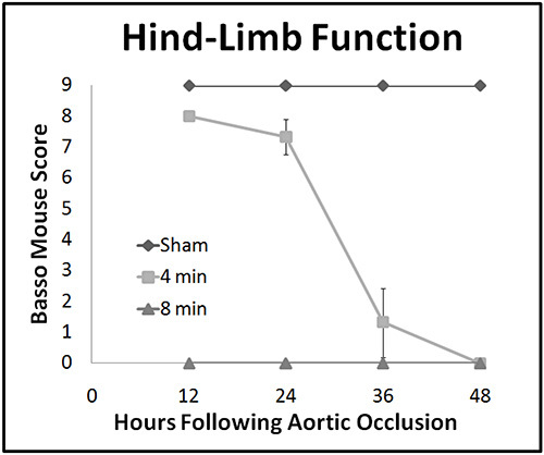
Results Mice that underwent sham surgery showed no observable hind limb deficit. Mice subjected to moderate ischemia for 4 min had minimal functional deficit at 12 hr followed by progression to complete paralysis at 48 hr. Mice subjected to prolonged ischemia had an immediate paralysis with no observable hind-limb movement at any point in the postoperative period. There was no observed intraoperative or post operative mortality.
Conclusion Reproducible lower extremity paralysis whether immediate or delayed can be achieved in a murine model. Additionally, by using a median sternotomy and careful dissection, high survival rates, and reproducibility can be achieved.
Keywords: Medicine, Issue 85, Spinal cord injury, thoracic aorta, paraplegia, Ischemia, reperfusion, murine model
Introduction
Lower extremity paralysis continues to complicate thoracoabdominal interventions. The injury, known as spinal cord ischemia-reperfusion injury (SCIR), results in paralysis in up to 20% of high risk patients1. Surgical adjuncts such as left heart bypass, lumbar cerbrospinal fluid drains, hypothermic circulatory arrest and intercostal artery reimplantation have reduced the incidence of this complication2; however, far too many patients continue to be affected.
Clinically, spinal cord ischemia and reperfusion injury is seen as either immediate or delayed paralysis following intervention3. However, our understanding of this injury has been stifled by a lack of mechanistic detail. As a result, few options are available to attenuate the injury once it has occurred.
We have thus enlisted a small animal, murine, model of spinal cord ischemia, and reperfusion injury to better characterize its pathogenesis. The majority of studies to date have used larger animal models to characterize this injury, namely rat4, rabbit5, and pig6 models. However, these are limited by their cost, complexity, variable reproducibility, and, most importantly, lack of available techniques for genetic manipulation. The most reliable of these published animal models involves infrarenal cross clamping of the abdominal aorta in rabbits. However, human anterior spinal neurons most often derive their vascular supply from more proximal branches7. Variable vascular anatomy of the spinal cord in these models adds to difficulty in transitioning their results into clinical use.
This manuscript presents a model for immediate or delayed paraplegia following thoracic aortic occlusion that is clinically relevant and easy to employ. Exposure of the aortic arch via mini sternotomy is less invasive and can elicit highly reproducible results with minimal morbidity and mortality. While this model in not without challenges and technical nuances, these can be overcome with careful dissection and tissue handling to produce a model of hind limb paralysis that can be easily implemented.
Protocol
1. Preoperative Preparation and Anesthesia
Be sure to observe sterile technique throughout the procedure. Lay out all instruments.
Turn on the temperature control bed prior to anesthetic induction so that it may warm to the appropriate temperature (36.5 °C). Power on the laser Doppler perfusion monitor so that it may boot during induction.
- Place the mouse in the induction chamber.
- Carefully monitor the respiratory rate of the mouse during induction.
- As soon as the respiratory rate has visually slowed, remove the mouse from the induction chamber.
- Perform toe pinch to assess adequacy of anesthetic.
With the mouse properly anesthetized, place mouse in supine position.
- Insert face into nose cone and secure all extremities to the heating table.
- Pay special attention to ensure that extremities are secured in anatomic position, with no deviation to one side. If the mouse is improperly positioned, it is difficult to avoid internal thoracic artery transection during sternotomy.
- Using clippers or commercially available hair removal cream, remove hair from the midline thorax and left lower extremity ventral surface.
- If using hair removal cream, avoid leaving cream in place for greater than 30 sec, as alkali burns can occur.
- Titrate volatile anesthetic vaporizer concentration to maintain adequate anesthesia.
- Expected vaporizer fractions are between 1-5% using isoflurane with high flow O2.
- Volatile anesthetic vaporizer concentration should be titrated to maintain anesthesia during surgical stimulation while maintaining spontaneous respirations.
2. Rectal Probe Laser Doppler Placement
Insert lubricated rectal probe into the mouse’s rectum. Secure in place to operating bed.
Adjust the heating bed for a target rectal temperature of 36.5 °C.
Make small incision over femoral artery of mouse and dissect skin away from subcutaneous tissue.
Insert laser Doppler probe over femoral artery.
- Adjust probe positions until the perfusion monitor registers greater than 800 perfusion units.
- Firmly secure probe in place. Improperly secured probes can have falsely low perfusion measurements.
3. Dissection of Aortic Arch/Subclavian Artery
Make a 2 cm skin incision above the sternal notch and gently dissect skin away from subcutaneous tissue.
- Dissect the submandibular gland free.
- If bleeding occurs, gentle pressure can be applied with a cotton swab.
- Divide the submandibular gland through midline in the avascular plane.
Gently lift sternum with forceps and using the scissors make 1 cm midline sternotomy through the midline of the sternum. Any deviation from midline can result in internal mammary artery hemorrhage which will be difficult to control.
Place 5-0 retraction sutures on each side at the edge of the sternum and retract sternum laterally securing sutures to the operating bed. Avoid placing retraction sutures too laterally to prevent pneumothoraces.
Using blunt dissection free strap muscles along trachea. The left strap muscle can be divided with scissors to improve exposure.
Dissect free the thymus from surrounding tissue. Continue blunt dissection until the great vessels are visualized. Use extreme caution to prevent entrance into the pleural space.
Place vascular clamps on aortic arch and left subclavian artery.
- Verify distal flow has appropriately disrupted. This will be seen as a >90% reduction in perfusion units.
- Continue occlusion for desired for 4-8 min.
Remove vascular clamp and verify hemostasis before closure of chest.
4. Closure of Sternotomy and Skin
Remove the retraction suture on the left side of the mouse.
- Close the sternotomy with the right retraction suture.
- A single sternal suture (using the previously placed retraction stitch) is adequate for sternal closure. Placing another stitch is unnecessary and increases risks of pneumothorax and hemorrhage.
Close skin with running 5-0 stitch.
5. Recovery and Postoperative Assessment
Transfer the mouse to recovery cage. Cage should be placed on a heating pad to increase the ambient temperature of the recovery chamber and reduce heat loss to the environment.
- Closely monitor the mouse for signs respiratory distress or seizure activity. Administer analgesia per institution guidelines. Euthanize mice immediately if seizure or respiratory distress is observed.
- CO2 chamber euthanasia is our preferred method. Cervical dislocation is another option if CO2 is not available.
- Complete recovery can be expected in 1-2 hr, depending on the length of volatile anesthetic and concentration used.
Return the mouse to normal cage. Place food and water place on the floor of the cage.
Assess neurologic status at 12 hr intervals using Basso Mouse Scale for Locomotion8.
Representative Results
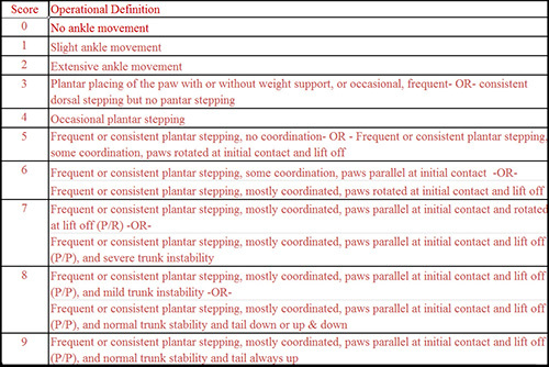
Mice underwent sham surgery (n=3) or aortic occlusion for 4 (n=3) to 8 min (n=3). Postoperatively mice were graded by the Basso Mouse Score (Figure 1). Mice that underwent sham surgery had no observable functional deficits at any point postoperatively. Mice subjected to moderate ischemia (4 min) had near normal hind-limb function at 12 hr with progressive functional decline to complete paralysis by 48 hr. Mice in prolonged ischemia group (8min) had complete paralysis following surgery without any recoverable function (Figure 2).
 Figure 1. Basso Score for Hind Limb Motor Function8. Scoring system for hind limb neurologic dysfunction graded from 0 (no function) to 9 (normal function). Click here to view larger image.
 Figure 2.Postoperative Hind-Limb Function. The severity of neurologic deficit was graded using the Basso Mouse Scale at 12 hr intervals postoperatively until 48 hr. Click here to view larger image.
Discussion
Paraplegia secondary to spinal cord ischemia reperfusion is the result of a complex of poorly understood pathologies9. While this is most commonly seen after thoracoabdominal aortic surgery, a variety of other insults such as aortic dissections, trauma, embolic phenomena, vasculitis, and systemic hypotension10 can result in paraplegia. To gain further understanding of this injury and provide future targets to eliminate this injury, animal models have become a necessity.
Patients afflicted with this complication exhibit either an immediate or delayed paralysis. This model and others adequately parallel11 the bimodal distribution of paralysis seen clinically. While moderate ischemia (4 min) produced a delayed paralysis, prolonged ischemia (8 min) resulted in an immediate and permanent paralysis.
The presented model has advantages compared to those requiring a lateral thoracotomy12,13. The median sternotomy exposure leaves the pleural cavity intact and eliminates the need for tracheal intubations or reestablishing negative pressure in the pleural space. Clinically, the sternotomy is associated with much less postoperative pain in than the lateral thoracotomy and might reduce postoperative analgesic requirements.
This model is not without limitations. As with other models, murine surgery has a learning curve and good surgical technique is imperative. Mice can quickly succumb if dissection is not done with care. The most common causes of intraoperative mortality are hemorrhage or pneumothorax. To avoid potentially fatal hemorrhage the sternotomy must be made midline. If the sternotomy is made too laterally a transaction of internal mammary arteries and subsequent hemorrhage may occur. Additionally, rough dissection of descending aorta and subclavian artery can result in hemorrhage the will be difficult or impossible to control. Pneumothoraces are another potentially fatal complication. These typically occur during the placement of retraction sutures or dissection of the aorta. If retraction sutures are place too laterally or the aortic arch is not dissected with care a pneumothorax can occur and are universally fatal.
Further steps need to be employed to ensure reproducibility. Normothermia is critical. During surgery mice are kept as close to 36.5 °C as possible. Even mild hypothermia has been shown in to have significant neuroprotective effects in spinal cord and stroke models14,15. Furthermore, laser Doppler measurements should be closely monitored and mice with only 90% reduction in distal flow should be included for comparison. Incomplete occlusion of the aorta or subclavian arteries can results in higher distal flows and results that are highly variable.
In conclusion, the murine model of lower extremity paralysis presented can be easily adopted, yielding highly reproducible results. Applications of this model can provided the investigator with a means to study the immediate and delayed paralysis. Moreover, adoption of this model might prove beneficial in combating this devastating complication.
Disclosures
The authors declare no competing financial interests.
Acknowledgments
We would like to thank the Thoracic Surgery Foundation for Research and Education for their Financial Support of this project.
References
- Conrad MF, Ye JY, Chung TK, Davison JK, Cambria RP. Spinal cord complications after thoracic aortic surgery: long-term survival and functional status varies with deficit severity. J. Vasc. Surg. 2008;48:47–53. doi: 10.1016/j.jvs.2008.02.047. [DOI] [PubMed] [Google Scholar]
- Okita Y. Fighting spinal cord complication during surgery for thoracoabdominal aortic disease. Gen. Thorac. Cardiovasc. Surg. 2011;59:79–90. doi: 10.1007/s11748-010-0668-x. [DOI] [PubMed] [Google Scholar]
- Wong DR, et al. Delayed spinal cord deficits after thoracoabdominal aortic aneurysm repair. Ann. Thorac. Surg. 2007;83:1345–1355. doi: 10.1016/j.athoracsur.2006.11.035. [DOI] [PubMed] [Google Scholar]
- Taira Y, Marsala M. Effect of proximal arterial perfusion pressure on function, spinal cord blood flow, and histopathologic changes after increasing intervals of aortic occlusion in the rat. Stroke. 1996;27:1850–1858. doi: 10.1161/01.str.27.10.1850. [DOI] [PubMed] [Google Scholar]
- Naslund TC, Hollier LH, Money SR, Facundus EC, Skenderis BS. Protecting the ischemic spinal cord during aortic clamping. The influence of anesthetics and hypothermia. Ann. Surg. 1992. pp. 409–515. [DOI] [PMC free article] [PubMed]
- Qayumi AK, Janusz MT, Lyster DM, Gillespie KD. Animal model for investigation of spinal cord injury caused by aortic cross-clamping. J. Invest. Surg. 1997;10:47–52. doi: 10.3109/08941939709032125. [DOI] [PubMed] [Google Scholar]
- Lang-Lazdunski L, Matsushita K, Hirt L, Waeber C, Vonsattel JP, Moskowitz MA, Dietrich WD. Spinal Cord Ischemia: Development of a model in the mouse. Stroke. 2000;31:208–213. doi: 10.1161/01.str.31.1.208. [DOI] [PubMed] [Google Scholar]
- Basso DM, Fisher LC, Anderson AJ, Jakeman LB, McTigue DM, Popovich PG. Basso Mouse Scale for locomotion detects differences in recovery after spinal cord injury in five common mouse strains. J. Neurotrauma. 2006;23:635–659. doi: 10.1089/neu.2006.23.635. [DOI] [PubMed] [Google Scholar]
- Kwon BK, Tetzlaff W, Grauer JN, Beiner J, Vaccaro AR. Pathophysiology and pharmacologic treatment of acute spinal cord injury. Spine. J. 2004;4:451–464. doi: 10.1016/j.spinee.2003.07.007. [DOI] [PubMed] [Google Scholar]
- Cheshire WP, Santos CC, Massey EW, Howard JF. Spinal cord infarction: etiology and outcome. Neurology. 1996;47:321–330. doi: 10.1212/wnl.47.2.321. [DOI] [PubMed] [Google Scholar]
- Kakinohana M, et al. Delayed paraplegia after spinal cord ischemic injury requires caspase-3 activation in mice. Stroke. 2011;42(8):2302–2307. doi: 10.1161/STROKEAHA.110.600429. [DOI] [PMC free article] [PubMed] [Google Scholar]
- Wang Z, Yang W, Britz GW, Lombard FW, Warner DS, Sheng H. Development of a simplified spinal cord ischemia model in mice. J. Neurosci. Methods. 2010;189:246–251. doi: 10.1016/j.jneumeth.2010.04.003. [DOI] [PMC free article] [PubMed] [Google Scholar]
- model of ischemic spinal cord injury with delayed paralysis caused by aortic cross-clamping. Anesthesiology. 2010;113:880–891. doi: 10.1097/ALN.0b013e3181ec61ee. [DOI] [PMC free article] [PubMed] [Google Scholar]
- Kang J, et al. The effects of systemic hypothermia on a murine model of thoracic aortic ischemia reperfusion. J. Vasc. Surg. 2010;52:435–443. doi: 10.1016/j.jvs.2010.03.021. [DOI] [PubMed] [Google Scholar]
- Li J, Benashski S, McCullough LD. Post-stroke hypothermia provides neuroprotection through inhibition of AMP-activated protein kinase. J. Neurotrauma. 2011;28(7):1281–1288. doi: 10.1089/neu.2011.1751. [DOI] [PMC free article] [PubMed] [Google Scholar]


