Fig. 5.
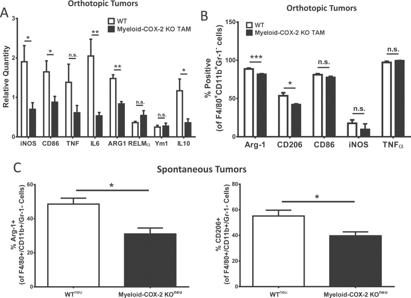
TAM in myeloid-COX-2 KO mice display an altered macrophage phenotype. (A) Q-PCR for mRNA levels of several M1 (iNOS, CD86, IL-6) and M2 (arginase-1 and IL-10) markers were lower in tumor tissue isolated from myeloid-COX-2 KO hosts compared with WT (n = 4–6). (B) Flow cytometric analysis of live-gated TAMs (F4/80+CD11b+Gr-1−; see Supplementary Figure 4) revealed a lower proportion of M2 (arginase-1 or mannose receptor, CD206, positive) TAMs with no change in M1 (CD86, iNOS or tumor necrosis factor α positive) TAMs in myeloid-COX-2 KO orthotopic tumors compared with WT (n = 3–6). (C) The proportion of arginase-1 and CD206 positive TAM was also reduced in spontaneous neu-driven tumors (n = 5–6). Data are mean ± SEM. *P < 0.05, **P < 0.01, ***P < 0.001, n.s. = not significant.

