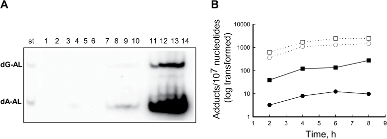
Fig. 3.
Murine renal and hepatic cytosols activate AL-I-NOH and AL-II-NOH in the presence of PAPS, leading to AL-DNA adduct formation. 0.8mg/ml of ssDNA was incubated with 300 μM of AL-I-NOH or AL-II-NOH and 1mg/ml of mouse cytosolic extracts in the presence of PAPS in a total volume of 500 μl. DNA was extracted and 20 μg of used for the adduct analysis. (A) Fragment of polyacrylamide gel showing results of 32P-post-labeling analysis; St—Mixture of 24mer oligonucleotides (30fmol) containing a single dG-AL-II or dA-AL-II, represented by the upper and lower band, respectively. Lanes 1–6, the following were incubated for 6h in a reaction buffer; 1—DNA; 2—DNA and AL-I-NOH; 3—DNA, AL-I-NOH and PAPS; 4—DNA, AL-I-NOH and acetyl-CoA; 5—DNA, AL-I-NOH and renal cytosol; 6—DNA, AL-I-NOH and hepatic cytosol; Lanes 7–10, DNA, AL-I-NOH, PAPS and renal cytosol, incubated for 2, 4, 6 and 8h, respectively; Lanes 11–14, DNA, AL-I-NOH, PAPS and hepatic cytosol, incubated for 2, 4, 6 and 8h, respectively. (B) Time dependence of AL-DNA adduct formation. Filled and open circles correspond to renal and hepatic SULTs activities towards AL-I-NOH. Filled and open squares correspond to renal and hepatic SULTs activities towards AL-II-NOH. Results are shown as mean values for three independent experiments; standard deviations are <30%.

