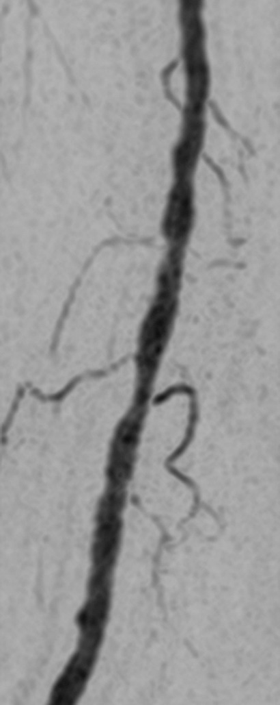
Figure 3c:

(a) Coronal MIP and sagittal MIP of left leg in a healthy 81-year-old male volunteer (subject 4) generated by using final abdomen-pelvis and thigh station time frames and third calf-foot time frame. Dwell time at thigh station was only 5.0 seconds. (b–d) Targeted coronal MIPs of the boxed regions in a highlight arterial detail. Although this subject was recruited as a healthy volunteer, multiple low-grade stenoses are seen in thigh and calf arteries, and anterior tibial arteries are partially occluded. Contrast material bolus was imaged at four abdomen-pelvis and two thigh station time frames, and venous contamination was avoided at calf-foot station. (Reprinted, with permission, from reference 17.) Also see Figure E1 (online) and Movie 1 (online).
