Abstract
Background:
Frozen shoulder has always been considered important because of the impact on the quality-of-life and long period of illness. Therefore, the use of noninvasive and safe techniques that can speed up the healing process of the disease is important.
Methods:
This study was a randomized clinical trial study on patients suffering from frozen shoulder who were referred to Isfahan University of Medical Sciences hospitals in 2011 and 2012. A total of 36 patients were enrolled in the study. Eligible patients were allocated into two groups. Intervention group received extracorporeal shockwave therapy (ESWT) once a week for 4 weeks. The control group received sham shockwave therapy once a week for 4 weeks. On the follow-up period, changes in individual performance and the amount of pain and disability were assessed by the Shoulder Pain and Disability Index (SPADI) questionnaire and the range of motion changes were assessed by a goniometer. Data obtained were analyzed using SPSS software.
Results:
Variance analysis revealed a difference in the mean pain and disability score of the SPADI questionnaire, flexion, extension, and abduction, external rotation of involved shoulder between two groups before and after the shockwave therapy (P < 0.05). Improvement was more satisfactory in the intervention group, but the mean internal rotation did not differ significantly in two groups (P > 0.05).
Conclusions:
The use of ESWT seems to have positive effects on treatment, quicker return to daily activities, and quality-of-life improvement on frozen shoulder.
Keywords: Adhesive capsulitis, extracorporeal shockwave therapy, frozen shoulder, Shoulder Pain and Disability Index questionnaire
INTRODUCTION
Frozen shoulder is an idiopathic and progressive disease, which is identified by pain and decreased range of motion (ROM) of the shoulder and shoulder joint capsule fibrosis. Adhesive capsulitis occurs in approximately 2-5% of the general population which is 2-4 times more common in women than men, and is most frequently seen in individuals between 40 and 60 years of age the disease is more common in patients with diabetes mellitus, rotator cuff lesions, thyroid disorders, Chronic Obstructive Pulmonary Diseases, cerebrovascular accident, myocardial infarction, inflammatory arthritis, trauma, and prolonged immobilization. Frozen shoulder has many complications such as sleep disturbance, impaired performance in daily activities, and personal grooming. The disease has three phases: In the first phase (pain phase), synovitis, thickening of the joint capsule, synovial fluid loss, and decreased ROM are observed. The duration of this phase is approximately 10-36 weeks. In the second phase (frozen phase), pain is decreased and joint capsule fibrosis, thickening of the rotator cuff tendons, and loss of joint space are seen. The duration of this phase is approximately 4-12 months. In the third phase (resolution phase), joint ROM again increases gradually and the patient gradually returns to normal daily activities. The duration of this phase is approximately 12-42 months. The disease is treatable although treatment is usually long and creates many problems for the patients. Intra-articular corticosteroid injections, physical therapy, supraclavicular nerve block, acupuncture, daily activities modification, etc., are traditionally used in this condition.[1,2,3,4] In physical therapy, various modalities such as joint mobilization,[5] heat, transcutaneous electrical nerve stimulation, exercise, distension arthrography and manipulation under anesthesia, arthroscopic and open surgical release are used early in the rehabilitation process; however, passive joint glides and nonpainful passive ROM exercises might be beneficial. Early scapular stability exercises and closed chain rotator cuff exercises can be instituted. As the patient's symptoms improve, active-assisted and active ROM activities can be added, along with open chain and proprioceptive exercises. The Pendulum and University of Washington (Jackins) exercises are useful for improving the shoulder's ROM.[6,7,8,9] Shockwave through generating low-energy waves and electromagnetic excitation could be effective in this condition due to increasing the regional blood flow, neovascular changes, enzymes release, reduction of inflammatory cytokines, and increasing the flexibility of the collagen fibers and tendons in that area. This type of treatment has been used in other orthopedic conditions such as tendinitis,[10,11,12,13,14,15] plantar fasciitis,[15,16,17] and lateral epicondilitis[18,19] the healing process of wounds[20] and fractures[21] and satisfactory results have been achieved. There is also limited number of investigations to evaluate the usefulness of shockwave therapy in frozen shoulder. This study aimed to determine the effect of extracorporeal shockwave therapy (ESWT) in the treatment of patients with frozen shoulder admitted to Isfahan University of Medical Sciences hospitals in 2011 and 2012.
METHODS
Study design and participants
This clinical randomized trial study was performed in Kashani Hospital, Isfahan, Iran in 2011 and 2012.
The study population was the patients with frozen shoulder who were admitted to Isfahan Medical University hospital. The patients were examined by a hand surgeon sub-specialist. After providing the necessary X-ray and injection of 40 mg triamcinolone (Hexal AG) into the involved shoulder joint, the patients were referred to the physical medicine and rehabilitation clinic. Exclusion criteria were history of previous surgery on the shoulder, history of shoulder fracture, cancer, inflammatory disorders, bleeding disorders, and unwillingness to participate in the study. Then, the patients completed a consent form to participate in the research. After that, the enrolled patients were randomized into two groups using random allocation software.
Proscure and variable assessment
All the patients received analgesics (meloxicam 15 mg daily), and activity modification to reduce pain, and were advised to perform Pendulum exercises (swinging arm forward and back, side to side, and around in circles for 5-10 times) and stretching the back of the involved shoulder 30 s twice a day. If the patient tolerated it progressed by wall walking and University of Washington (Jackins) exercises, the patients were blind to the study. The patients in the intervention group received shock wave therapy once a week for 4 weeks. The focus probe sets were used and in each session, patients received ESWT from anterior and posterior directions (on the average 1200 shocks between 0.1 and 0.3 mJ/mm2) up to the maximum threshold of pain tolerance in the shoulder. The control group received sham shockwave therapy once a week for 4 weeks while the device was turned off and placed on the patient's shoulder for the same period of time. The device used in this study was the standard electromagnetic shockwave (Doulith SD1). This procedure was performed with patients in the sitting position on a chair after applying the coupling gel. Pain and disability score were assessed with the Shoulder Pain and Disability Index (SPADI) questionnaire before the treatment, immediately after the intervention, and 2 and 5 months after the intervention. The SPADI questionnaire was completed for each patient by the physician in each session. The SPADI questionnaire's is a self-administered instrument developed to measure pain (five items) and disability (eight items) associated with shoulder complaints. For the five pain items, “no pain” scored zero and “the worst imaginable pain” scored 10; for the eight disability items, “no difficulty” received zero score while “difficulty requiring assistance” received 10 scores. The SPADI demonstrates good internal consistency, test-retest reliability and criterion and construct validity. It also appears to be able to detect change in patient status over time. The SPADI should therefore prove to be a useful instrument both in clinical practice and in clinical research.[22] The ROM of the involved shoulder was measured by a goniometer in flexion, abduction, extension, internal, and external rotation.
Statistical analyses
The obtained data were analyzed with the Statistical Package for the Social Sciences version 20.0 (SPSS Inc., Chicago, IL, USA), using independent t-test, paired t-test, Chi-square test, and repeated measures ANOVA.
RESULTS
Figure 1 shows a total of 40 patients with frozen shoulder were randomly divided into two groups. The intervention groups received ESWT and the control group received sham ESWT. Four patients (one in the intervention group and three in the control group) did not complete treatment sessions due to unwillingness; therefore, 19 patients in the intervention group (13 women and 6 men, 68.4% vs. 31.6%) and 17 patients in the control group (12 women and 5 men, 70.5% vs. 29.5%) completed the study. The mean age of the intervention and control groups was 56.1 ± 10.6 and 60.3 ± 4.8 years respectively and t-test revealed no significant difference between the two groups in this regard (P > 0.05). As for gender distribution in the intervention and control groups, there were 13 females and 6 males. Chi-square test showed no significant difference in gender distribution between the two groups (P > 0.05). Furthermore, the right shoulder was involved in 7 patients in the intervention group and 9 patients in the control group and the rest of the patients had their left shoulders involved; Chi-square test showed no significant difference in the involved shoulder between the two groups (P > 0.05). During the study, all the patients completed the primary medical therapy and the recommended exercises and no significant complications were noted. The mean and standard deviation of pain and disability scores, flexion, extension, and abduction, internal, and external rotation are shown in Table 1.
Figure 1.
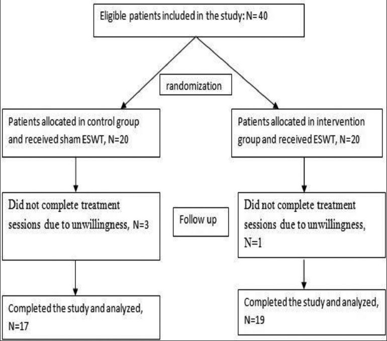
Patient's enrollment, allocation, follows up and analysis in both study groups
Table 1.
Mean and standard deviation of pain and disability scores, flexion, extension, and abduction, internal and external rotation
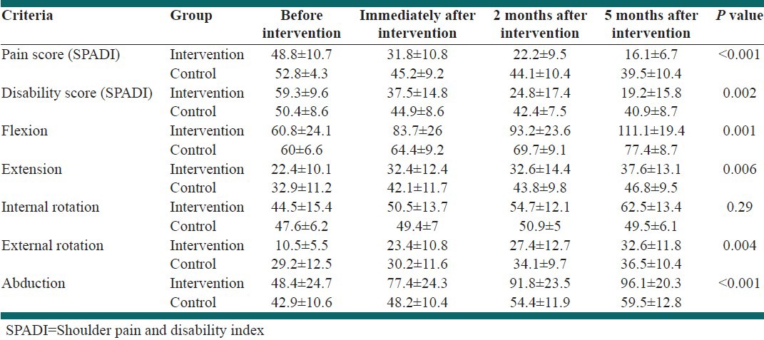
Table 1 shows mean and standard deviation of pain and disability scores, flexion, extension, abduction, internal, and external rotation.
Independent t-test was performed on the data which revealed a difference in the mean pain and disability score of the SPADI questionnaire, flexion, extension, and abduction, internal, and external rotation of involved shoulder between two groups before and after the ESWT. Improvement was more satisfactory in the intervention group (P < 0.05). Although, the mean internal rotation in both groups was similar and no significant difference was observed in this variable between the two groups (P > 0.05). The fastest recovery in all mentioned values was obtained within 2 months after the intervention. After this period of time, the healing process slowed down. Minimal response to treatment was seen in patients with uncontrolled diabetes. Variable factors in the treatment period in both groups are shown in Figures 2-9.
Figure 2.
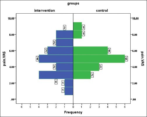
Distribution of the pain score according to the Shoulder Pain and Disability Index score before the intervention
Figure 9.
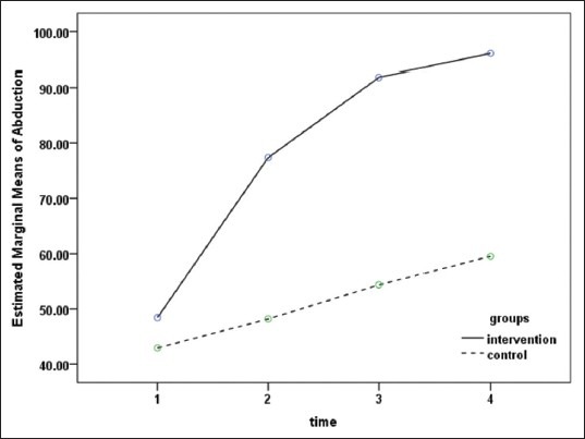
Changes in the abduction score in both groups
Figure 3.
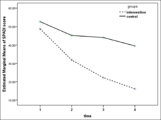
Changs in the pain score according to the Shoulder Pain and Disability Index score in both groups
Figure 4.
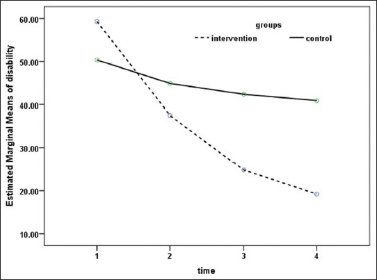
Changes in the disability score in both groups
Figure 5.
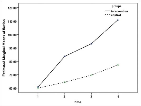
Changes in the flexion score in both groups
Figure 6.
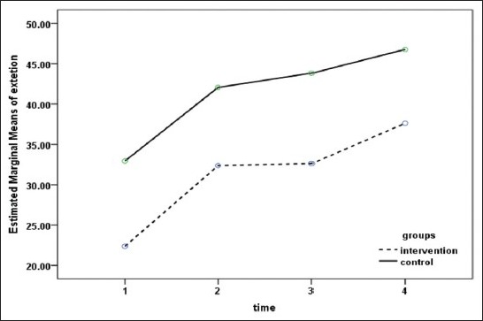
Changes in the extension score in both groups
Figure 7.
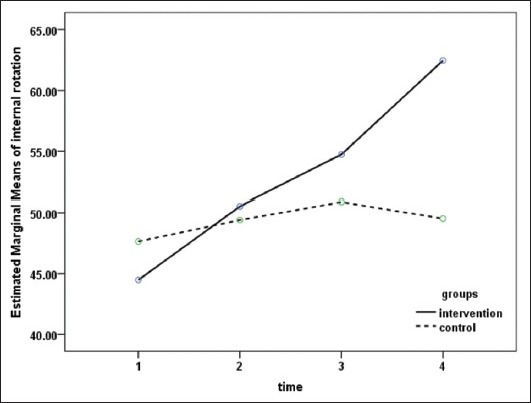
Changes in the internal rotation score in both groups
Figure 8.
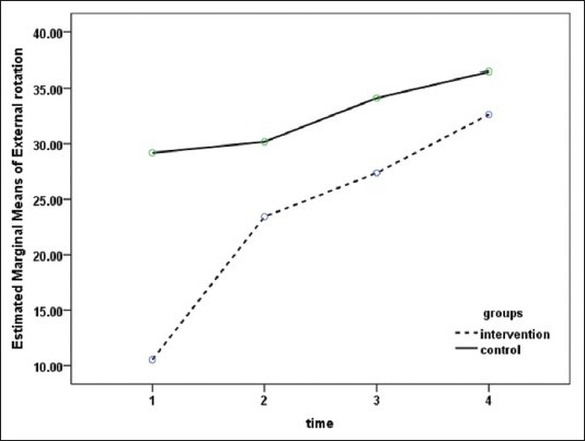
Changes in the external rotation score in both groups
DISCUSSION
There are different types of treatments for frozen shoulder such as medical therapy, physical therapy, surgery, etc. However, these methods are expensive and time consuming and require several sessions for physical therapy; therefore, some patients may leave treatment. Other methods such as distension arthroscopy and manipulation under anesthesia are recommended and although, the risk of general anesthesia is considered for all of these conditions. Since some patients have diabetes and cardiac problems, surgery methods have more complications. ESWT has positive results in orthopedic conditions such as tendinitis, plantar fasciitis, lateral epicondilitis and rotator cuff disease. Since the rotator cuff problem is one of the predisposing factors for frozen shoulder and ESWT is effective in this condition, ESWT may also be useful in frozen shoulder. Durante et al. (2006) studied patients with similar conditions. One group of the patients were treated with extracorporeal shockwave combined with physiokinesiotherapy and the second group were treated with physiokinesiotherapy alone.[23] Patients received 2500 shocks from anterior and lateral directions with energy between 0.07 and 0.11 mJ/mm2 two sessions per week 2 weeks. All patients were followed for 6 months. About 30% of the patients showed excellent response, while 30% of patients showed poor response, as well. 10% of patients in the second group showed complete response. According to arthroscopic findings, adhesions occur more in the descendent fold and surrounding synovium, therefore, stimulation from anterior and posterior directions is more effective rather than the lateral direction. In the current study, the amount of the energy was higher than the study by Durante; other researches on the rotator cuff tendonitis have reported more effective responses with higher energy; therefore, it is better to increase the level of energy and decrease the number of shocks. In the present study, the better response to treatment was likely due to proper session intervals.
CONCLUSIONS
According to the findings of this study and in comparison with similar studies, ESWT has positive effects on acceleration of the healing process of frozen shoulder. Considering the significant side-effects of other therapies such as surgery, patients with frozen shoulder can take advantage of ESWT because of its noninvasive, safe nature, low costs, no need for hospitalization, fewer visits of patient in the hospital, and the lack of significant adverse events during the treatment.
RECOMMENDATION
Since systematic reviews on plantar fasciitis have revealed effective response to the use of the radial shockwave probe, the authors suggest that the efficacy of the radial probe in comparison with the probe focused on the treatment of frozen shoulder be evaluated in future studies.
ACKNOWLEDGMENT
This study was supported by a grant from Isfahan University of Medical Sciences (Grant no: 391061). The authors are grateful to the authorities of Kashani Hospital for their cooperation.
Footnotes
Source of Support: This study was supported by a grant from Isfahan University of Medical Sciences (grant No: 391061)
Conflict of Interest: None declared
REFERENCES
- 1.Frontra G. Essentials of Physical Medicine and Rehabilitation. 2nd ed. Philadelphia: Saunders, Elsevier; 2008. Adhesive capsulitis; pp. 49–54. [Google Scholar]
- 2.Braddom RL. 4th ed. Philadelphia: WB Saunders; 2011. Handbook of Physical Medicine and Rehabilitation; p. 825. [Google Scholar]
- 3.Dalton SE. The shoulder. In: Hochberg M, Silman AJ, Smolen JS, editors. Rheumatology. 3rd ed. Toronto: Mosby; 2003. pp. 615–30. [Google Scholar]
- 4.Dias R, Cutts S, Massoud S. Frozen shoulder. BMJ. 2005;331:1453–6. doi: 10.1136/bmj.331.7530.1453. [DOI] [PMC free article] [PubMed] [Google Scholar]
- 5.Yang JL, Chang CW, Chen SY, Wang SF, Lin JJ. Mobilization techniques in subjects with frozen shoulder syndrome: Randomized multiple-treatment trial. J Am Phys Ther Assoc Phys Med. 2013;93:1307–15. doi: 10.2522/ptj.20060295. [DOI] [PubMed] [Google Scholar]
- 6.Basford JR. Therapeutic physical agents. In: Delisa JA, editor. Physical Medicine and Rehabilitation. Principles and Practice. Philadelphia: Lippincott Williams and Wilkins; 2005. pp. 251–70. [Google Scholar]
- 7.Berghs BM, Sole-Molins X, Bunker TD. Arthroscopic release of adhesive capsulitis. J Shoulder Elbow Surg. 2004;13:180–5. doi: 10.1016/j.jse.2003.12.004. [DOI] [PubMed] [Google Scholar]
- 8.Lynch SA. Surgical and nonsurgical treatment of adhesive capsulitis. Curr Opin Orthop. 2002;13:271–4. [Google Scholar]
- 9.Miller MD. 3rd ed. Philadelphia: WB Saunders; 2000. Review of Orthopedics; pp. 218–25. [Google Scholar]
- 10.Loew M, Daecke W, Kusnierczak D, Rahmanzadeh M, Ewerbeck V. Shock-wave therapy is effective for chronic calcifying tendinitis of the shoulder. J Bone Joint Surg Br. 1999;81:863–7. doi: 10.1302/0301-620x.81b5.9374. [DOI] [PubMed] [Google Scholar]
- 11.Wilson M, Stacy J. Shock wave therapy for Achilles tendinopathy. Curr Rev Musculoskelet Med. 2010;4:6–10. doi: 10.1007/s12178-010-9067-2. [DOI] [PMC free article] [PubMed] [Google Scholar]
- 12.Huisstede BM, Gebremariam L, van der Sande R, Hay EM, Koes BW. Evidence for effectiveness of Extracorporal Shock-Wave Therapy (ESWT) to treat calcific and non-calcific rotator cuff tendinosis – A systematic review. Man Ther. 2011;16:419–33. doi: 10.1016/j.math.2011.02.005. [DOI] [PubMed] [Google Scholar]
- 13.Harniman E, Carette S, Kennedy C, Beaton D. Extracorporeal shock wave therapy for calcific and noncalcific tendonitis of the rotator cuff: A systematic review. J Hand Ther. 2004;17:132–51. doi: 10.1197/j.jht.2004.02.003. [DOI] [PubMed] [Google Scholar]
- 14.Schmitt J, Haake M, Tosch A, Hildebrand R, Deike B, Griss P. Low-energy extracorporeal shock-wave treatment (ESWT) for tendinitis of the supraspinatus. A prospective, randomised study. J Bone Joint Surg Br. 2001;83:873–6. doi: 10.1302/0301-620x.83b6.11591. [DOI] [PubMed] [Google Scholar]
- 15.Vahdatpour B, Sajadieh S, Bateni V, Karami M, Sajjadieh H. Extracorporeal shock wave therapy in patients with plantar fasciitis. A randomized, placebo-controlled trial with ultrasonographic and subjective outcome assessments. J Res Med Sci. 2012;17:834–8. [PMC free article] [PubMed] [Google Scholar]
- 16.Rompe JD, Cacchio A, Weil L, Jr, Furia JP, Haist J, Reiners V, et al. Plantar fascia-specific stretching versus radial shock-wave therapy as initial treatment of plantar fasciopathy. J Bone Joint Surg Am. 2010;92:2514–22. doi: 10.2106/JBJS.I.01651. [DOI] [PubMed] [Google Scholar]
- 17.Rompe JD, Schoellner C, Nafe B. Evaluation of low-energy extracorporeal shock-wave application for treatment of chronic plantar fasciitis. J Bone Joint Surg Am. 2002;84-A:335–41. doi: 10.2106/00004623-200203000-00001. [DOI] [PubMed] [Google Scholar]
- 18.Speed CA, Nichols D, Richards C, Humphreys H, Wies JT, Burnet S, et al. Extracorporeal shock wave therapy for lateral epicondylitis – A double blind randomised controlled trial. J Orthop Res. 2002;20:895–8. doi: 10.1016/S0736-0266(02)00013-X. [DOI] [PubMed] [Google Scholar]
- 19.Ko JY, Chen HS, Chen LM. Treatment of lateral epicondylitis of the elbow with shock waves. Clin Orthop Relat Res. 2001;387:60–7. doi: 10.1097/00003086-200106000-00008. [DOI] [PubMed] [Google Scholar]
- 20.Kuo YR, Wang CT, Wang FS, Chiang YC, Wang CJ. Extracorporeal shock-wave therapy enhanced wound healing via increasing topical blood perfusion and tissue regeneration in a rat model of STZ-induced diabetes. Wound Repair Regen. 2009;17:522–30. doi: 10.1111/j.1524-475X.2009.00504.x. [DOI] [PubMed] [Google Scholar]
- 21.Biedermann R, Martin A, Handle G, Auckenthaler T, Bach C, Krismer M. Extracorporeal shock waves in the treatment of nonunions. J Trauma. 2003;54:936–42. doi: 10.1097/01.TA.0000042155.26936.03. [DOI] [PubMed] [Google Scholar]
- 22.Habermeyer P, Magosch P, Lichtenberg S. Heidelberg: Springer; 2006. Classifications and Scores of the Shoulder; pp. 251–2. [Google Scholar]
- 23.Durante C, Corrado B, Galasso O, Carillo MR. 4thInternational Congress of the ISMST. Berlin, Germany: 2001. May, The frozen shoulder: Indications for extracorporeal shockwave therapy; pp. 16–7. [Google Scholar]


