Figure 7.
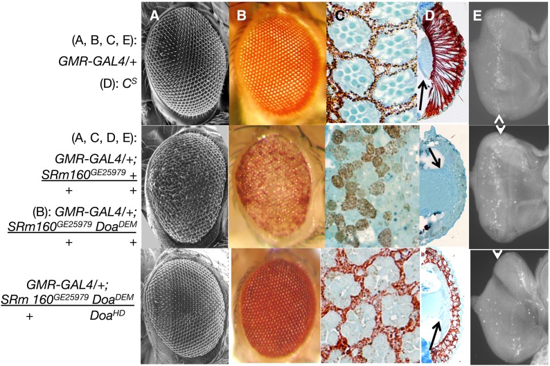
Overexpression of SRm160 in the eye induces loss of differentiation, disorganization, and apoptosis. (A) Scanning electron microscopy; (B) light microscopy; (C) semithin saggital sections, (D) Transverse sections; (E) eye imaginal discs stained with anti-activated caspase-3. Genotypes are as indicated. GMR > SRm160 DoaDEM/+ + and GMR > SRm160 DoaDEM/+ DoaHD samples were from female siblings, raised at 25°. GMR > SRm160 eyes (middle row) show significant eye aberrations, including disorganized facets in A and B, disrupted ommatidial pigment cell lattice and photoreceptor organization (C), loss of the elongation of photoreceptors and pigment cells, as well as the lamina (D), and higher levels of activated caspase (E), all as compared with GMR–GAL4 or CS as indicated (top row). The arrows in D indicate the lamina, present in Cs (top), but eliminated in GMR > SRm160 (second from top). Higher relative fluorescent signal (E, arrowheads) of activated caspase 3 in GMR > SRm160 (second from top) relative to GMR–GAL4/+ alone (top) (arrow). Simultaneous mutation of Doa (GMR > SRm160; DoaDEM/DoaHD, bottom row), almost completely suppresses the smooth glassy eye of GMR > SRm160 (A and B), restoring organization and pigment cell differentiation to the retina (C and D), and even to the lamina (D, arrow) and also strongly reduces caspase 3 activation (arrow, bottom). GMR–GAL4 as a homozygote is capable of autonomously inducing eye roughness and apoptosis, but has neither effect as a heterozygote.

