Abstract
A 54-year-old woman was admitted to our hospital with the complaint of right upper quadrant pain. Upon physical examination the vital signs of the patient were within normal ranges. Ultrasonography and computed tomography (CT) examination of the abdomen was obtained, which demonstrated a large dilatated cystic structure, measuring approximately 68.6 mm × 48.6 mm, with marked distension and inflammation. Additionally, the enhanced CT was characterized by the non-enhanced wall of the gallbladder. As the third examination in this study, magnetic resonance imaging (MRI), namely coronal MRI and magnetic resonance cholangio-pancreatography (MRCP), were performed. The MRCP demonstrated a dilatation of the gallbladder but detected no neck of the gallbladder. Simple cholecystectomy was performed. Macroscopic findings included a distended and gangrenous gallbladder, and closer examination revealed a counterclockwise torsion of 360 degrees on the gallbladder mesentery. Coronal MRI and MRCP showing characteristic radiography may be useful in making a definitive diagnosis.
Keywords: Volvulus of the gallbladder, Computed tomography, Magnetic resonance imaging, Magnetic resonance cholangio-pancreatography
INTRODUCTION
Gallbladder volvulus is a relatively rare disease and well recognized in the elderly people. It has been reported in only about 300 cases in the literature ranging in age from 2 to 100 years old. Preoperative diagnosis of gallbladder volvulus has always been considered difficult. Although recent advances in radiographic finding have helped in the diagnosis of many diseases, abdominal computed tomography (CT) and Ultrasonography (US) remain nonspecific in diagnosing volvulus of the gallbladder. However, we could make definitive radiographic imaging by coronal magnetic resonance imaging (MRI) and magnetic resonance cholangio-pancreatography (MRCP).
CASE REPORT
A 54-year-old woman was admitted to our hospital with the complaint of right upper quadrant pain of approximately 5 hours in duration. Pain was accompanied by anorexia and nausea without vomiting and was not preceded by jaundice. She had no relevant past history. Upon physical examination the vital signs of the patient were within normal ranges. Right upper quadrant tenderness and Murphy’s sign were detected in the abdominal examination. Laboratory data showed a leukocyte count of 11 800/ mm3 and C-reactive protein (CRP) of 2.57 mg/dL. Initial treatment consisted of administering intravenous fluids and broad-spectrum antibiotics. Abdominal ultrasound US demonstrated a distended, fluid-filled neck of abnormal swelling, a normal-walled gallbladder with surrounding ascites and edema (Figure 1), but no stones. Secondary computed tomography (CT) examination of the abdomen was obtained. CT demonstrated a large dilatated cystic structure, measuring approximately 68.6 mm × 48.6 mm, with marked distension and inflammation. Additionally, the enhanced CT was characterized by the non-enhanced wall of the gallbladder (Figure 2). As the third examination in this study, magnetic resonance imaging (MRI), namely coronal MRI and magnetic resonance cholangio-pancreatography (MRCP), were performed. The MRCP demonstrated a dilatation of the gallbladder but detected no neck of the gallbladder (Figure 3). The coronal MRI revealed a dilatated gallbladder, and additionally an imagination that identified the neck of the gallbladder (Figure 4). She was diagnosed with acute cholecystitis and volvulus of the gallbladder according to the findings of US, CT, MRI and MRCP and underwent percutaneous transhepatic gallbladder aspiration (PTGBA) for one day. Following the treatment, a hemobilia discharge from the PTGBA was noted, and she was diagnosed with necrotic gallbladder. Her pain was slightly improved, but she still had tenderness and high fever. She underwent open laparotomy, and simple cholecystectomy was performed. Macroscopic findings included a distended and gangrenous gallbladder, and closer examination revealed a counterclockwise torsion of 360 degrees on the gallbladder mesentery. De-torsion and cholecystectomy were easily performed (Figure 5). The histopathology report showed necrosis and hemorrhage of the gallbladder without evidence of lithiasis. Nine days after the operation she was discharged with no complications.
Figure 1.
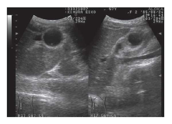
Abdominal ultrasonography revealed an abnormally large floating gallbladder without gallstones, and a thickened gallbladder wall.
Figure 2.
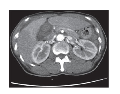
Abdominal enhanced computed tomography revealed a dilatated gallbladder, but a non-enhanced gallbladder wall.
Figure 3.
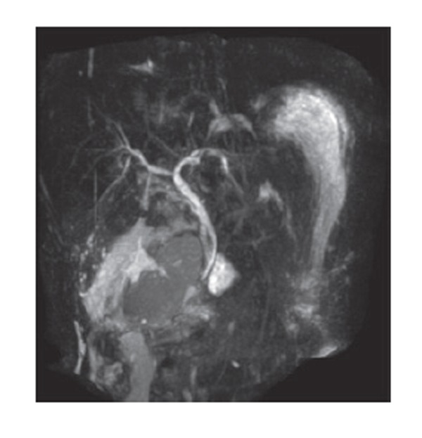
MRCP demons-trated dilatation of the gall-bladder, but its image identified no gallbladder neck.
Figure 4.
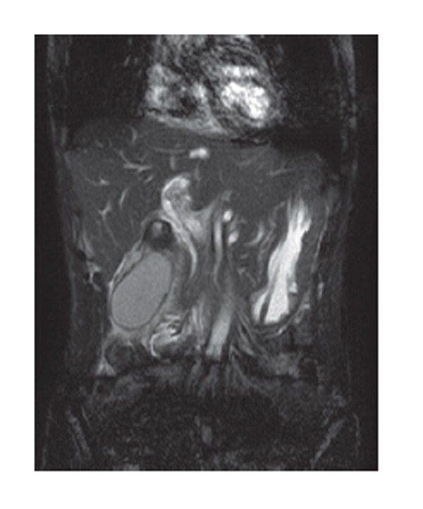
Coronal MRI revealed a dilatated gallbladder, and its invagination-like image identified the neck of the gallbladder.
Figure 5.
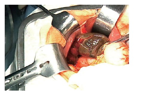
At laparotomy, macroscopic findings showed a distended and gangrenous gallbladder along with a counterclockwise torsion of 360 degrees of the gallbladder mesentery.
DISCUSSION
First reported by Wendel in 1898[1], volvulus of the gallbladder is a relatively uncommon phenomenon, with no more than 300 cases reported in the literature. It occurs in all age groups, with the highest incidence in elderly women, and a female-to-male ratio of 3:1. Perhaps the incidence would increase with a longer life expectancy rate[2,3]. Gallstones are unlikely to be the cause of gallbladder torsion, as gallstones are not uniformly present in all patients reported with torsion. One study of 245 patients found stones in only 24.4%; 51% developed a clockwise torsion rotation[4]. Although supportive evidence is lacking, inferences have been made in the literature linking gastric peristalsis to clockwise gallbladder torsion and colonic peristalsis to counterclockwise torsion[5]. Because volvulus of the gallbladder is a relatively uncommon phenomenon, preoperative diagnosis of gallbladder torsion remains difficult. Therefore, most cases are diagnosed intraoperatively at present. Also, laboratory evaluations are often nonspecific. For example, white blood cell count (WBC), CRP and creatine phosphokinase (CPK) are frequently elevated, while liver function tests are commonly normal. In our case, WBC, CRP and CPK were elevated. Although recent advances in radiographic studies have helped in the diagnosis of many diseases, radiographic studies remain nonspecific in diagnosing volvulus of the gallbladder. Fewer than a dozen cases have been reported in the literature where a preoperative diagnosis was made. Ultrasound studies often reveal a large floating gallbladder without gallstones, and a thickened gallbladder wall. Specific ultrasound signs seen with gallbladder torsion include the presence of the gallbladder outside its normal anatomic fossa, inferior to the liver or in a transverse orientation with an echogenic conical structure[6]. Additionally, CT findings are also nonspecific. A few cases of CT diagnosis of gallbladder torsion commented on radiographic findings of marked enlargement of the gallbladder with an unusual shape and configuration[7,8]. MRI has been used to establish a diagnosis preoperatively. MRI findings include high signal intensity within the gallbladder wall on T1 weighted images, suggesting necrosis and hemorrhage consistent with gallbladder torsion. In the present case, MRCP revealed anatomic details of the neck of the gallbladder and cystic duct.
We consider the characteristics of these radiography images to be useful for differential diagnosis of torsion of the gallbladder from gallbladder stone and gallbladder cancer. Usui et al have reported that only MRCP made it possible to determine the relationships between the distorted bile ducts, the interrupted cystic duct, and the enlarged gallbladder, and it was a relatively non-invasive procedure[9]. In addition, Shaikh et al have reported that the presence of a redundant mesentry was a prerequisite for torsion[10]. Although torsion of the gallbladder is a rare occurrence, the diagnosis should be considered in all patients presenting with right upper quadrant pain. If diagnosed early and treated, it remains a benign condition; however, a delay in diagnosis and management may lead to sequelae associated with gallbladder rupture and biliary peritonitis. US, CT, and magnetic resonance techniques, especially coronal MRI and MRCP, are useful in diagnosing volvulus of the gallbladder. About 300 cases have been described in the literature so far, but only a minor portion (putatively less than 50 cases) have preoperative imaging studies such as CT and MRI. Ours is the first case in the medical literature in English to report on volvulus of the gallbladder diagnosed by coronal MRI and MRCP showing characteristic radiography.
Footnotes
S- Editor Wang J L- Editor Zhu LH E- Editor Liu WF
References
- 1.Wendel AV. VI. A Case of Floating Gall-Bladder and Kidney complicated by Cholelithiasis, with Perforation of the Gall-Bladder. Ann Surg. 1898;27:199–202. [PMC free article] [PubMed] [Google Scholar]
- 2.Short AR, Paul RG. Torsion of gall-bladder. Brit J Surg. 1934;22:301–309. [Google Scholar]
- 3.Gross RE. Congenital anomalies of gallbladder: review of 148 cases with report of double gallbladder. Arch Surg. 1936;32:131–162. [Google Scholar]
- 4.Nakao A, Matsuda T, Funabiki S, Mori T, Koguchi K, Iwado T, Matsuda K, Takakura N, Isozaki H, Tanaka N. Gallbladder torsion: case report and review of 245 cases reported in the Japanese literature. J Hepatobiliary Pancreat Surg. 1999;6:418–421. doi: 10.1007/s005340050143. [DOI] [PubMed] [Google Scholar]
- 5.Marks RM, Shedd CG, Locke AW. Volvulus of the gall bladder associated with acute myocardial infarction; report of a case and review of the literature. N Engl J Med. 1954;251:95–97. doi: 10.1056/NEJM195407152510304. [DOI] [PubMed] [Google Scholar]
- 6.Yeh H, Weiss M, Green C. Torsion of the gallbladder: the ultrasonographic diagnosis of the gallbladder torsion. J Ultrasound Med. 1989;5:296–298. [Google Scholar]
- 7.Merine D, Meziane M, Fishman EK. CT diagnosis of gallbladder torsion. J Comput Assist Tomogr. 1987;11:712–713. doi: 10.1097/00004728-198707000-00032. [DOI] [PubMed] [Google Scholar]
- 8.Aibe H, Honda H, Kuroiwa T, Yoshimitsu K, Irie H, Shinozaki K, Mizumoto K, Nishiyama K, Yamagata N, Masuda K. Gallbladder torsion: case report. Abdom Imaging. 2002;27:51–53. doi: 10.1007/s00261-001-0050-7. [DOI] [PubMed] [Google Scholar]
- 9.Usui M, Matsuda S, Suzuki H, Ogura Y. Preoperative diagnosis of gallbladder torsion by magnetic resonance cholangiopancreatography. Scand J Gastroenterol. 2000;35:218–222. doi: 10.1080/003655200750024425. [DOI] [PubMed] [Google Scholar]
- 10.Shaikh AA, Charles A, Domingo S, Schaub G. Gallbladder volvulus: report of two original cases and review of the literature. Am Surg. 2005;71:87–89. [PubMed] [Google Scholar]


