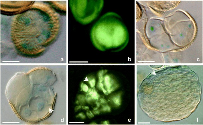
Fig. 4.

Local auxin distribution in the prolonged heat-treated microspores and MDEs of B. napus. a The microspore with non-polar activity of the DR5 promoter. b Two-celled pro-embryo with the DR5rev activity in the both symmetrical cells. c–e Multicellular structures emerging from the exine. The polar DR5 (c, d) or DR5rev (e) activities at the few-celled stage. The arrow indicates the stronger DR5rev activity on the one pole (e). (f) The globular embryo stage with the DR5 activity in the protoderm (arrow). The blue and light green colors show the expression of the reporter gus and gfp genes driven by the DR5 or DR5rev promoters (respectively). Bar = 20 μm
