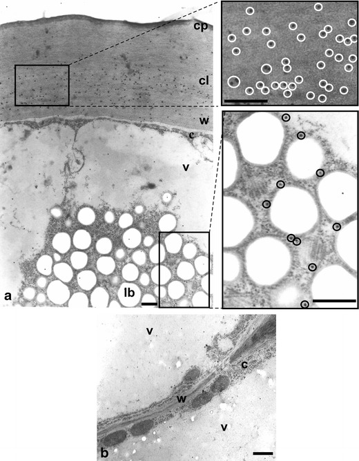
Fig. 3.

Cross-sections of O. umbellatum ovary epidermis and parenchyma cells after immunogold reaction with anti-cutinsome antibody. a Part of an epidermal cell with lipotubuloid, the cuticular layer and lipotubuloid labelled with gold particles. Fragments of images in windows are shown to the right in higher magnification. Gold particles are highlighted with circles. b Fragment of parenchyma cells with cell wall lacking gold particles. c cytoplasm, cl cuticular layer, cp cuticle proper, lb lipid bodies, v vacuole, w cell wall. Scale bars, 0.5 μm (a), 1 μm (b)
