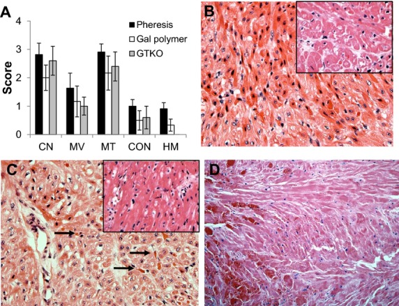
Fig. 3.

Histopathologic features of DXR in the absence of the effects of anti-Gal antibody. Data from three treatment groups are shown. (i) Recipient treated by plasmapheresis (Pheresis) to deplete anti-Gal antibody pre- and post-transplant; (ii) Chronic Gal-polymer-treated recipient to block anti-Gal antibody in vivo; and (iii) Transplantation of a GTKO donor heart. A. Histologic features of DXR in the absence of acute anti-Gal antibody. The intensity of major histopathologic features at explant (mean histology score ± standard error of the mean) are shown. (Abbreviations: CN, coagulative necrosis; MV, myocyte vacuolization; MT, microvascular thrombosis; CON, congestion; HM, hemorrhage.) B–D. Progressive development of DXR (H&E 400×). B. Cardiac biopsy from an apheresis-treated recipient (day 13 of 53) showing early (stage 1) DXR characterized by myocyte vacuolization with minimal microvascular thrombosis or systemic release of cardiac troponin. Insert shows a stage 1 biopsy (day 47 of 71) from a GTKO/CD55 heart (H&E 200×). C. Interim biopsy (day 15 of 21) of a heart from an apheresis-treated recipient showing progressive (stage 2) DXR, characterized by increased levels of microvascular thrombosis (arrows) and developing coagulative necrosis. Insert shows a stage 2 biopsy (day 14 of 26) of a GTKO/CD55 heart (H&E 200×). D. Representative histopathology of grafts at explant in all three groups (Portions of this figure adapted from data in Tazelaar HD, Byrne GW, McGregor CG. Comparison of Gal and non-Gal-mediated cardiac xenograft rejection. Transplantation. 2011: 91: 968–975).
