The 11th affiliation for the 20th author is incorrect. Shin Takasawa’s institution is not located in the Wakayama prefecture, but in the Nara prefecture. The correct affiliation should read:
Department of Biochemistry, Nara Medical University, Kashihara, Nara, Japan.
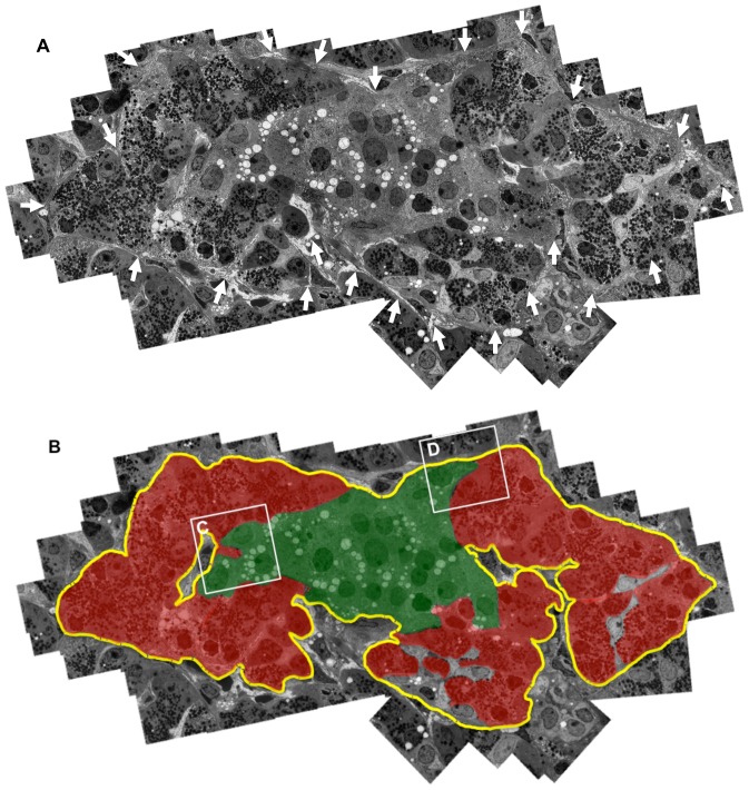
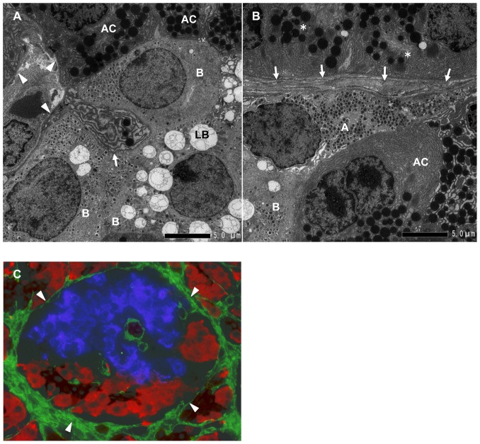
The last sentence of the Figure 1B legend, LB: lipofuscin body, should be the last sentence of the Figure 2A legend. Please see the figures and their corrected legends below.
Figure 1. Demonstration of pancreatic acinar-like cell clusters touching islet-cell clusters that are covered by a common capsule.
A: Continuous basement membranes (BMs) and extracellular matrix (ECM) (arrows) cover the two cell clusters. A combined figure of 65 electron microscopic photos is shown. B: Schematic demonstration of Figure 1A. The yellow line indicates continuous BMs and ECM surrounding islet cell (green) and acinar-like cell (red) clusters.
Figure 2. The interface between acinar-like cell clusters and islet cell clusters.
A: Magnified view of the interface between acinar-like cell clusters and islet cell clusters shown in Figure 1B (inset C). Acinar-like cells (AC) contact beta cells (B). Note that the acinar-like cell has a process (arrow) containing vesicles that protrude to the beta cell cytoplasm. BMs and ECM (arrowheads) surround beta cells (B) and acinar-like cells (AC). LB: lipofuscin body. B: Magnified view of the interface between acinar-like cell clusters and islet cell clusters shown in Figure 1B (inset D). Alpha cell (A) and beta cell (B) touching an acinar-like cell (AC) and the covering BMs and ECM (arrows) and pancreatic acinar cells (*) separated by BMs and ECM (arrows). C: Immunohistological demonstration of BMs and ECM stained for fibronectin (arrowheads, green), surrounding the islet beta cells stained for insulin (blue), and acinar-like cells (red) stained for amylase and the ductal marker cytokeratin 19 (brown).
Reference
- 1. Aida K, Saitoh S, Nishida Y, Yokota S, Ohno S, et al. (2014) Distinct Cell Clusters Touching Islet Cells Induce Islet Cell Replication in Association with Over-Expression of Regenerating Gene (REG) Protein in Fulminant Type 1 Diabetes. PLoS ONE 9(4): e95110 doi:10.1371/journal.pone.0095110 [DOI] [PMC free article] [PubMed] [Google Scholar]


