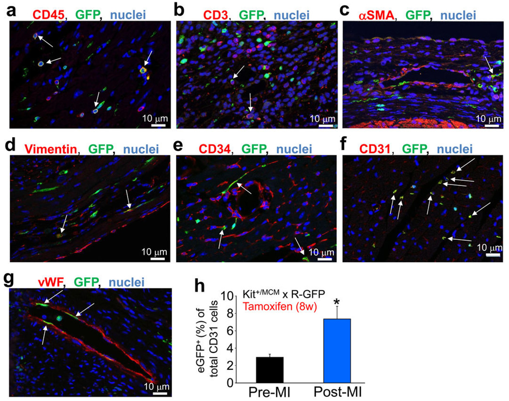
Extended Data Figure 5. Analysis of eGFP+ non-myocytes in the hearts of Kit+/MCM × R-GFP mice at baseline or after MI injury.
a–g,Tamoxifen was given to Kit+/MCM × R-GFP mice for 1 day – 6 months of age (a,e,f) or in mice given tamoxifen and MI injury (b,c,d,g), followed by harvesting the hearts for immunohistochemistry with antibodies for GFP (green), or the indicated antibodies in red; (a) CD45, (b) CD3, (c) smooth muscle α-actin (αSMA), (d) vimentin, (e) CD34, (f) CD31, (g) von Willebrand factor (vWF). Nuclei are shown in blue. The white arrows show cells with coincident green and red reactivity for each of the markers, although sometimes the red marker is membrane localized while the green (eGFP) is always cytoplasmic. The most overlapping activity with GFP expression was observed for CD31 (endothelial cells), then CD34, followed by CD45 (hematopoietic cells). h, Quantitation from FACS plots of total CD31 cells (antibody) in the heart that are also eGFP+ from Kit+/MCM × R-GFP mice (Pre-MI, n=3) after 8 weeks of tamoxifen in early adulthood at either baseline or 4 weeks after MI injury (Post-MI, n=3). The data show about a doubling in the number of CD31 cells that are eGFP+ after MI (*P<0.05 vs pre-MI).

