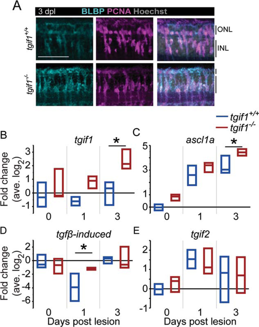
FIGURE 4.
After acute light lesion, Müller glia in tgif1−/− fish express a dedifferentiation marker similar to tgif1+/+ but misre-gulate Smad2/3 target genes. (A) BLBP and PCNA immunoreac-tivity in tgif1+/+ and tgif1−/− fish at 3 dpl in the lesioned area, as determined by the absence of cone nuclei. (B–E) qRT-PCR of whole retina extracts (retinas pooled from individual fish, n=3 replicates). We normalized gene expression to gpia and calculated fold change relative to the average tgif1+/+ 0 dpl level. * P<0.05, Student t-test comparing genotypes at the same lesion interval. Abbreviations as in Fig. 1.

