Live imaging of heart cell function in mice

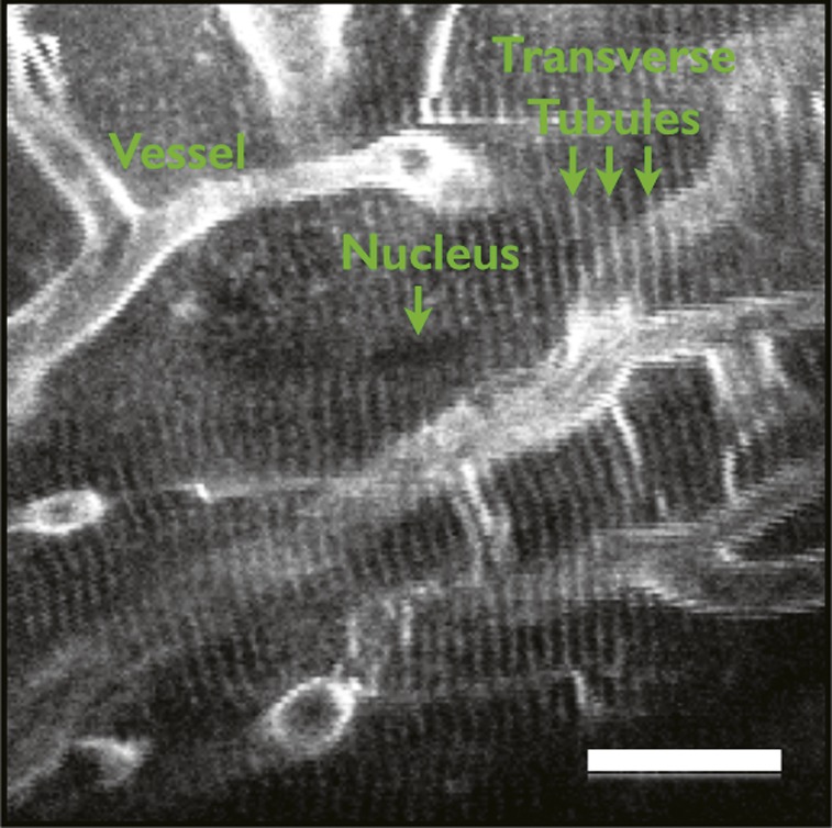
Motion artifact-free imaging of cardiomyocyte contractile cycle in mice.
Advanced technologies such as MRI and CT scans have transformed cardiac imaging, yet measuring the contractile function of individual cardiomyocytes, the building blocks of heart muscle, at high resolution in the beating hearts of animal models remains a challenge, partly due to contraction- and respiration-related heart movement. Using a fluorescent dye to stain cardiomyocytes, tissue stabilizers to reduce movement artifact, and an improved gating and image acquisition algorithm that incorporates cardiac pacing, Aaron Aguirre et al. (pp. 11257–11262) developed a microscopy based method to visualize contracting cardiomyocytes in the hearts of anesthetized and mechanically ventilated mice. The method, based on a confocal and multiphoton imaging system, helped acquire images precisely synchronized with the cardiac cycle at 7–10 frames per second, corresponding to the physiological heart rate of 400–600 bpm in mice. Within seconds, the authors sampled the individual cardiomyocyte contractile cycle—an indicator of heart function—with millisecond time resolution and visualized transverse tubules, nuclei, microvasculature, and endothelial cells at high spatial resolution with minimal motion artifact. According to the authors, the method, suitable for use with commercial microscopes and any mouse strain, might help researchers study variations in cardiomyocyte function in healthy and diseased hearts. — P.N.
Parenting, socioeconomic status, and inflammation in children
Children of low socioeconomic status (SES) often experience poorer health than children of higher SES at all stages of life, including lower birth weights at infancy, higher rates of obesity and diabetes during childhood, and higher rates of age-related cardiovascular disease and cancer. Gregory Miller et al. (pp. 11287–11292) investigated whether improvements in parents’ nurturing skills may counteract the effects of low SES on inflammation, a process that underlies many health problems. The authors recruited 272 African American mother–child pairs of low SES from rural Georgia in which the child was 11 years old. Of the mother–child pairs, 173 participated in 14 hours of training on parenting techniques, setting goals and expectations, and issues surrounding sex, racism, and alcohol abuse. Control families received leaflets on the same topics but no in-person training. Eight years later, the authors collected blood samples from the children and measured the levels of six proinflammatory cytokines, markers of inflammation. Children who had participated in the training displayed lower levels of the six cytokines than the control group. According to the authors, the results suggest that family-oriented interventions may have the potential to counteract the increased incidence of inflammation in children of low SES. — J.P.J.
Potential drug target for fatty liver disease
Nonalcoholic fatty liver disease (NAFLD) is a chronic disorder that affects at least one out of 10 individuals worldwide, potentially leading to liver scarring or failure. To uncover the mechanism of NAFLD, Wen Su et al. (pp. 11437–11442) used mass spectrometry to analyze proteins in liver biopsies from three humans with normal livers as well as three patients with simple steatosis—an NAFLD hallmark characterized by the build-up of lipids in liver cells. The authors identified 89 lipid droplet-associated proteins with altered levels in liver samples from NAFLD patients, compared with normal liver samples. One abundant protein in the lipid droplets was 17β-hydroxysteroid dehydrogenase 13 (17β-HSD13), the levels of which were much higher in biopsies from NAFLD patients than in normal liver samples. Similarly, 17β-HSD13 levels were higher in the livers of mice with fatty liver, compared with mice without fatty liver. Mice genetically manipulated to express high 17β-HSD13 levels in the liver developed a greater number of lipid droplets than did mice with normal 17β-HSD13 levels. According to the authors, the findings suggest that 17β-HSD13 and possibly other lipid droplet-associated proteins may contribute to the development of NAFLD and could represent potential targets for the treatment of fatty liver. — J.W.
Host-specificity in bee gut microbiota

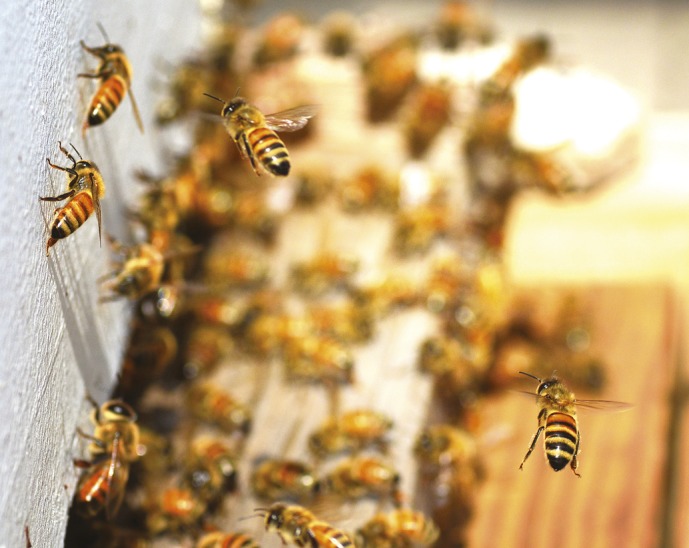
Honey bees returning to hive.
The honey bee (Apis spp.) and its cousin, the bumble bee (Bombus spp.), possess well-defined microbiota and social transmission routes similar to mammalian systems. Waldan Kwong et al. (pp. 11509–11514) examined the genomic characteristics of the bee gut microbiota and found that it is host-specific, with coresident species playing complementary roles in energy metabolism. Microbes Gilliamella apicola and Snodgrassella alvi occupy distinct metabolic niches in the bee gut and may form a mutually dependent network for partitioning metabolic resources. The authors found that strains of S. alvi were able to colonize their native bee, but not bees of another genus. Additionally, the authors generated complete genomes of two dominant bee gut microbes, G. apicola type wkB1T and S. alvi type wkB2T, and draft genomes of four other strains. A comparative genomic analysis revealed minimal gene flow between Apis and Bombus gut symbionts, but uncovered evidence of gene transfer within single bee hosts. The results underscore the importance of multiple forces shaping gut community evolution—including direct host–microbe interactions, microbe–microbe interactions, and social transmission. According to the authors, the findings might have implications in agriculture and applied sciences, such as in the engineering and application of probiotics. — A.G.
Histamine and appetite suppression

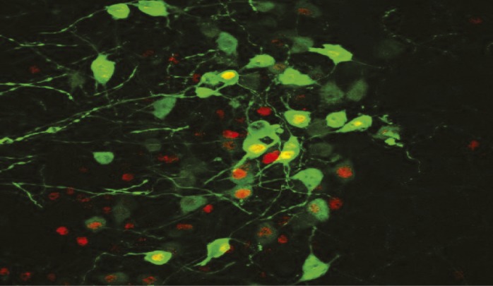
Oxytocin-producing neurons (green) in the hypothalamus.
Feelings of hunger and satiety are regulated by the interplay of food stimuli and neurotransmitter systems within the body. Consumption of dietary fats, for example, is known to induce release of the chemical oleoylethanolamide (OEA) from the small intestine, stimulating production of the neurotransmitter oxytocin and inhibiting food intake. Gustavo Provensi et al. (pp. 11527–11532) investigated whether OEA also recruits the histamine neurotransmitter system, known to influence eating behavior, to regulate food intake. The authors measured food intake and behavioral indicators of satiety in mice with abnormally low levels of histamine and observed reduced appetite suppression in response to OEA release, compared with normal mice. Pharmacologically induced histamine release in normal mice increased appetite suppression that was induced by OEA. The authors also observed that OEA directly increased histamine release in the cortex region of the mouse brain and increased c-Fos gene expression in neuron clusters rich in oxytocin-producing neurons. The results suggest that normal appetite suppression depends on the interaction of OEA from the intestines with the brain’s histamine system and that this interaction occurs in the oxytocin-producing neurons of the hypothalamus. — J.P.J.


