
FIG 1 .
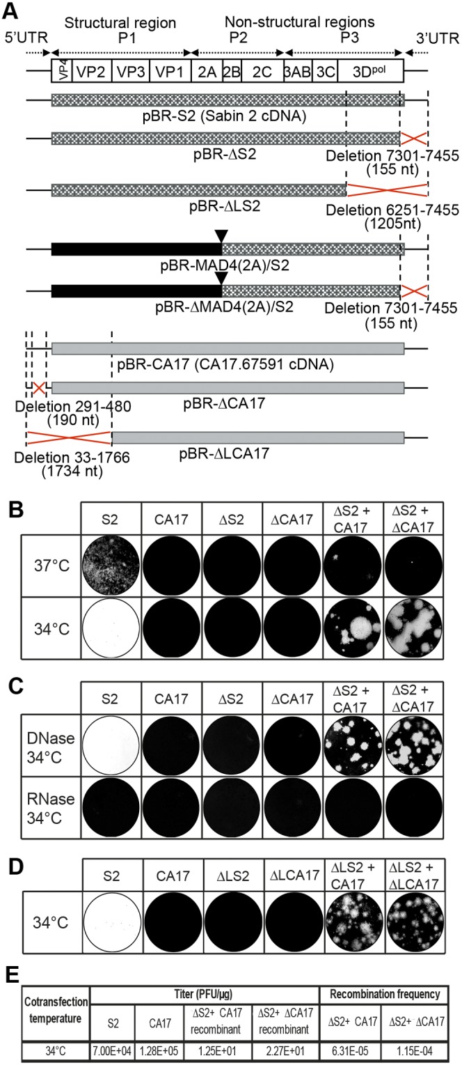
RNA-based cotransfection assay. (A) Genomic structure of the parental viral cDNAs and the viral cDNAs with deletions inserted into plasmids for in vitro RNA synthesis. The genetic organization of the PV genome is shown (at the top). The plasmids carrying the full-length cDNA sequences of Sabin 2 and CA17 (pBR-S2 and -CA17, respectively) and those of the cVDPV/Sabin 2 recombinant generated in vitro [pBR-MAD4(2A)/S2] have been described elsewhere (24, 25). Red crosses indicate the genomic regions in which the deletions were made. The nucleotide positions and the lengths [numbers of nucleotides deleted are shown in parentheses, including the poly(A) tail of 16 nucleotides] of these deletions are also indicated. Solid triangles indicate in vitro recombination sites. (B to E) Cotransfections were performed in L20B cells, in semisolid medium, with 2.5 µg of each RNA. The overlay was added, and cells were incubated at 34°C or 37°C, as indicated, for 4 days before staining. Cells transfected with Sabin 2 RNA (S2) and incubated at 34°C were entirely lysed after 4 days (wells appear white). (B and C) Before cotransfections, genomic RNAs were either left untreated (B) or treated with DNase or RNase for 1 h at 37°C (C) and cells were incubated at 34°C after cotransfection. (D) Cotransfections were performed with RNAs with large deletions. (E) The in vitro recombination frequencies after cotransfection with ΔS2 plus CA17 RNAs and ΔS2 plus ΔCA17 RNAs at 34°C are indicated (see Materials and Methods for calculation).