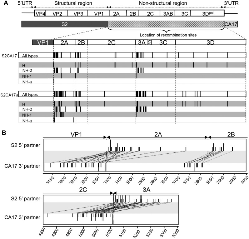
FIG 3 .
Genomic analysis of recombinant viruses. (A) Distribution of the recombination sites identified in recombinant viruses S2CA17 and S2CA17Δ, isolated at passage P1, following cotransfection with deleted S2 and either CA17 (for S2CA17) or defective ΔCA17 (for S2CA17Δ) RNA. The names of the recombinants are indicated on the left, together with the type of recombination. Black vertical lines indicate recombination sites according to S2 numbering. For NH-2 recombinants, the locations of the recombination sites in the S2 5′ partner are shown. (B) Sites of recombination for NH-2 recombinants, for each RNA partner. Regions with recombination sites are enlarged. The names of the partners are indicated on the left. Vertical lines indicate the sites of recombination for each partner. The dotted lines represent the bond between each partner and a given NH-2 recombinant. Site locations (nucleotide numbering) are indicated at the bottom.

