Abstract
Stress can exert long-lasting changes on the brain that contribute to vulnerability to mental illness, yet mechanisms underlying this long-term vulnerability are not well understood. We hypothesized that stress may alter the production of oligodendrocytes in the adult brain, providing a cellular and structural basis for stress-related disorders. We found that immobilization stress decreased neurogenesis and increased oligodendrogenesis in the dentate gyrus (DG) of the adult rat hippocampus, and that injections of the rat glucocorticoid stress hormone corticosterone (cort) were sufficient to replicate this effect. The DG contains a unique population of multipotent neural stem cells (NSCs) that give rise to adult newborn neurons, but oligodendrogenic potential has not been demonstrated in vivo. We used a nestin-CreER/YFP transgenic mouse line for lineage tracing and found that cort induces oligodendrogenesis from nestin-expressing NSCs in vivo. Using hippocampal NSCs cultured in vitro, we further showed that exposure to cort induced a pro-oligodendrogenic transcriptional program and resulted in an increase in oligodendrogenesis and decrease in neurogenesis, which was prevented by genetic blockade of glucocorticoid receptor (GR). Together, these results suggest a novel model in which stress may alter hippocampal function by promoting oligodendrogenesis, thereby altering the cellular composition and white matter structure.
Introduction
Stress is a risk factor for a variety of mood and anxiety disorders, including depression and post-traumatic stress disorder (PTSD), which can manifest years after the stressful event1. The mechanisms that account for this persistent vulnerability are not fully understood. While many effects of stress on the brain are relatively transient – involving the actions of glucocorticoid (GC) stress hormones on mineralcorticoid and glucocorticoid receptors (MR and GR) to regulate short-term physiological responses 2, 3 – long-term effects have also been identified. For example, stress affects long-term potentiation (LTP), and epigenetically regulates the hypothalamic-pituitary-adrenal axis 4, 5. However, mechanisms by which stress may lead to long-lasting structural changes in the brain have not been thoroughly explored. There are hints that GCs may directly affect white matter structure by regulating oligodendrogenesis. In oligodendrocyte precursor cell (OPC) culture, GCs are potent inducers of pro-oligodendrogenic transcription factors and increase oligodendrogenesis 6-8 and myelination 9-14. GCs also dysregulate myelination in utero15. In the periphery, myelinating Schwann cells similarly show responsiveness to GCs, including increased myelination in vitro and in vivo 16, 17. Interestingly, changes in white matter have been documented in many brain regions in a variety of stress-related mental illnesses, including PTSD, schizophrenia, autism and depression18-23. Together, these data suggest the unexplored possibility that stress may cause dysregulation of oligodendrogenesis, which may create a persistent, white matter structural vulnerability to mental illness.
The hippocampus is a key structure regulating memory and emotion, and plays a role in a variety of emotional disorders 24. Changes in hippocampal cell composition therefore could have dramatic implications for vulnerability to and progression of mental illness. In particular, the DG contains a unique population of neural stem cells (NSCs) that proliferate throughout adulthood, and have functions in regulating negative affective states including stress response, fear, anxiety, and depression25-28. NSCs are multipotent, producing neurons and glia, but their oligodendrogenic potential is debated, with a recent report that they do not produce oligodendrocytes in vivo29. Resolving this ambiguity may reveal new insight into NSC function, since alteration of the oligodendrogenic component of NSC output could provide a novel pathway through which stress affects white matter composition and hippocampal function. In this study, we sought to determine whether stress and GCs affect oligodendrogenesis in the adult hippocampus, and specifically if the oligodendrogenic potential of NSCs is modulated by GC exposure.
Materials and methods
Cell culture conditions
Preparation of NSC culture was as previously described30, 31. Cells were cultured (37°C, 5% CO2) on poly-ornithine (Sigma, St. Louis MO) and laminin (Invitrogen, Carlsbad CA) coated plates in N2-supplemented (Invitrogen) Dulbecco’s modified Eagle medium (DMEM)/F-12 (1:1) (Invitrogen) with 20ng/mL recombinant human FGF-2 (PeproTech, Rocky Hill NJ). For differentiation studies, cells were cultured in low FGF-2 (0-5ng/mL) for 12-18h (200,000-300,000 cells per 6cm dish for RT-qPCR and 12,000-15,000 cells per coverslip in 24 well plates for immunocytochemistry (ICC), and treated with vehicle (EtOH, 0.1%), 1 μM corticosterone (cort), or 1 μM dexamethasone (DEX). Cells were treated every 24h for 75h with no media replacement for ICC, once for 48h for RT-qPCR, and once for 72h for Western blot. For dominant negative GR (dnGR) experiments, dnGR or GFP vector was added to cultures after cells were in differentiating conditions for 6-10h, with cort treatment 12h later. For GR ICC, NSCs were incubated with BrdU (30μM) 12h before fixation.
Amplicon construction of dnGR
dnGR was generated as previously described32. Briefly, a truncated rat GR (Genbank M14053; amino acids 1-745) was PCR amplified and ligated to the first 24 base pairs of the human GR-β isoform then transfected into E5 cells. Superinfection occured 24 hours later with the helper virus d120 at a multiplicity of infection (MOI) of 0.3. Amplicons were harvested by freeze-thaw lysis followed by PEG-IT (System Biosciences, Mountain View, CA) precipitation as per manufacturer’s instructions. Pelleted viruses were resuspended in dH2O. Vector titers were 2-3×10⁶ infectious particles/mL and d120 helper virus titers of 0.4 – 1.0×10⁷ plaque forming units (PFU)/mL.
Real-time PCR
Real-time PCR was performed with an iCycler/iQ detection system (Bio-Rad, Hercules, CA). Primer pairs (Supplementary Table 1) were used with SYBR green. Data were analyzed with the Relative Expression Software Tool. The ribosomal subunit, 18S, was used as an internal control.
Immunocytochemistry and in vitro quantification
NSCs were fixed in 4% paraformaldehyde (PFA) and immunostained with rabbit anti-GR (1:500, Thermo Scientific, Rockford IL), mouse anti-Nestin (1:500, BD Biosciences, San Jose CA), and rat anti-BrdU (1:500) overnight at 4°C, and then treated with Cy3 anti-mouse, FITC anti-rat, and Cy5 anti-rabbit (1:500, Jackson ImmunoResearch, West Grove PA) for 2h.
For differentiation studies, cells were immunostained (overnight, 4°C) with mouse anti-β-III tubulin (Tuj1; 1:1000, Covance, Princeton NJ) or rat anti-MBP (1:100, Abcam, Cambridge MA) followed by 2h incubation with Cy3 or FITC-conjugated secondary antibodies (1:500, Jackson). The nuclear stain DAPI was added prior to mounting coverslips in 1,4-diazabicyclo[2.2.2]octane (DABCO). Images were obtained with an inverted fluorescence microscope (20× objective; Zeiss, Oberkochen, Germany), and blind cell counts were taken using Metamorph software. 20 random visual fields were analyzed per coverslip. Data are the percentage of positive cells in relation to the total number of DAPI-stained nuclei present in the culture. All experiments were independently replicated at least 3 times.
Polyacrylamide gel electrophoresis (PAGE) and immunoblotting
Whole-cell proteins were extracted using Radio-Immunoprecipitation Assay (RIPA) buffer. Nuclear and cytoplasmic proteins were extracted using NE-PER Nuclear and Cytoplasmic Extraction Reagents (ThermoSci). Protein concentrations for each sample were determined using bicinchoninic acid (BCA) Protein Assay Kit (ThermoSci). Proteins were separated in 8% or 10% polyacrylamide denaturing gels (for GR and olig1, respectively) and transferred to Immuno-Blot polyvinyl difluoride (PVDF) membrane (Bio-Rad), then incubated (overnight, 4°C) with anti-GR monoclonal antibody (PA1-510A; ThermoSci) at 1:500 dilution, followed by anti-mouse or anti-rabbit conjugated to horseradish peroxidase, or with primary anti-olig1 (rabbit anti Olig-1, 1:25000; from Charles Stiles, Harvard University), followed by horseradish peroxidase-conjugated goat anti-rabbit IgG antibody (1:2000, Jackson; 1h, RT). Membranes were detected using a chemiluminescence kit (Western lightning Plus ECL, Perkin Elmer, Waltham MA) and quantified by densitometry. Membranes were stripped and reprobed using ß-actin antibody (1:3000; Sigma) to control for equal loading.
Animal treatments
Experiments used adult male Sprague-Dawley rats (2-3 mo; Charles River) pair housed, or C57Bl/6J transgenic nestin-Cre ERT2/RosaYFP mice33 (2-2.5 mo; 25g) housed 5 per cage, with ad libitum access to food and water and a 12/12 hr light/dark cycle. Animals were acclimated to the facility for at least one week before handling. Procedures were approved by UC Berkeley and UC Davis animal care committees.
Rats were stressed by restraint in plastic decapicone bags for 3 hours each day for 1 week (stress n = 6; unstressed controls, n = 5). For cort studies, rats received 7 daily subcutaneous (s.c.) injections of cort (40mg/kg) or vehicle oil, a dose that reciprocates many aspects of stress treatments 34-36, and were sacrificed on day 7 (vehicle, n=6; cort, n=6) or on day 14 (vehicle, n=7; cort, n=6). Intraperitoneal (i.p.) injections of BrdU (200mg/kg) were given on the first 3 days of stress or cort treatments.
Nestin-Cre ERT2/RosaYFP mice33 were given tamoxifen injections (180 mg/kg, i.p.) twice per day for 5 days to induce maximal recombination. 15 days later, mice were injected with cort (25 mg/kg, s.c., n=8) or vehicle (n=8) for 10 days, and then sacrificed 7 days after the end of cort treatment. BrdU (50 mg/kg, i.p.) was injected twice per day on the first 3 days of cort treatment.
At endpoints, animals were transcardially perfused with saline followed by cold 4% PFA.
Histology
Brains were post-fixed in 4% PFA, cryoprotected and cryosectioned (40μm coronal slices) through the dentate gyrus. Immunohistochemistry was performed with rat anti-BrdU (1:500; Accurate, Westbury, NY) and mouse anti-TuJ1 (1:5000; Covance), mouse anti-RIP (1:1000; Millipore, Billerica MA), or rat anti-MBP (1:100; Abcam) for 48h at 4°C; or rabbit anti-GST-π (1:5000; MBL, Woburn MA) and goat anti-GFP (targeting YFP; 1:500, Rockland Immunochemicals, Gilbertsville PA) for 18h at 4°C. Secondary incubations were FITC-conjugated donkey anti-mouse (1:500, Jackson) for 2 hours, and biotinylated anti-rat (1:500; Jackson) for 2 hours followed by Streptavidin-Alexa 568 (1:1000; Invitrogen) for 1 hour. For Olig1 labeling, sections were immunostained with rabbit Olig1 (1:5000, from Charles Stiles, Harvard University) and anti-rabbit Cy3 or anti-rabbit Cy5 (1:500, Jackson). For combined BrdU and MBP labeling, sections were incubated in mouse anti-BrdU (1:500, BD) and rat anti-MBP (1:100, Abcam), followed by incubation with FITC donkey anti-rat (1:500, Jackson) and then biotinylated anti-mouse (1:500, Jackson), and visualized with Streptavidin-Alexa 568 (1:1000, Jackson). Sections were coverslipped with DABCO anti-fading medium in TBS.
BrdU-labeled cells were visualized using a Zeiss 510 AxioImager confocal laser-scanning microscope with LSM 510 software. At least 25 BrdU-labeled cells were examined per rat, and co-labeling with cell fate markers was quantified. Confocal and z-stacked images were used in coordination to determine the percentage of BrdU+ cells co-labeling with cell fate markers. For cytoplasmic markers, fluorescence signals surrounding BrdU+ nuclei were considered positive. BrdU counts were quantified as total BrdU cells per tissue section, or as density measurements, in which the areas of the GCL and hilus were measured using Metamorph and multiplied by section thickness (40μm). Total BrdU-labeled cells within each ROI were then normalized to the total volume per animal. For transgenic lineage tracing, all YFP positive cells were examined in every sixth serial section throughout the mouse dentate gyrus (40μm section thickness), and scored for co-localization with GST-π.
Statistical analyses
Means and SEMs were determined for the above variables. For statistical comparisons, these values were subjected to unpaired two-tailed Students t-tests or one-way ANOVA. P-values ≤ 0.05 were considered significant.
Results
Stress and GCs increase oligodendrogenesis in the hippocampus
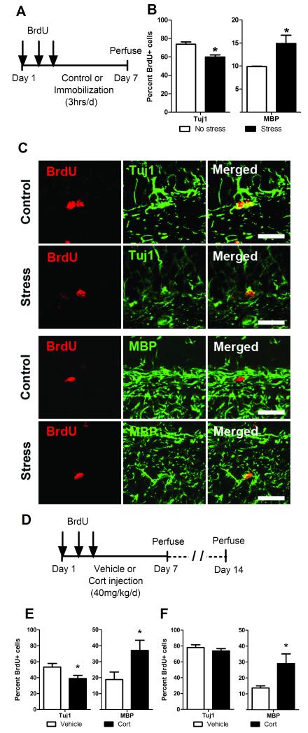
Studies investigating the effects of stress on NSCs in the adult hippocampus have primarily focused on the extent to which new neurons are generated. However, new glial cells are also generated in the hippocampus and may similarly be susceptible to environmental factors and hormones. Here we investigated whether stress and stress hormones (GCs) alter the oligodendrogenic potential in the adult hippocampus. To assess these effects, we subjected rats to 1 week of immobilization restraint stress and subsequently quantified the percentage of newborn cells (marked by BrdU, Figure 1A) in the hippocampal DG that adopted a neuronal or oligodendrocytic cell fate. In agreement with previous studies37, 38, stress significantly decreased the percentage of BrdU+ cells that co-labeled with the early neuronal marker Tuj1 (Figure 1B and C). Interestingly, stress significantly increased the percentage of BrdU+ cells that co-labeled with the mature oligodendrocyte marker MBP (Figure 1B and C), as well as the oligodendrocyte marker RIP (Supplemental Figure 1), revealing that stress increases oligodendrogenesis in the DG. To determine if an increase in circulating GCs is sufficient to upregulate hippocampal oligodendrogenesis, we next subjected rats to daily injections of cort (40 mg/kg, comparable to elevated serum GC levels induced by immobilization stress34-36) or vehicle for 1 week (Figure 1D) and assessed the neurogenic and oligodendrogenic potential within the DG. Similar to stressed rats, cort-injected rats showed a decrease in the percentage of new neurons, and an increase in the percentage of new oligodendrocytes, relative to controls (Figure 1E). When rats were allowed to recover from cort injections for 1 week, the percentage of new neurons reached control levels and did not differ between cort and vehicle injected rats (Figure 1F), consistent with prior studies on the long-term changes in neurogenesis following recovery from GC exposure39, 40. In contrast, the percentage of newborn oligodendrocytes in cort vs. vehicle injected rats remained significantly higher and persisted even following recovery from cort injections (Figure 1F).
Figure 1.
Immobilization stress or cort injections increase hippocampal oligodendrogenesis. (a) BrdU-injected adult male rats were subjected to either 1 week of daily immobilization stress or no stress (n=5 no stress control, n=6 stress). (b) IHC analysis of cell fate, quantified as the percentage of BrdU positive cells that co-label as neurons (Tuj1) or oligodendrocytes (MBP) shows that stress decreases neurogenesis and increases oligodendrogenesis. (c) Representative images of confocal analysis represents cells identified as positive for co-localization of BrdU and Tuj1 or MBP; scale bar=10 μM. (d) BrdU-injected adult male rats received daily cort or vehicle injections for 1 week and were perfused on day 7 (n=6 vehicle injected, n=6 cort injected) or day 14 (n=7 vehicle injected, n=6 cort injected). (e) IHC analysis of cell fate at day 7 shows that exposure to stress hormones (cort) decreases neurogenesis and increases oligodendrogenesis. (f) IHC analysis of cell fate at day 14 (1 week after recovery from cort treatment) shows that while neurogenesis is restored to control levels, the effects of increased oligodendrogenesis persist in cort injected animals. *p < 0.05 (mean ± SEM).
GCs increase oligodendrogenesis from NSCs in vivo
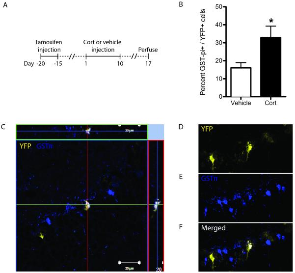
While NSCs produce neurons, astrocytes, and oligodendrocytes in vitro41, a recent study reported that they do not produce oligodendrocytes in vivo29. If true, this would suggest that hippocampal oligodendrogenesis arises exclusively from a distinct population of OPCs. An alternate possibility is that NSCs generate new oligodendrocytes at a level that is low under “baseline” conditions but higher in response to stress. To investigate this, we used a transgenic mouse (nestin-Cre ERT2/RosaYFP)33 expressing an inducible (fused to estrogen receptor variant ERT2) cre recombinase enzyme driven by the promoter for the NSC marker nestin. In this system, induction with the ERT2 agonist tamoxifen causes recombination leading to constitutive expression of the YFP marker in nestin-expressing NSCs, allowing for unambiguous analysis of their cell fate. After tamoxifen induction to initiate nestin-driven NSC labeling, mice were injected with cort (25 mg/kg) or vehicle for 10 days and assessed for oligodendrogenic potential 7 days later (Figure 2A). Quantification of the percent of NSC derived cells (YFP tagged) that co-labeled with the oligodendrocyte marker GST-π showed that NSCs produced a low number of new oligodendrocytes under control conditions, and that stress significantly increased oligodendrogenesis from NSCs (Figure 2B-F, Supplemental Figure 2).
Figure 2.
Cort increases hippocampal oligodendrogenesis from nestin lineage NSCs in vivo. (a) Nestin-Cre ERT2/RosaYFP mice were injected with tamoxifen to induce YFP reporter gene expression, injected with cort or vehicle for 10 days, and perfused 7 days later for IHC analysis. (b) Cort injection increased the percentage of YFP-labeled cells that co-labeled with the oligodendrocytic marker GST-π; n=8. (c) Representative image of an orthogonal slice of a YFP+/GST-π+ co-labeled cell as well as 3D reconstruction of an image stack of the same cell (d-f). *p < 0.05 (mean ± SEM).
GCs increase oligodendrogenesis from NSCs in vitro
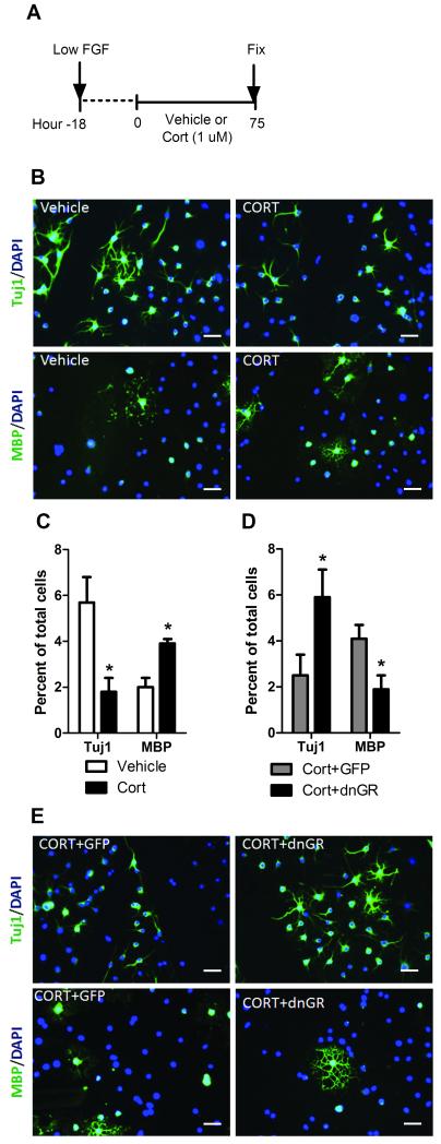
Adult hippocampal NSCs can be cultured in a proliferative state in vitro, and can be induced to terminally differentiate by withdrawal of fibroblast growth factor 2 (FGF-2) from the culture media41, thus providing a useful system to investigate the direct effects of cort on NSC fate. We first confirmed that NSCs in culture have functional GR. NSCs expressed GR mRNA and protein, and cort treatment induced GR translocation to the nuclear fraction (Supplemental Figure 3). Next, we treated NSCs with 1 μM cort or vehicle for 75 hrs (under low FGF conditions that permit unbiased differentiation) and determined the total number of new neurons (Tuj1+) and oligodendrocytes (MBP+) (Figure 3A). Compared to vehicle controls, cort-treated NSCs produced significantly fewer neurons (Figure 3B-C). Cort also caused a significant increase in the production of oligodendrocytes from NSCs, suggesting that cort can directly induce NSC oligodendrogenesis (Figure 3B-C). There was no significant difference in total cell numbers between cort and vehicle treated cultures (vehicle, 334.0±66.9; cort, 432.7±88.7; p = 0.4). To determine if cort-induced oligodendrogenesis is GR mediated, we blocked GR signaling by transfecting NSC cultures with the dominant negative GR (dnGR) viral vector32. Compared to NSCs infected with a control GFP vector, NSCs infected with dnGR and treated with cort showed significantly more neurons and fewer oligodendrocytes (Figure 3D and E), similar to baseline levels seen in vehicle-treated cultures. Thus, blocking GR function prevented the effect of cort on NSC oligodendrogenesis.
Figure 3.
Cort treatment increases the oligodendrogenic potential of NSCs. (a) NSC cultures were treated with cort or vehicle for 75 h. (b) Representative images of ICC analysis, scale bar = 50 μm. (c) ICC analysis of cell fate, quantified as total number of cells labeling as neurons (Tuj1) or oligodendrocytes (MBP). (d, e) ICC analysis of cell fate of NSCs transfected with dnGR or GFP (control) and treated with cort. *p < 0.05 (mean ± SEM); n≥3.
GCs promote an oligodendrogenic transcriptional program in NSCs
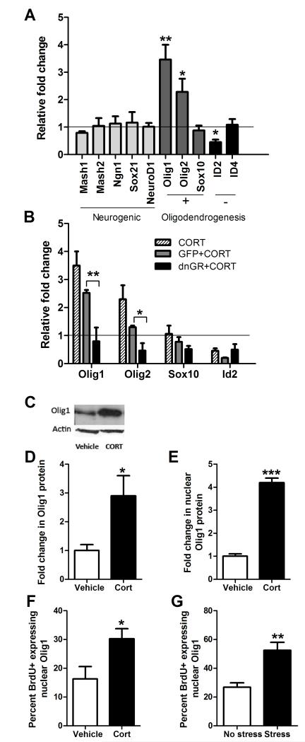
Cell fate programming is known to be regulated by complex interactions of multiple transcription factors. Oligodendrocytic fate is regulated by the pro-oligodendrogenic transcription factors Olig1 and Olig2 42-48, which are inhibited by Inhibitor of Differentiation 2 and 4 (Id2, Id4) 49. To determine if GCs promote a pro-oligodendrogenic transcriptional program we performed RT-qPCR on NSC cultures treated with cort or vehicle for 48 hrs. There was no change in the expression of any neurogenic factors analyzed following cort treatment (Mash1 and 2; Neurogenin1, Ngn1; Sox21; and NeuroD1; Figure 4A). In contrast, Olig1 and Olig2 mRNA levels were dramatically increased and Id2 mRNA was significantly decreased following cort treatment, relative to control. The levels of Sox10 and Id4 mRNA did not change following cort exposure. Cort treatment also resulted in a significant increase in MBP mRNA expression (1.66± 2.2 fold change; p<0.01), while no difference was observed for β-tubulin III (Tuj1) mRNA levels (1.16 ± 1.8; Supplemental Figure 4).
Figure 4.
Cort treatment induces a pro-oligodendrogenic transcriptional program in NSCs. (a) Fold change in mRNA expression of genes regulating neurogenic and oligodendrogenic fate in cort-treated NSCs, relative to vehicle-treated controls, including genes that promote (+) and inhibit (−) oligodendrogenesis. (b) Fold change in mRNA expression of oligodendrogenic regulatory genes in NSCs transfected with dnGR or GFP (control) vectors, or no vector, and treated with cort, relative to vehicle-treated controls; n≥3. (c) Representative image of Western blot for Olig1 protein in NSCs treated with cort or vehicle. (d) Densitometric analysis of Western blot for total protein fraction and (e) the isolated nuclear fraction of treated NSCs; n=3. (f) IHC analysis of the percent of BrdU positive cells expressing nuclear-localized Olig1 in the hilus of adult rats after 1 week of daily cort injections; n=6 vehicle injected, n=6 cort injected (g) or after 1 week of daily immobilization stress; n=5 no stress control, n=6 stress. *p < 0.05, **p<0.005, ***p < 0.0001 (mean ± SEM).
Treatment of cultured NSCs with dexamethasone (a synthetic GC and selective GR-agonist) similarly promoted a pro-oligodendrogenic transcriptional program and increase in Olig1 protein (Supplemental Figure 5). Additionally, blocking GR signaling by transfection with dnGR prior to cort treatment eliminated the GC-induced increases in Olig1 and 2 transcripts, and suppression of Id2, relative to GFP transfected controls (Figure 4B). These results demonstrate that the Id-Olig network is modulated by cort via a GR-dependent mechanism, suggesting that this transcriptional program is involved in GC-induced oligodendrogenesis.
Olig1 is a key regulator of oligodendrogenesis that acts as a nuclear-localized transcription factor when specifying oligodendrocyte differentiation and progressively localizes in the cytoplasm in mature oligodendrocytes 43, 50, 51. Olig1 Nuclear localization is inhibited by the binding of Id2 and Id4 49, therefore the decrease in Id2 would predict an increase in functionally active nuclear Olig1. We assessed the level of Olig1 protein in total protein and in nuclear fractions of NSCs treated with cort, and found an increase in Olig1 protein in both total (Figure 4C-D) and nuclear (Figure 4E) fractions following cort treatment. We further assessed nuclear co-localization of Olig1 with BrdU in vivo following 1 week of GC injections or immobilization stress treatment. Both GC injections (Figure 4F) and stress (Figure 4G) resulted in an increase in nuclear co-localization of Olig1 and BrdU, relative to respective controls, in the DG. These results indicate that stress and GCs cause an increase in Olig1 protein levels in the nucleus both in vitro and in vivo, implicating Olig1 in GC-induced oligodendrogenesis.
Discussion
We demonstrate that stress and GCs increase hippocampal oligodendrogenesis in vivo and show that new oligodendrocytes can arise from nestin positive NSCs in the DG. Furthermore, we demonstrate a direct, GR-dependent effect of GCs on NSC differentiation into oligodendrocytes, which involves an upregulation of pro-oligodendrogenic genes, and increased nuclear localization of Olig1 protein. Current models for stress-induced emotional disorders suggest that previous stress experience can create a persistent vulnerability to mental illness that lasts many years beyond the stressful experience. Such models require mechanisms by which stress can cause long term changes in brain structure and function. Here we describe a novel mechanism of stress-induced oligodendrogenesis which may contribute to persistent changes in brain structure. In turn, increases in oligodendrogenesis could affect cognition in at least two ways: by altering the ratio of oligodendrocytes to neurons, and by altering myelination (white matter tracts).
Alteration of the oligodendrocyte:neuron ratio could affect cognition due to oligodendrocytic roles in synapse formation. Oligodendrocytes inhibit axon growth cones 52-56, and OPCs are both repulsive and nonpermissive for growing axons 57, 58. While the effects of changes in oligodendrocyte abundance on synaptic formation and plasticity are unknown, it seems likely that increased abundance would result in suppression of synaptogenesis. This suppressive effect, combined with the well-documented effects of GCs in reducing neurogenesis and new neuron survival37, 38,59, would be predicted to dramatically impair hippocampal function. In terms of myelination, increased stress reactivity in aging 2 correlates with increase in white matter in both humans 60-62, and monkeys 63. Age-related cognitive decline also correlates with white matter increases in the brain 63, 64. Beyond cognition, a variety of mental health conditions 18, including depression, schizophrenia, PTSD, and suicide 19-22, are linked to changes in white matter abundance23. Interestingly, Cushing’s syndrome and other diseases involving elevated cortisol levels are associated with alterations in affective and hippocampal function65-67, though possible white matter contributions to these symptoms have not been investigated.
As the first report that stress hormones increase oligodendrogenesis in the adult hippocampus, this study opens avenues for additional investigation. Many species differences have been observed in hippocampal neurogenesis; for example rats produce more new neurons than mice, and those neurons mature more rapidly68, while humans may produce fewer new neurons69. We found stress induced-oligodendrogenesis in both rats and mice, though the different labeling techniques used in these species preclude direct comparison of oligodendrogenic rate. Key questions for future study involve whether effects of stress induced oligodendrogenesis can be generalized to other brain regions and other mammals (especially primates70), and if so whether there are important species differences; if increased oligodendrogenesis causes increased myelination; whether these effects persist and accumulate with stress load, and in particular whether stress-induced epigenetic changes4, 5, 71 could target oligodendrocytic genes to persistently dysregulate white matter; and ultimately how changes in oligodendrogenesis affect neural function and behavior. Given that the effects reported in this study occur within the hippocampus, our results suggest a possible link between GC-induced oligodendrogenesis and memory function. Long term alterations in oligodendrogenesis and myelination could regulate memory retention and have important implications for disorders like PTSD. Stress has been shown to induce long-lasting changes on cholinergic neurotransmission and neuronal hypersensitivity as a result of genetic and epigenetic changes72-74. Whether stress-induced oligodendrogenesis plays a role in regulating neurotransmission signaling systems remains to be investigated and could potentially offer new targets for therapeutic interference. Furthermore, our findings raise the possibility that stress is an underlying factor in unexplained changes in white matter (leukoaraiosis) that are frequently observed in older patients and, in some studies, correlated with cognitive deficits75, 76.
There is also considerable current interest in understanding the functional role of adult hippocampal neurogenesis from NSCs. We show that cort increased oligodendrogenesis in NSC cultures in addition to decreasing neurogenesis, and used lineage tracing of nestin-expressing NSCs to show that cort also increases NSC oligodendrogenesis in vivo. These findings challenge the current thinking that hippocampal NSCs do not generate oligodendrocytes in vivo, and suggest that while NSCs generate few oligodendrocytes under basal conditions, exposure to stress and GCs redirects the cell fate of differentiating NSCs towards oligodendrogenesis via a GR-mediated mechanism. The differences observed in the increases in oligodendrogenesis between exogenous and endogenous GCs (cort vs. stress treatment) may be due to differential GR/MR activation and/or variations in the rate of maturation of oligodendrocytes. Since Olig1 translocates to the cytoplasm as oligodendrocytes mature, the increased nuclear localization of Olig1 in BrdU+ cells in stressed animals relative to cort-treated animals may indicate variations in the rate of maturation of new oligodendrocytes. Overall, the data shows that GCs mimic the effects of stress and supports the conclusion that both immobilization stress or cort hormone injection produce an increase in oligodendrogenesis. The possibility that a GR-induced oligodendrogenic transcriptional program could override neural fate is supported by studies that have found that Olig1 promotes oligodendrogenesis in embryonic NSCs 77. Our results suggest that, at the step of NSC differentiation and fate choice, neurogenesis may be inversely related to oligodendrogenesis. This finding advocates a shift in perspective on adult neurogenesis, which has largely focused on the proliferation, survival, and function of new neurons as a basis for cognitive changes associated with altered neurogenesis. We propose that changes in oligodendrogenesis, or in the ratio of glia and neurons, may contribute to alterations in hippocampal-dependent behavior. Possible roles for oligodendrocytes should be considered in previous and future studies that implicate neurogenesis in behavioral outputs. In particular, changes in neurogenesis have recently been implicated in emotional dysfunction, altered stress response, and depression26, 27, 78-80. However, potential contributions of concurrent altered gliogenesis to these emotional perturbations have not been considered. Overall, our results suggest a new model in which stress may contribute to persistent vulnerability to mental illness by inducing oligodendrogenesis. We speculate that accumulated stress load across the lifespan may alter the rate or extent of myelination to create a persistent, structural vulnerability to mental illness.
Supplementary Material
Acknowledgements
We thank David V. Schaffer for feedback; Charles Stiles and David Rowitch for feedback and Olig1 antibody; Amelia Eisch for the Nestin-Cre ERT2/RosaYFP mice; and Ashmi Ullal and Jennifer Shih for research assistance. SC, EDK and DC were supported by CIRM pre-doctoral fellowships; EDK was supported by a DoD NDSEG fellowship. ARF was supported by a NSF fellowship. CM was supported by a NRSA postdoctoral fellowship (1F32AG029733-01A1). This work was supported by NIMH BRAINS (R01 MH087495), Berkeley Stem Cell Center Seed Grant, Hellman Family Foundation, and NARSAD awards to DK.
Support: SC, DC and EDK were supported by CIRM pre-doctoral fellowships; EDK was supported by a DoD NDSEG Fellowship. ARF was supported by a NSF fellowship. CM was supported by a NRSA postdoctoral fellowship (1F32AG029733-01A1). This work was supported by NIMH BRAINS (R01 MH087495), Berkeley Stem Cell Center Seed Grant, Hellman Family Foundation, and NARSAD awards to DK.
REFERENCES
- 1.Heim C, Nemeroff CB. The role of childhood trauma in the neurobiology of mood and anxietydisorders: preclinical and clinical studies. Biol Psychiatry. 2001;49(12):1023–1039. doi: 10.1016/s0006-3223(01)01157-x. [DOI] [PubMed] [Google Scholar]
- 2.McEwen BS. Physiology and neurobiology of stress and adaptation: central role of the brain. Physiol Rev. 2007;87(3):873–904. doi: 10.1152/physrev.00041.2006. [DOI] [PubMed] [Google Scholar]
- 3.de Kloet ER, Karst H, Joels M. Corticosteroid hormones in the central stress response: quick-and-slow. Front Neuroendocrinol. 2008;29(2):268–272. doi: 10.1016/j.yfrne.2007.10.002. [DOI] [PubMed] [Google Scholar]
- 4.Gillespie CF, Phifer J, Bradley B, Ressler KJ. Risk and resilience: genetic and environmental influences on development of the stress response. Depress Anxiety. 2009;26(11):984–992. doi: 10.1002/da.20605. [DOI] [PMC free article] [PubMed] [Google Scholar]
- 5.Francis D, Diorio J, Liu D, Meaney MJ. Nongenomic transmission across generations of maternal behavior and stress responses in the rat. Science. 1999;286(5442):1155–1158. doi: 10.1126/science.286.5442.1155. [DOI] [PubMed] [Google Scholar]
- 6.Almazan G, Honegger P, Du Pasquier P, Matthieu JM. Dexamethasone stimulates the biochemical differentiation of fetal forebrain cells in reaggregating cultures. Dev Neurosci. 1986;8(1):14–23. doi: 10.1159/000112237. [DOI] [PubMed] [Google Scholar]
- 7.Barres BA, Lazar MA, Raff MC. A novel role for thyroid hormone, glucocorticoids and retinoic acid in timing oligodendrocyte development. Development. 1994;120(5):1097–1108. doi: 10.1242/dev.120.5.1097. [DOI] [PubMed] [Google Scholar]
- 8.Mann SA, Versmold B, Marx R, Stahlhofen S, Dietzel ID, Heumann R, et al. Corticosteroids reverse cytokine-induced block of survival and differentiation of oligodendrocyte progenitor cells from rats. J Neuroinflammation. 2008;5:39. doi: 10.1186/1742-2094-5-39. [DOI] [PMC free article] [PubMed] [Google Scholar]
- 9.Cheng JD, de Vellis J. Oligodendrocytes as glucocorticoids target cells: functional analysis of the glycerol phosphate dehydrogenase gene. J Neurosci Res. 2000;59(3):436–445. doi: 10.1002/(SICI)1097-4547(20000201)59:3<436::AID-JNR19>3.0.CO;2-Z. [DOI] [PubMed] [Google Scholar]
- 10.Desarnaud F, Bidichandani S, Patel PI, Baulieu EE, Schumacher M. Glucocorticosteroids stimulate the activity of the promoters of peripheral myelin protein-22 and protein zero genes in Schwann cells. Brain Res. 2000;865(1):12–16. doi: 10.1016/s0006-8993(00)02130-2. [DOI] [PubMed] [Google Scholar]
- 11.Kumar S, Cole R, Chiappelli F, de Vellis J. Differential regulation of oligodendrocyte markers by glucocorticoids: post-transcriptional regulation of both proteolipid protein and myelin basic protein and transcriptional regulation of glycerol phosphate dehydrogenase. Proc Natl Acad Sci U S A. 1989;86(17):6807–6811. doi: 10.1073/pnas.86.17.6807. [DOI] [PMC free article] [PubMed] [Google Scholar]
- 12.Masters JN, Finch CE, Nichols NR. Rapid increase in glycerol phosphate dehydrogenase mRNA in adult rat brain: a glucocorticoid-dependent stress response. Neuroendocrinology. 1994;60(1):23–35. doi: 10.1159/000126716. [DOI] [PubMed] [Google Scholar]
- 13.Tsuneishi S, Takada S, Motoike T, Ohashi T, Sano K, Nakamura H. Effects of dexamethasone on the expression of myelin basic protein, proteolipid protein, and glial fibrillary acidic protein genes in developing rat brain. Brain Res Dev Brain Res. 1991;61(1):117–123. doi: 10.1016/0165-3806(91)90121-x. [DOI] [PubMed] [Google Scholar]
- 14.Zhu W, Wiggins RC, Konat GW. Glucocorticoid-induced upregulation of proteolipid protein and myelin-associated glycoprotein genes in C6 cells. J Neurosci Res. 1994;37(2):208–212. doi: 10.1002/jnr.490370206. [DOI] [PubMed] [Google Scholar]
- 15.Antonow-Schlorke I, Helgert A, Gey C, Coksaygan T, Schubert H, Nathanielsz PW, et al. Adverse effects of antenatal glucocorticoids on cerebral myelination in sheep. Obstetrics and gynecology. 2009;113(1):142–151. doi: 10.1097/AOG.0b013e3181924d3b. [DOI] [PubMed] [Google Scholar]
- 16.Morisaki S, Nishi M, Fujiwara H, Oda R, Kawata M, Kubo T. Endogenous glucocorticoids improve myelination via Schwann cells after peripheral nerve injury: An in vivo study using a crush injury model. Glia. 2010;58(8):954–963. doi: 10.1002/glia.20977. [DOI] [PubMed] [Google Scholar]
- 17.Chan JR, Phillips LJ, 2nd, Glaser M. Glucocorticoids and progestins signal the initiation and enhance the rate of myelin formation. Proc Natl Acad Sci U S A. 1998;95(18):10459–10464. doi: 10.1073/pnas.95.18.10459. [DOI] [PMC free article] [PubMed] [Google Scholar]
- 18.Fields RD. White matter in learning, cognition and psychiatric disorders. Trends Neurosci. 2008;31(7):361–370. doi: 10.1016/j.tins.2008.04.001. [DOI] [PMC free article] [PubMed] [Google Scholar]
- 19.Ehrlich S, Breeze JL, Hesdorffer DC, Noam GG, Hong X, Alban RL, et al. White matter hyperintensities and their association with suicidality in depressed young adults. J Affect Disord. 2005;86(2-3):281–287. doi: 10.1016/j.jad.2005.01.007. [DOI] [PubMed] [Google Scholar]
- 20.Jackowski AP, Douglas-Palumberi H, Jackowski M, Win L, Schultz RT, Staib LW, et al. Corpus callosum in maltreated children with posttraumatic stress disorder: a diffusion tensor imaging study. Psychiatry Res. 2008;162(3):256–261. doi: 10.1016/j.pscychresns.2007.08.006. [DOI] [PMC free article] [PubMed] [Google Scholar]
- 21.Regenold WT, Phatak P, Marano CM, Gearhart L, Viens CH, Hisley KC. Myelin staining of deep white matter in the dorsolateral prefrontal cortex in schizophrenia, bipolar disorder, and unipolar major depression. Psychiatry Res. 2007;151(3):179–188. doi: 10.1016/j.psychres.2006.12.019. [DOI] [PubMed] [Google Scholar]
- 22.Schneiderman JS, Buchsbaum MS, Haznedar MM, Hazlett EA, Brickman AM, Shihabuddin L, et al. Age and diffusion tensor anisotropy in adolescent and adult patients with schizophrenia. Neuroimage. 2009;45(3):662–671. doi: 10.1016/j.neuroimage.2008.12.057. [DOI] [PMC free article] [PubMed] [Google Scholar]
- 23.Nave KA. Myelination and support of axonal integrity by glia. Nature. 2010;468(7321):244–252. doi: 10.1038/nature09614. [DOI] [PubMed] [Google Scholar]
- 24.Fanselow MS, Dong HW. Are the dorsal and ventral hippocampus functionally distinct structures? Neuron. 2010;65(1):7–19. doi: 10.1016/j.neuron.2009.11.031. [DOI] [PMC free article] [PubMed] [Google Scholar]
- 25.Santarelli L, Saxe M, Gross C, Surget A, Battaglia F, Dulawa S, et al. Requirement of hippocampal neurogenesis for the behavioral effects of antidepressants. Science. 2003;301(5634):805–809. doi: 10.1126/science.1083328. [DOI] [PubMed] [Google Scholar]
- 26.Saxe MD, Battaglia F, Wang JW, Malleret G, David DJ, Monckton JE, et al. Ablation of hippocampal neurogenesis impairs contextual fear conditioning and synaptic plasticity in the dentate gyrus. Proc Natl Acad Sci U S A. 2006;103(46):17501–17506. doi: 10.1073/pnas.0607207103. [DOI] [PMC free article] [PubMed] [Google Scholar]
- 27.Snyder JS, Soumier A, Brewer M, Pickel J, Cameron HA. Adult hippocampal neurogenesis buffers stress responses and depressive behaviour. Nature. 2011;476(7361):458–461. doi: 10.1038/nature10287. [DOI] [PMC free article] [PubMed] [Google Scholar]
- 28.David DJ, Samuels BA, Rainer Q, Wang JW, Marsteller D, Mendez I, et al. Neurogenesis-dependent and -independent effects of fluoxetine in an animal model of anxiety/depression. Neuron. 2009;62(4):479–493. doi: 10.1016/j.neuron.2009.04.017. [DOI] [PMC free article] [PubMed] [Google Scholar]
- 29.Bonaguidi MA, Wheeler MA, Shapiro JS, Stadel RP, Sun GJ, Ming GL, et al. In vivo clonal analysis reveals self-renewing and multipotent adult neural stem cell characteristics. Cell. 2011;145(7):1142–1155. doi: 10.1016/j.cell.2011.05.024. [DOI] [PMC free article] [PubMed] [Google Scholar]
- 30.Gage FH. Mammalian neural stem cells. Science. 2000;287(5457):1433–1438. doi: 10.1126/science.287.5457.1433. [DOI] [PubMed] [Google Scholar]
- 31.Reynolds BA, Weiss S. Generation of neurons and astrocytes from isolated cells of the adult mammalian central nervous system. Science. 1992;255(5052):1707–1710. doi: 10.1126/science.1553558. [DOI] [PubMed] [Google Scholar]
- 32.Kaufer D, Ogle WO, Pincus ZS, Clark KL, Nicholas AC, Dinkel KM, et al. Restructuring the neuronal stress response with anti-glucocorticoid gene delivery. Nat Neurosci. 2004;7(9):947–953. doi: 10.1038/nn1296. [DOI] [PubMed] [Google Scholar]
- 33.Lagace DC, Whitman MC, Noonan MA, Ables JL, DeCarolis NA, Arguello AA, et al. Dynamic contribution of nestin-expressing stem cells to adult neurogenesis. J Neurosci. 2007;27(46):12623–12629. doi: 10.1523/JNEUROSCI.3812-07.2007. [DOI] [PMC free article] [PubMed] [Google Scholar]
- 34.Cameron HA, Gould E. Adult neurogenesis is regulated by adrenal steroids in the dentate gyrus. Neuroscience. 1994;61(2):203–209. doi: 10.1016/0306-4522(94)90224-0. [DOI] [PubMed] [Google Scholar]
- 35.Sousa N, Madeira MD, Paula-Barbosa MM. Effects of corticosterone treatment and rehabilitation on the hippocampal formation of neonatal and adult rats. An unbiased stereological study. Brain Res. 1998;794(2):199–210. doi: 10.1016/s0006-8993(98)00218-2. [DOI] [PubMed] [Google Scholar]
- 36.Johnson SA, Fournier NM, Kalynchuk LE. Effect of different doses of corticosterone on depression-like behavior and HPA axis responses to a novel stressor. Behav Brain Res. 2006;168(2):280–288. doi: 10.1016/j.bbr.2005.11.019. [DOI] [PubMed] [Google Scholar]
- 37.Wong EY, Herbert J. The corticoid environment: a determining factor for neural progenitors’ survival in the adult hippocampus. Eur J Neurosci. 2004;20(10):2491–2498. doi: 10.1111/j.1460-9568.2004.03717.x. [DOI] [PMC free article] [PubMed] [Google Scholar]
- 38.Wong EY, Herbert J. Raised circulating corticosterone inhibits neuronal differentiation of progenitor cells in the adult hippocampus. Neuroscience. 2006;137(1):83–92. doi: 10.1016/j.neuroscience.2005.08.073. [DOI] [PMC free article] [PubMed] [Google Scholar]
- 39.Malberg JE, Duman RS. Cell proliferation in adult hippocampus is decreased by inescapable stress: reversal by fluoxetine treatment. Neuropsychopharmacology. 2003;28(9):1562–1571. doi: 10.1038/sj.npp.1300234. [DOI] [PubMed] [Google Scholar]
- 40.Tanapat P, Hastings NB, Rydel TA, Galea LA, Gould E. Exposure to fox odor inhibits cell proliferation in the hippocampus of adult rats via an adrenal hormone-dependent mechanism. J Comp Neurol. 2001;437(4):496–504. doi: 10.1002/cne.1297. [DOI] [PubMed] [Google Scholar]
- 41.Palmer TD, Takahashi J, Gage FH. The adult rat hippocampus contains primordial neural stem cells. Mol Cell Neurosci. 1997;8(6):389–404. doi: 10.1006/mcne.1996.0595. [DOI] [PubMed] [Google Scholar]
- 42.Marshall CA, Novitch BG, Goldman JE. Olig2 directs astrocyte and oligodendrocyte formation in postnatal subventricular zone cells. J Neurosci. 2005;25(32):7289–7298. doi: 10.1523/JNEUROSCI.1924-05.2005. [DOI] [PMC free article] [PubMed] [Google Scholar]
- 43.Ligon KL, Kesari S, Kitada M, Sun T, Arnett HA, Alberta JA, et al. Development of NG2 neural progenitor cells requires Olig gene function. Proc Natl Acad Sci U S A. 2006;103(20):7853–7858. doi: 10.1073/pnas.0511001103. [DOI] [PMC free article] [PubMed] [Google Scholar]
- 44.Lu QR, Cai L, Rowitch D, Cepko CL, Stiles CD. Ectopic expression of Olig1 promotes oligodendrocyte formation and reduces neuronal survival in developing mouse cortex. Nat Neurosci. 2001;4(10):973–974. doi: 10.1038/nn718. [DOI] [PubMed] [Google Scholar]
- 45.Menn B, Garcia-Verdugo JM, Yaschine C, Gonzalez-Perez O, Rowitch D, Alvarez-Buylla A. Origin of oligodendrocytes in the subventricular zone of the adult brain. J Neurosci. 2006;26(30):7907–7918. doi: 10.1523/JNEUROSCI.1299-06.2006. [DOI] [PMC free article] [PubMed] [Google Scholar]
- 46.Stolt CC, Rehberg S, Ader M, Lommes P, Riethmacher D, Schachner M, et al. Terminal differentiation of myelin-forming oligodendrocytes depends on the transcription factor Sox10. Genes Dev. 2002;16(2):165–170. doi: 10.1101/gad.215802. [DOI] [PMC free article] [PubMed] [Google Scholar]
- 47.Wegner M. Expression of transcription factors during oligodendroglial development. Microsc Res Tech. 2001;52(6):746–752. doi: 10.1002/jemt.1059. [DOI] [PubMed] [Google Scholar]
- 48.Zhou Q, Anderson DJ. The bHLH transcription factors OLIG2 and OLIG1 couple neuronal and glial subtype specification. Cell. 2002;109(1):61–73. doi: 10.1016/s0092-8674(02)00677-3. [DOI] [PubMed] [Google Scholar]
- 49.Samanta J, Kessler JA. Interactions between ID and OLIG proteins mediate the inhibitory effects of BMP4 on oligodendroglial differentiation. Development. 2004;131(17):4131–4142. doi: 10.1242/dev.01273. [DOI] [PubMed] [Google Scholar]
- 50.Kitada M, Rowitch DH. Transcription factor co-expression patterns indicate heterogeneity of oligodendroglial subpopulations in adult spinal cord. Glia. 2006;54(1):35–46. doi: 10.1002/glia.20354. [DOI] [PubMed] [Google Scholar]
- 51.Arnett HA, Fancy SP, Alberta JA, Zhao C, Plant SR, Kaing S, et al. bHLH transcription factor Olig1 is required to repair demyelinated lesions in the CNS. Science. 2004;306(5704):2111–2115. doi: 10.1126/science.1103709. [DOI] [PubMed] [Google Scholar]
- 52.Fawcett JW, Rokos J, Bakst I. Oligodendrocytes repel axons and cause axonal growth cone collapse. J Cell Sci. 1989;92(Pt 1):93–100. doi: 10.1242/jcs.92.1.93. [DOI] [PubMed] [Google Scholar]
- 53.Morganti MC, Taylor J, Pesheva P, Schachner M. Oligodendrocyte-derived J1-160/180 extracellular matrix glycoproteins are adhesive or repulsive depending on the partner cell type and time of interaction. Exp Neurol. 1990;109(1):98–110. doi: 10.1016/s0014-4886(05)80012-3. [DOI] [PubMed] [Google Scholar]
- 54.Pesheva P, Gennarini G, Goridis C, Schachner M. The F3/11 cell adhesion molecule mediates the repulsion of neurons by the extracellular matrix glycoprotein J1-160/180. Neuron. 1993;10(1):69–82. doi: 10.1016/0896-6273(93)90243-k. [DOI] [PubMed] [Google Scholar]
- 55.Pesheva P, Spiess E, Schachner M. J1-160 and J1-180 are oligodendrocyte-secreted nonpermissive substrates for cell adhesion. J Cell Biol. 1989;109(4 Pt 1):1765–1778. doi: 10.1083/jcb.109.4.1765. [DOI] [PMC free article] [PubMed] [Google Scholar]
- 56.Bandtlow C, Zachleder T, Schwab ME. Oligodendrocytes arrest neurite growth by contact inhibition. J Neurosci. 1990;10(12):3837–3848. doi: 10.1523/JNEUROSCI.10-12-03837.1990. [DOI] [PMC free article] [PubMed] [Google Scholar]
- 57.Chen ZJ, Ughrin Y, Levine JM. Inhibition of axon growth by oligodendrocyte precursor cells. Mol Cell Neurosci. 2002;20(1):125–139. doi: 10.1006/mcne.2002.1102. [DOI] [PubMed] [Google Scholar]
- 58.Chen ZJ, Negra M, Levine A, Ughrin Y, Levine JM. Oligodendrocyte precursor cells: reactive cells that inhibit axon growth and regeneration. J Neurocytol. 2002;31(6-7):481–495. doi: 10.1023/a:1025791614468. [DOI] [PubMed] [Google Scholar]
- 59.Balu DT, Lucki I. Adult hippocampal neurogenesis: regulation, functional implications, and contribution to disease pathology. Neurosci Biobehav Rev. 2009;33(3):232–252. doi: 10.1016/j.neubiorev.2008.08.007. [DOI] [PMC free article] [PubMed] [Google Scholar]
- 60.Bartzokis G, Beckson M, Lu PH, Nuechterlein KH, Edwards N, Mintz J. Age-related changes in frontal and temporal lobe volumes in men: a magnetic resonance imaging study. Arch Gen Psychiatry. 2001;58(5):461–465. doi: 10.1001/archpsyc.58.5.461. [DOI] [PubMed] [Google Scholar]
- 61.Courchesne E, Chisum HJ, Townsend J, Cowles A, Covington J, Egaas B, et al. Normal brain development and aging: quantitative analysis at in vivo MR imaging in healthy volunteers. Radiology. 2000;216(3):672–682. doi: 10.1148/radiology.216.3.r00au37672. [DOI] [PubMed] [Google Scholar]
- 62.Jernigan TL, Fennema-Notestine C. White matter mapping is needed. Neurobiol Aging. 2004;25(1):37–39. doi: 10.1016/j.neurobiolaging.2003.06.002. [DOI] [PubMed] [Google Scholar]
- 63.Lyons DM, Yang C, Eliez S, Reiss AL, Schatzberg AF. Cognitive correlates of white matter growth and stress hormones in female squirrel monkey adults. J Neurosci. 2004;24(14):3655–3662. doi: 10.1523/JNEUROSCI.0324-04.2004. [DOI] [PMC free article] [PubMed] [Google Scholar]
- 64.Ziegler DA, Piguet O, Salat DH, Prince K, Connally E, Corkin S. Cognition in healthy aging is related to regional white matter integrity, but not cortical thickness. Neurobiol Aging. 2010;31(11):1912–1926. doi: 10.1016/j.neurobiolaging.2008.10.015. [DOI] [PMC free article] [PubMed] [Google Scholar]
- 65.Maheu FS, Merke DP, Schroth EA, Keil MF, Hardin J, Poeth K, et al. Steroid abnormalities and the developing brain: declarative memory for emotionally arousing and neutral material in children with congenital adrenal hyperplasia. Psychoneuroendocrinology. 2008;33(2):238–245. doi: 10.1016/j.psyneuen.2007.11.006. [DOI] [PMC free article] [PubMed] [Google Scholar]
- 66.Grillon C, Smith K, Haynos A, Nieman LK. Deficits in hippocampus-mediated Pavlovian conditioning in endogenous hypercortisolism. Biol Psychiatry. 2004;56(11):837–843. doi: 10.1016/j.biopsych.2004.09.003. [DOI] [PubMed] [Google Scholar]
- 67.Starkman MN, Giordani B, Berent S, Schork MA, Schteingart DE. Elevated cortisol levels in Cushing’s disease are associated with cognitive decrements. Psychosomatic medicine. 2001;63(6):985–993. doi: 10.1097/00006842-200111000-00018. [DOI] [PubMed] [Google Scholar]
- 68.Snyder JS, Choe JS, Clifford MA, Jeurling SI, Hurley P, Brown A, et al. Adult-born hippocampal neurons are more numerous, faster maturing, and more involved in behavior in rats than in mice. J Neurosci. 2009;29(46):14484–14495. doi: 10.1523/JNEUROSCI.1768-09.2009. [DOI] [PMC free article] [PubMed] [Google Scholar]
- 69.Spalding KL, Bergmann O, Alkass K, Bernard S, Salehpour M, Huttner HB, et al. Dynamics of hippocampal neurogenesis in adult humans. Cell. 2013;153(6):1219–1227. doi: 10.1016/j.cell.2013.05.002. [DOI] [PMC free article] [PubMed] [Google Scholar]
- 70.Katz M, Liu C, Schaer M, Parker KJ, Ottet MC, Epps A, et al. Prefrontal plasticity and stress inoculation-induced resilience. Dev Neurosci. 2009;31(4):293–299. doi: 10.1159/000216540. [DOI] [PMC free article] [PubMed] [Google Scholar]
- 71.Roozendaal B, Hernandez A, Cabrera SM, Hagewoud R, Malvaez M, Stefanko DP, et al. Membrane-associated glucocorticoid activity is necessary for modulation of long-term memory via chromatin modification. J Neurosci. 2010;30(14):5037–5046. doi: 10.1523/JNEUROSCI.5717-09.2010. [DOI] [PMC free article] [PubMed] [Google Scholar]
- 72.Kaufer D, Friedman A, Seidman S, Soreq H. Acute stress facilitates long-lasting changes in cholinergic gene expression. Nature. 1998;393(6683):373–377. doi: 10.1038/30741. [DOI] [PubMed] [Google Scholar]
- 73.Meshorer E, Erb C, Gazit R, Pavlovsky L, Kaufer D, Friedman A, et al. Alternative splicing and neuritic mRNA translocation under long-term neuronal hypersensitivity. Science. 2002;295(5554):508–512. doi: 10.1126/science.1066752. [DOI] [PubMed] [Google Scholar]
- 74.Sailaja BS, Cohen-Carmon D, Zimmerman G, Soreq H, Meshorer E. Stress-induced epigenetic transcriptional memory of acetylcholinesterase by HDAC4. Proc Natl Acad Sci U S A. 2012;109(52):E3687–3695. doi: 10.1073/pnas.1209990110. [DOI] [PMC free article] [PubMed] [Google Scholar]
- 75.Pantoni L, Garcia JH. The significance of cerebral white matter abnormalities 100 years after Binswanger’s report. A review. Stroke. 1995;26(7):1293–1301. doi: 10.1161/01.str.26.7.1293. [DOI] [PubMed] [Google Scholar]
- 76.Jokinen H, Schmidt R, Ropele S, Fazekas F, Gouw AA, Barkhof F, et al. Diffusion changes predict cognitive and functional outcome: the LADIS study. Ann Neurol. 2013;73(5):576–583. doi: 10.1002/ana.23802. [DOI] [PubMed] [Google Scholar]
- 77.Balasubramaniyan V, Timmer N, Kust B, Boddeke E, Copray S. Transient expression of Olig1 initiates the differentiation of neural stem cells into oligodendrocyte progenitor cells. Stem Cells. 2004;22(6):878–882. doi: 10.1634/stemcells.22-6-878. [DOI] [PubMed] [Google Scholar]
- 78.Drew MR, Denny CA, Hen R. Arrest of adult hippocampal neurogenesis in mice impairs single-but not multiple-trial contextual fear conditioning. Behavioral neuroscience. 2010;124(4):446–454. doi: 10.1037/a0020081. [DOI] [PMC free article] [PubMed] [Google Scholar]
- 79.Arruda-Carvalho M, Sakaguchi M, Akers KG, Josselyn SA, Frankland PW. Posttraining ablation of adult-generated neurons degrades previously acquired memories. J Neurosci. 2011;31(42):15113–15127. doi: 10.1523/JNEUROSCI.3432-11.2011. [DOI] [PMC free article] [PubMed] [Google Scholar]
- 80.Sahay A, Hen R. Adult hippocampal neurogenesis in depression. Nat Neurosci. 2007;10(9):1110–1115. doi: 10.1038/nn1969. [DOI] [PubMed] [Google Scholar]
Associated Data
This section collects any data citations, data availability statements, or supplementary materials included in this article.


