Abstract
BACKGROUND:
In the perspective of traditional Chinese medicine, few studies have focused on the compound preparations though there are many investigations. The present study was undertaken to investigate the effect of Zhenwu Tang Granule on chronic pressure-overloaded left ventricular hypertrophy in rats.
METHODS:
The study was performed at the laboratory of Guangzhou Institute of Respiratory Disease. Male SD rats were divided randomly into 3 groups: sham operation group (n=8), operation group (n=15) and traditional Chinese medicine (TCM) group (n=15). The model of myocardial hypertrophy was made by gradually constricting the abdominal aorta. Sixteen weeks later, cardiac ultrasonography was performed in all groups in order to ascertain post-operational left ventricular (LV) hypertrophy. And Zhenwu Tang Granule was added at a dose of 12 g/kg in the mixed feedstuff for 8 weeks in the TCM group. In the 24th week, weight, structure as well as function of the heart in each group were measured by high-frequency ultrasonography, and Masson’s staining was performed on the cardiac muscles. Meanwhile, total collagen volume fraction (CVF-T) and non-coronary vessel collagen volume fraction (CVF-NV) were analyzed.
RESULTS:
There was an increase in the weight of the heart in the operation group, with the left ventricule dominated (P<0.05). The heart was enlarged, with diastolic interventricular septal distance (IVSd) and left ventricular posterior wall distance (LVPWd) dominated (P<0.01). There was a significant decrease in the cardiac function (P<0.05). The weight (P<0.01) and volume of the heart decreased in the TCM group compared with the operation group, with IVSd and systolic left ventricular posterior wall dominated (P<0.01). And the cardiac function was improved (P<0.05). Significant interstitial and collagen hyperplasia was shown in the operation group based on pathological analysis, and various improvements were proved in the TCM group, i.e. there was a significant decrease in CVF-T and CVF-NV (P<0.01) compared with the operation group; but no difference (P>0.05) was found when compared with the pseudo-operation group.
CONCLUSION:
Zhenwu Tang Granule could reduce the weight and volume of the heart, improve the cardiac function, inhibit hyperplasia of collagen, and reverse myocardial hypertrophy in rats with pressure-overloaded left ventricular hypertrophy.
KEY WORDS: Zhenwu Tang Granule, Heart failure, Ventricular remodeling, Hypertrophy, Pressure load, Masson stain, Myocardial collagenous fiber, Abdominal aorta constriction
INTRODUCTION
The basic mechanism of both development and progression of chronic heart failure is ventricular remodeling, which is the vital pathological process of cardiovascular diseases. In the perspective of traditional Chinese medicine, few studies have focused on the compound preparations though many studies were performed.[1,2] In this study, the intervention of Zhenwu Tang Granule was adopted so that the impact to myocardial hypertrophy could be understood clearly
METHODS
Materials and instruments
Thirty-eight male SPF-graded SD rats weighing 100-120 g from the Medical Experimental Animal Center of Guangdong Province were studied at the laboratory of Guangzhou Institue of Respiratory Disease. The Zhenwu Tang granule is made from Chinese herbal materials including Monkshood, Atractylodes macrocephala, Paeoniae Alba, Poria cocos and ginger (Guangdong Yifang Pharmaceutical Co. Ltd). Both the microscopic image capturing system (Olympus BX41) and ultrasonic echocardiogram (GE Vivid7) were also used in the study.
Animal models
Abdominal aortic constriction was utilized according to Anversca’s approach:[3] 10% chloral hydrate (0.3 ml/100g) was used for abdominal anesthesia. The abdominal aorta was localized following the left renal artery, and the head of a pin with the caliber of 0.7 mm was placed following the pathway of the vessel. Then the pin should be ligated together with the abdominal aorta using the sucturing thread (size 0). After pulling out the head of the pin, a residual cavity with the caliber of 0.7 mm was left inside the abdominal aorta. The identical method was utilized in the Sham group except that ligation should be aborted after dissection of the abdominal aorta.
Grouping of animals
In the rats randomized, 8 rats were included in the Sham group, and 15 in the operation group, and 15 in the TCM group, respectively. After myocardial hypertrophy was confirmed by ultrasonography in the 16th week, the TCM group was fed with the mixture of Zhenwu Tang Granule. The granule was prepared with Monkshood, Paeoniae Alba, Poria cocos, Ginger and Atractylodes macrocephala in the proportion of 3:3:3:3:2 and the feedstuff. And the rats were fed with the preparation at a dose of 12 g/kg.
Ultrasonic echocardiogram
The structure and function of the heart were assessed after 8-week feeding of Zhenwu Tang Granule according to the method reported.[4] The feeding frequency adopted in the study can be selected flexibly only if the structure of the left ventricle can be observed clearly. The measurement was performed under the guidance of M-shaped curves of two-dimensional ultrasonic echocardiogram. Every 3 continuous means of the cardiac cycle were selected as the test value. There were a series of parameters designed for the test: diastolic and systolic inter-ventricular septum (IVSd, IVSs), thickness of diastolic and systolic left ventricular posterior wall (LVPWd, LVPWs), diastolic and systolic left ventricular diameter (LVDd, LVDs), fraction of shortening of the left ventricle (LV-FS), ejection fraction (EF), cardiac output (CO) and stroke volume (SV).
Processing of specimens
All rats were weighed before opening the chest. The heart was weighed after being taken out, leaving the left ventricle (including the inter-ventricular septum) for further weighing. And the weight index of the left ventricle (weight of the left ventricle/the body weight) was calculated.
Analysis of myocardial collagen
Myocytes were fixed with formaldehyde and imbedded with solid paraffin. After Masson’s staining the specimen was cut into sections and analyzed with a color pathological graphic-context analyzing system under the 200×microscopic vision. Each section was selected from its respective specimen (the 6 visions with and without coronary arterioles were selected, respectively), and the fraction of collagen tissue in the vision was calculated. The mean represented myocardial collagen volume fraction (CVF). The CVF under the coronary arteriole-rich vision was regarded as the fraction of collagen tissue of the left ventricle (CVF-T), and the CVF under the coronary arteriole-free vision was recorded as CVF-NV.
Statistical analysis
Quantitative data were expressed as mean±standard deviation. The data were compared using SNK’s method after the variance equality test. All the data were processed by SPSS 13.0.
RESULTS
Weight index
There was an increase in the cardiac (left ventricle) weight (P<0.05), while there was a decrease in the weight with significance (P<0.01) in the TCM group compared with the operation group (Table 1).
Table 1.
The weight index of the left ventricle

Measurement of cardiac structure and function using ultrasonic echocardiogram
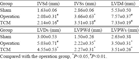
Both myocardial hypertrophy and ventricular cavity enlargement were present in the operation group and TCM group in the 16th week (Table 2).
Table 2.
The cardiac structure in the 16th week

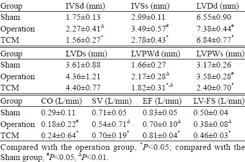
In the 24th week, both significant myocardial hypertrophy and ventricular cavity enlargement were present in the operation group, and there was statistical significance when compared with the Sham group. There was certain improvement in both myocardial hypertrophy and ventricular cavity enlargement after the intervention of TCM, with statistical significance compared with the operation group.
There was an apparent decrease in each parameter (CO, SV, EF, LV-FS) of cardiac function in the operation group. There was a significant improvement in the TCM group (Table 3).
Table 3.
The cardiac structure and function in the 24th week

Content of myocardial collagen
In the operation group, myocyte hypertrophy and fiber derangement were found with lots of collagens piling in the intercellular space. Formation of microscopic scars was also seen with the perivescular areas dominated. The extent of myocyte hypertrophy and interstitial fiber hyperplasia was lower in the TCM group compared with the operation group. From the perspective of semi-quantitative analysis, CVF was much higher with significant variance in the operation group compared with the Sham group; however, CVF was much lower in the TCM group after the intervention of traditional Chinese medicine, with a decrease in CVF-NV dominated (Figure 1, Table 4).
Figure 1.

Histopathological changes of cardiomyocytes (Masson’s stain, original magnification×200)
Table 4.
The weight index of the left ventricle

DISCUSSION
In the present study, the model of myocardial hypertrophy was established through aortic constriction, which was consistent with the pathogensis of clinical diseases. Therefore it was the simpliest and most effective model. There are various studies on this model. In our study the extent of myocardial hypertrophy was found to be positively correlated with the duration of the disease, as reportd in the literature, thereby confirming the successful establishment of the model.
Myocardial hypertrophy is a vital constituent in cardiac remodeling. It was generally reported that myocardial hypertrophy was closely related to the systems of catecholamine and rennin axis. Briest et al[6] discovered that norepinephrine could assume the time-dependance in inducing left ventricular hypertrophy and matrix fibrosis in rats, which could trigger an increase in collagen synthesis; however, the expression of collagen would be reduced and myocardial hypertrophy would be inhibited when the activity of norepinephrine was inhibited. Large scale randomized, double-blind control trials, such as the COPERNICUS[7] study, have also revealed the protective action of beta-receptor blockers on cardiomyocytes. Angiotensin converting enzyme inhibitor (ACEI) is a sort of drug that has confirmed therapeutic effect on myocardial hypertrophy. Cai et al[8] discovered that ACEI could block the action of the rennin-angiotensin-aldosterone system (RAS), thereby suppressing the expression of type I and III collagen.
There were many studies on traditional Chinese medicine, for instance, the studies on the protective action of Shenfu injection[9] and Luomaishutong[10] on cardiomyocytes. There were also a number of studies on Zhenwu Tang. Chen et al[11] discovered a significant decrease in the level of rennin and angiotensin after the intervention of Zhenwu Tang in the heart failure model of rabbits. Zhu et al[12] reported that there was an increase in endothelin and a decrease in calcitonin gene-related peptide (CGRP) in the heart failure model induced by doxorubicin; however, both indices showed certain improvement after the treatment. In the study on the model of Yang deficiency, Wang et al[13] found that hydrogen peroxide dismutase activity was reduced and the immunity was elevated in rats, indicating the anti-radical action of Zhenwu Tang.
However, little has been studied on the action of Zhenwu Tang on the cardiac structure in pressure-overloaded myocardial hypertrophy. Through constricting the aorta, we discovered that the cardiac weight of rats was reduced and the cardiac function could be improved by Zhenwu Tang. Thus hyperplasia of collaginous fibers was inhibited, thereby reversing the trend of myocardial hypertrophy. From the perspective of the cardiac weight and thickness of interventricular septum, we found non-statistical significance between the TCM group and Sham group, possibly because of insufficient sampling size. It is imperative to increase the sampling size in the future studies in order to diminish the error. There are few studies on the dosage-efficacy and time-efficacy relation of Zhenwu Tang, but the therapeutic outcome might be enhanced through increasing the dosage as well as prolonging the duration of administration of the drug. Wang et al[14] discovered that myocardial remodeling could be inhibited by the Monkshood decoction, as the microscopic structure of the left ventricule and cardiomyocyte was improved by the rennin-angiotensin-aldosterone system. In the orthogonal experiment on each component of Zhenwu Tang, Wang et al[15] explored the summit cardiotonic and diuretic action when the components were constituted in proportion. All these pharmacologic studies were beneficial. Further studies are needed on the specific cause of the distinction between the two groups.
Footnotes
Funding: The study was supported by a grant from the Natural Science Foundation of Guangdong Province (06022688).
Ethical approval: Not needed.
Conflicts of interest: No benefits in any form have been received or will be received from a commercial party related directly or indirectly to he subject of this article.
Contributors: Xie ZX proposed the study and wrote the first draft of the paper. All authors read and approved the final version.
REFERENCES
- 1.Zheng Z, Xiong W, Gong LY, Liang QS. Effects of tanshinone II A on angiotensin II-induced hypertrophy of cardiomyocytes. Chin J Emerg Med. 2007;16:167–169. [Google Scholar]
- 2.Zheng Z, Liang QS, Feng J. Effects of tanshinoneii on the expression of c-fos and c-jun in angiotensin II–induced hypertrophy of cardiomyocytes. Chin J Emerg Med. 2007;16:583–586. [Google Scholar]
- 3.Anversea P, Hagopian M, Alder V. Quantitative radioautographic localization of protein synthesis in experimental cardiac hypertrophy. Lab Invest. 1973;29:282. [PubMed] [Google Scholar]
- 4.Xu F, Wang JQ, Bai XJ, Yang J. Serial high-frequency ultrasound assessment of progressive changes in left ventricular structure and function in rats with chronic pressure overload. Chin Med J (Engl) 2002;115:487–490. [PubMed] [Google Scholar]
- 5.Liang ZJ, Zeng LB, Ye Z, Chen GQ, Wang PK, Lu MJ. The influence of selective cyclooxygenase 2 inhibitor on left ventricular reconstitution in rats with pressure overloaded myocardial hypertrophy. Chin J Emerg Med. 2005;14:479–481. [Google Scholar]
- 6.Briest W, Hölzl A, Rassler B, Deten A, Leicht M, Baba HA, et al. Cardiac remodeling after long term norepinephrine treatment in rats. Cardiovasc Res. 2001;52:265–273. doi: 10.1016/s0008-6363(01)00398-4. [DOI] [PubMed] [Google Scholar]
- 7.Fowler MB. Carvedilol prospective randomized cumulative survival (COPERNICUS) trial: carvedilol in severe heart failure. Am J Cardiol. 2004;93:35B–39B. doi: 10.1016/j.amjcard.2004.01.004. [DOI] [PubMed] [Google Scholar]
- 8.Cai H, Hu WY, Wang YJ. Mechanism research on captopril regressing left ventricular remodeling in rat with pressure overload. J Chin Microcirc. 2003;7:89–92. [Google Scholar]
- 9.Zheng BJ, Wang YL, Wang CY. Effect of injectio codonopsitis pilosulae and aconiti carmicheaeli praeparata(ica) on activation of nf-κb and levels of TNF-α and IL-6 during hypoxia/ reoxygenation injury in cultured cardiomyocytes in neonatal rats. Chin J Emerg Med. 2007;16:42–45. [Google Scholar]
- 10.Sun Y, Chen YG, Zhang Y, Xu F, Li RJ, Lv RJ, et al. Protective effect of luomaishutong on acute myocardial ischemia reperfusion injury in rabbits. Chin J Emerg Med. 2007;16:1160–1162. [Google Scholar]
- 11.Chen JS, Yang DX, Jiang Y. The experimental effect of zhenwu decoction on pneumonic cardiopathy and the hormonal level of the right heart failure. J Chengdu Univ TCM. 2004;273:46–48. [Google Scholar]
- 12.Zhu BB, Guo W, Huang L, Yang J, Deng XX. The effect of zhenwu decoction on et and cgrp in rats with chronic congestive heart failure. Jiangsu J TCM. 2005;26:49–51. [Google Scholar]
- 13.Wang YX, Chen KM, Hao W, Wang W. The experimental effect of zhenwu decoction on the rats with yangyi reduced. Chin J Exp Tradit Med Formulae. 2001;7:48–49. [Google Scholar]
- 14.Wang SL, Dong YR. The effect of aconitic decoction on the hemodynamics of myocardial infarction in rats with heart failure. Shanxi J TCM. 2007;28:745–748. [Google Scholar]
- 15.Wang JM, Long ZJ, Wang QM. Experimental study on cardiotonic and diuretic actions of zhenwu decoction (radix aconiti praeparata, poria, etc.) and its simplified prescriptions. Chin Tradit Patent Med. 1997;19:27–29. [Google Scholar]


