Abstract
Injury of the head and neck region can result in substantial morbidity. Comprehensive management of such patients requires team work of several specialties, including dentists. A young female patient with extensive loss of cranium and associated pathological chewing was referred to the dental department. The lost cranium was replaced by a custom-made, hand-fabricated cranioplast. Trauma due to pathological mastication was reduced by usage of a custom-made mouthguard. Favorable results were seen in the appearance of the patient and after insertion of the mouthguard as evidenced in good healing response. The intricate role of a dental specialist in the team to manage a patient with post traumatic head injury has been highlighted. The take away message is to make the surgical fraternity aware of the scope of dentistry in the comprehensive management of patients requiring special care.
Keywords: Comatose, cranioplast, head injury, lip guard
INTRODUCTION
Trauma causing injury to the head and neck region can result in various degree of morbidity ranging from mild aesthetic disfigurement to complete comatose state. While a conscious patient can provide an advantage of reasonable cooperation during management, the same becomes a limiting factor when dealing with a comatose patient. Conventionally a Glasgow coma scale has been used to describe the level of consciousness in a patient with head injury.[1] Categorizing the patient in a particular scale provides an insight into the level of difficulty in managing the patient.
Scope of dental rehabilitation expands beyond the realm of replacement of missing teeth, when a patient with head and neck injury is rehabilitated. The current documentation describes a case of head injury due to trauma which had resulted in loss of part of cranium and pathologic mastication leading to severe lip biting. Usually a collaboration of trained specialists is required to rehabilitate such patients.[2] The primary aim in managing these patients is to protect the tissues which have undergone injury and lost their protection. The emphasis in the description of the management of this case will be the role of prosthodontists (dental specialist replacing missing structures with artificial substitutes) in protecting the soft tissues and improving the cosmetics together with details on the problems encountered during the rehabilitation procedure.
CASE REPORT
Problem
A case of 23-year-old non ambulatory female patient was referred to the department of oral and maxillofacial prosthodontics with loss of cranium on left side. The defect was due to trauma which had occurred one month back. It had resulted in substantial loss of neurological tissue [Figure 1a–d]. General examination revealed that, the patient was non ambulatory, had loss of motor control (hemiplegia of right side was noted) and loss of speech. Severe brain injury with Glasgow coma scale value less than 9 was noted (GCS 7 = E2V2M3). Local examination revealed a large ovoid defect of about 10 × 15 cm on left side. In anterior-posterior direction, the defect involved loss of posterior part of frontal bone, temporal bone, parietal bone and anterior portion of the occipital bone. Superiorly, the defect extended 2 cm lateral to the midline till 1.5 cm superior to the left ear.
Figure 1.
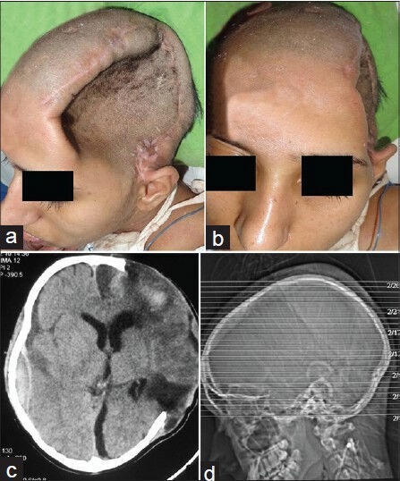
Pre operative view (a and b) Clinical and (c and d) Computer tomography scans
An involuntary lip biting activity (neuropathological chewing) had developed, due to which the lower lip showed an ulcer of about 1 × 1.5 cm. On observation, the activity repeated almost once in every three minutes causing frequent bleeding and an unhealed ulcer on the left side of lower lip. The probable reason for this activity was adduction of lower lip which was subsequently trapped between upper and lower teeth. The patient probably perceived the entrapped lip as a bolus and that lead to initiation of the pathologic chewing cycle.[2] Intra oral examination also revealed palatally placed left lateral incisor, which was aggravating the problem of lip biting.
Treatment planning
An acrylic cranial implant was planned to be fabricated for the loss of cranium. After fabrication, the cranioplast was planned to be surgically placed by neurosurgeon to cover the defect. For the ulcer caused by the pathologic chewing on lower lip, a lip guard was planned for continuous use to prevent lip injury. Coronoplasty of the palatally placed lateral incisor on left side was not performed. This was because, for sufficient reduction of the palatally placed lateral incisor to prevent lip injury, intentional endodontic treatment was indicated. This was not feasible as a part of bed side care in the intensive care unit set up. Also extraction was ruled out for the same reason. In order to provide immediate relief, topical application of a gel (lidocaine hydrochloride, choline salicylate and benzalkonium chloride) was advised three times a day on the lesion.
Fabrication of cranioplast
The steps involved in the fabrication of the cranioplast included impression making, wax-up of the cranioplast, trial of wax-up, processing and finishing of the cranioplast.
Impression making and analogue preparation
After initial examination, impression of the defect was made in irreversible hydrocolloid backed by quick setting plaster. The patient could not sit due to the medical problem; hence the impression was made with the patient in reclined position. Delineation of the defect was done using a glass marking pencil. Wax sheet was adapted around the delineated area to build vertical walls 3 cm high. Within these walls irreversible hydrocolloid was poured and wet gauze strips were embedded partially in the unset material. Soon after the irreversible hydrocolloid had set, quick setting plaster was added on top of the impression material. The two were then removed as one unit and the impression was verified. Type III gypsum was poured in the impression thus recorded to obtain a working analogue [Figure 2]. The margins of the defect were refined on the analogue.
Figure 2.
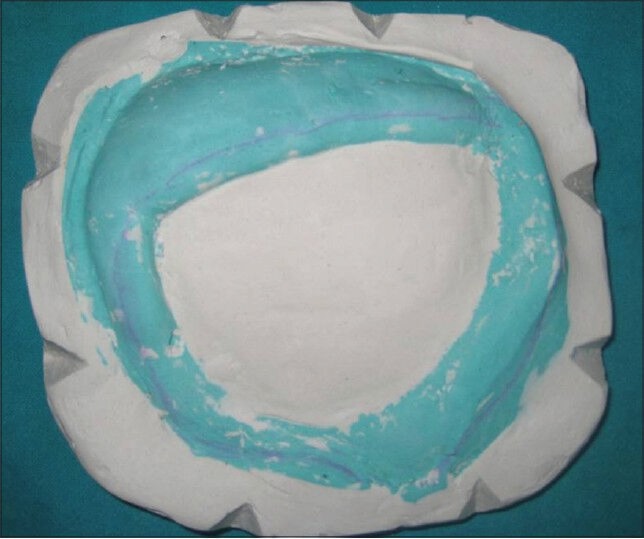
Analogue for fabrication of prosthesis.
Wax-up of cranioplast and trial
Two different thickness of modeling wax was adapted on the delineated area of the defect on the analogue. The shape, thickness, margins and surface of the wax-up were the chief features which were customized. The wax adapted was contoured to the shape of the cranium. The wax-up of the cranioplast was tried on the patient to confirm the continuity with remaining natural bone and symmetry in contour with the normal side. During the trial, bilateral symmetry was ensured along with complete coverage of the defect [Figure 3a and b]. The overall thickness of the wax-up was around 6-7 mm. The thickness was controlled to this amount so as to impart sufficient strength to the cranioplast as well as minimize the risk of weakening due to porosity attributed to the increased thickness. The margin of the cranioplast was beveled with a 5 mm overlap over the remaining cranium. This was done so as to provide scope for adjustment of margins if required (in case of overextension). This also provided sufficient material at the margin, where in holes were made and wire was used to secure the cranioplast to the remaining natural bone during surgical placement. On the remaining surface also, several holes of 1.5 mm diameter were made on the surface of the wax-up [Figure 3c]. Each of these holes was made at a distance of 1 cm from the other. The purpose of the holes was to allow reduction of edema and neo angiogenesis after placement of the cranioplast.
Figure 3.

Wax try in (a) Lateral View, (b) Frontal View (c) On the analog
Processing and finishing of the cranioplast
Since the size of the wax-up was extremely large it was not possible to invest the same in a regular brass flask. For investing, the wax-up (sealed on the analogue), was directly placed in a cardboard box into which dental stone was poured and the stone was leveled to keep the wax-up exposed. It was ensured that the stone base was approximately 4 inches thick. After the stone had set, the cardboard box was removed. Keys were cut up as V shaped notches on the borders of the stone base. The function of the keys was to allow reseating of the counter portion and enable easy escape of the excess resin during packing. In order to pour the counter portion, separating media was applied on the base (in which the analogue and wax-up were embedded). Another cardboard was placed above the base and dental stone was poured on the wax-up in to the cardboard box boundaries. The thickness of the counter portion was again maintained at approximately 4 inches and care was taken to include stone in the V shaped notches of the base to form keys. Once the dental stone had set, the cardboard box was removed. The function of the cardboard box in both the instances was to confine the dental stone.
After the base and counter portion were set, the customized investing was placed for dewaxing. After the wax was lost, clear heat polymerized resin was used to pack the cranioplast. Various precautions to minimize porosity were taken. These included maintaining adequate thickness of base and counter portion of the investing, maintaining adequate proportion of powder and liquid while mixing resin, packing resin in dough stage, bench curing for half an hour and following a long polymerization cycle for curing. After the polymerization was complete, the cranioplast was retrieved, finished and polished [Figure 4].
Figure 4.
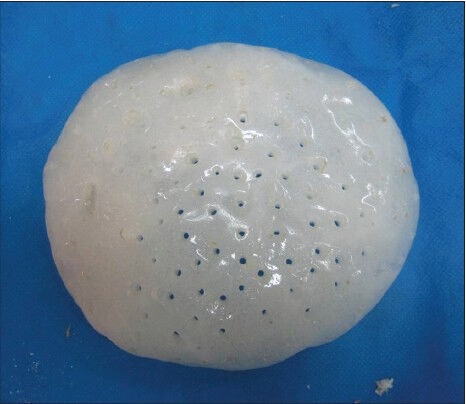
Fabricated cranioplast
FINAL RESULT
The cranioplast was surgically placed [Figure 5] over the defect. Minimal adjustment was required to adapt the cranioplast over the defect. The importance of wax trial cannot be ruled out to attain satisfactory fit. The margins of the cranioplast were sutured with wire on to the remaining natural bone. Initial postsurgical swelling was seen on the entire left side of head and face which reduced subsequently over a period of 20 days. The final improvement in contour was evident [Figure 6a and b] and [Figure 7a and b].
Figure 5.
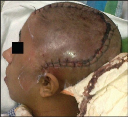
Post surgical view
Figure 6.
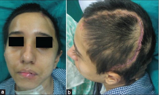
Final results (a) Frontal (b) Lateral
Figure 7.
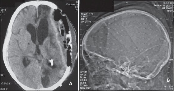
Final computer tomography scans
Fabrication of mouth guard
A single piece mouthguard made in heat polymerized resin was planned to be fabricated for treating the lip injury. The mouthguard was planned to extend from first premolar to first premolar in mesiodistal direction. A resin shield extending from maxillary labial vestibule to mandibular labial vestibule was made in continuity with resin covering the occlusal surface of the maxillary and mandibular teeth.
The mesiodistal extent was limited between the first premolars due to restriction in access to make the impression. A resin shield would extend between the vestibular sulcus which was designed to act as a barrier between lips and teeth. Occlusal surface of maxillary and mandibular teeth were covered so as to provide a vertical opening between the maxillary and mandibular teeth by separation and to reduce impact of bite force on teeth during the pathological chewing cycle due to the property of force dampening by resin.
Limiting factors
The clinical problem encountered was that before the impression material would set the patient would repeat the involuntary activity. Also since the patient did not follow any verbal or non-verbal commands, it was not possible to keep the patients mouth open for impression making. Hence, it was decided to make the impression under sedation and the mouth was forcibly opened by sequentially inserted tongue blades followed by stabilization of the open mouth by a mouth prop. Any attempts to open the mouth manually were futile due to extreme resistance of mandibular elevator muscle. Also such a procedure should not be practiced due to risk of injury to operator hands due to sudden clamping of mandible. The sedation was performed by the anesthetist in the intensive care unit and during the sedation procedure, electrocardiography, pulse oximetry, and blood pressure were monitored by the specialists.
Impression making and prosthesis designing
It was decided to provide a mouthguard in the anterior region extending from first premolar on right side to the first premolar on left side. The impression was made using hard consistency of quick setting addition silicone putty, after the mouth was stabilized with the mouth prop in open position [Figure 8a and b]. No tray was used and putty was directly adapted on the upper eight teeth, lower eight teeth. This recorded the occlusal and a part of lingual surface of teeth (occlusal-lingual impression). Additional layer of putty was made to merge with the already applied putty which would cover the labial surface of teeth and extend till the maxillary and mandibular labial vestibule (cameo portion of impression). This recorded the labial surface of teeth and maxillary and mandibular labial vestibule. A single piece mouthguard was designed to fit on the occlusal/incisal surface of maxillary teeth[Figure 9a]. The occlusal extension separated the maxillary and mandibular teeth. The separation of the 2 arches reduced the impact caused on the teeth when the maxillary and mandibular teeth would strike during pathologic masticatory movement. A shield of resin was made to extend from the occlusal surface between maxillary and mandibular vestibular sulcus. This shield would act as a barrier between the lower lip and the teeth. The recording of mandibular occlusal/incisal surface was done so that the mandibular teeth would be stabilized in the mouthguard at rest position. The putty impression was directly invested in a dental flask and single piece mouthguard was fabricated in clear heat polymerized resin.
Figure 8.
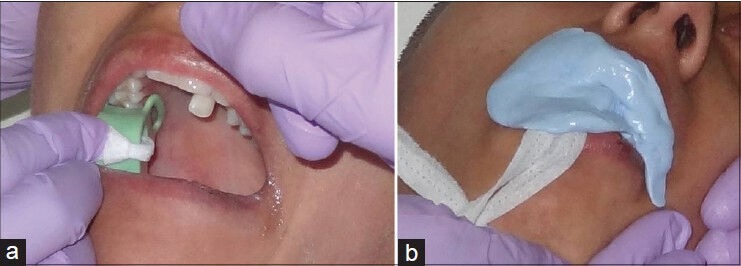
Impression making for lip guard
Figure 9.
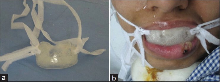
Fabricated lip guard
Since the mouthguard was of a small extent, its retention was a concern. It was decided to provide the mouthguard an extra oral retention feature. Two holes at two different corners of the mouth guard were made and threads for tying were passed through these holes [Figure 9b]. These threads were then tied behind the head and enabled to hold the mouthguard in place. Metal bows were avoided to prevent any accidental injury to the patient during placement and removal by the attendant.
After care
The attendants of the patient were instructed to remove the appliance three times a day followed by cleaning the appliance and the oral cavity. Healing of the ulcer was evident after consistent use of the mouthguard [Figure 10].
Figure 10.
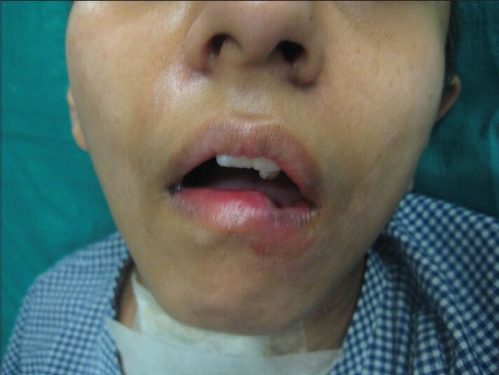
Final results showing healing after using lip guard
DISCUSSION
During the management of a comatose patient, a clinician can encounter several limiting factors. Inability of the patient to respond to any commands is one of the biggest clinical challenges. In the case managed by the authors neither verbal commands nor written commands could be followed. This limitation was not a deterrent when making impression for the cranial implant. For the mouthguard however sedation was used. The entire set up was done in the intensive care unit, so all efforts were made to keep minimal instrumentation and to be swift in the procedures to avoid risk of infection and contamination.
Cranial implants or cranioplasts can be prefabricated or fabricated at the time of surgery[3,4,5,6,7,8,9,10,11,12]. The extent of the defect in this case was tremendous; hence the authors chose to prefabricate the cranioplast. This was because fabrication of the cranioplast during surgery can add to increased exposure of vulnerable neurological tissue to infection, while the flap has been reflected. Also presurgical fabrication of contour is more desirable as reproduction of contour is easily possible, resulting in stronger prosthesis.
A customized cranioplast was fabricated so as to provide a suitable aesthetic contour and maintain the continuity of the cranium. A titanium metal implant was avoided to prevent thermal changes from being conducted to the tissues and thus imposing a risk of neurological tissue damage. Also with metal implants, there is a drawback of interference of electrical conduction which is often required in such patients to monitor their prognosis by Electroencephalogram (EEG). For making the analogue of the defect even though a more accurate analogue could have been made by rapid prototyping, the same was avoided for economical reasons. In order to record the tissues in a minimally distorted state, irreversible hydrocolloid was used in a thin consistency to record the defect and adjoining area. Care was taken to verify the contour and shape of the cranioplast during the wax try in. The margin of the try in was made to overlap the normal bone, so as to attain a better adaptation and provide scope for adjustment if required at the time of surgical positioning of cranial implant. Perforations were made all over the wax-up. The perforations at the margin were made so as to secure the implant to the normal bone with wires. The perforations all over the implant were made to permit the accumulated fluid to flow out of the subgaleal spaces, permit adhesion and migration of connective tissue which ensures better stabilization and to provide adequate blood supply to the overlying scalp. The main aim during the technical procedure was reducing the probability of incorporating porosity during polymerization of the polymethyl methacrylate material. The main drawback of incorporating porosity is reduction in strength, which makes the prosthesis prone to fracture with trauma. The extensive size of the cranioplast did not permit use of the conventional brass flasks hence direct investing in type III gypsum was done. Adequate thickness of the gypsum was ensured to prevent sudden and immense heat changes from surrounding water to bring about porosity. The heat polymerized resin was mixed in 3:1 volume of powder to liquid, packed in dough stage and polymerized using a long curing cycle to reduce the risk of incorporating porosity.
Myostatic masticatory reflex may be initiated in a comatose patient if the lips or tongue are trapped between the teeth, mimicking the placement of a bolus of food on the occlusal surfaces of the teeth.[2,13] Lack of control over the masticatory cycle in the comatose adult patient may sometimes result in neuropathologic chewing.[2,13,14,15,16,17]
The management of such patients depends on medical history, frequency and severity of injury and whether treatment required is long term or short term. Amongst the various treatment options bite planes, intermaxillary fixation, mouth props and tongue blades have often been suggested.[2,18,19,20] The management of such patients should however be immediate using an easily fabricated removable appliance in the short term. A more definitive solution should be sought as the level of consciousness improves.[2] An appropriate appliance must be simple to make and be well retained and easily repaired if the need arises. It must also deflect the tissues most likely to be damaged by involuntary movements of the mandible away from the occlusal table, permit a full range of mandibular movement, allow for daily oral care, withstand breakage and displacement forces over an indefinite period and most importantly allow healing of traumatized tissues.[2,21] In the case described, all attempts were made to fabricate a simple and easy to handle appliance which would prevent injury to the lower lip. The appliance design was effective in providing a barrier between the lower lip and the teeth. Also the trauma to the teeth by impact force of closure during masticatory cycle was reduced by the resin separating the occlusal surface of both maxillary and mandibular teeth. The only drawback in the appliance design was that retention in the appliance was obtained primarily from the front teeth, hence to enhance the same extra oral soft and broad (1 cm wide) tie threads were used to tie around the face without providing injury to the extra oral tissues.
CONCLUSION
Clinicians often face a situation that is far outside the realm of usual treatment conditions.[17] Management of a comatose patient is one such situation. The main emphasis in management of such patients is to comprehend the underlying altered anatomy and physiology, to cause minimum trauma during the management and to be quick in providing relief. No doubt the improvement in quality of life is marginal, yet the main aim in providing a cranioplast is protection of remaining brain tissue and to make the patient socially more acceptable. The main aim in providing the mouthguard was to prevent further injury to the lower lip and enable healing of the previous one. Both the prostheses were successful in fulfilling the primary purpose for which they were fabricated.
Footnotes
Source of Support: Department of Neurosurgery and Anaesthesiology, G. B. Pant Hospital, New Delhi.
Conflict of Interest: None declared.
REFERENCES
- 1.Teasdale G, Jennett B. Assessment of coma and severity of brain damage. Anesthesiology. 1978;49:225–6. doi: 10.1097/00000542-197809000-00023. [DOI] [PubMed] [Google Scholar]
- 2.Kobayashi T, Ghanem H, Umezawa K, Mega J, Kawara M, Feine JS. Treatment of self-inflicted oral trauma in a comatose patient: A case report. J Can Dent Assoc. 2005;71:661–4. [PubMed] [Google Scholar]
- 3.Raja AI, Linskey ME. In situ cranioplasty with methylmethacrylate and wire lattice. Br J Neurosurg. 2005;19:416–9. doi: 10.1080/02688690500390250. [DOI] [PubMed] [Google Scholar]
- 4.Dumbrigue HB, Arcuri MR, LaVelle WE, Ceynar KJ. Fabrication procedure for cranial prostheses. J Prosthet Dent. 1998;79:229–31. doi: 10.1016/s0022-3913(98)70222-7. [DOI] [PubMed] [Google Scholar]
- 5.Beumer J, 3rd, Firtell DN, Curtis TA. Current concepts in cranioplasty. J Prosthet Dent. 1979;42:67–77. doi: 10.1016/0022-3913(79)90332-9. [DOI] [PubMed] [Google Scholar]
- 6.Martin JW, Ganz SD, King GE, Jacob RF, Kramer DC. Cranial implant modification. J Prosthet Dent. 1984;52:414–6. doi: 10.1016/0022-3913(84)90457-8. [DOI] [PubMed] [Google Scholar]
- 7.Blum KS, Schneider SJ, Rosenthal AD. Methyl methacrylate cranioplasty in children: Long-term results. Pediatr Neurosurg. 1997;26:33–5. doi: 10.1159/000121158. [DOI] [PubMed] [Google Scholar]
- 8.Ducic Y. Titanium mesh and hydroxyapatite cement cranioplasty: A report of 20 cases. J Oral Maxillofac Surg. 2002;60:272–6. doi: 10.1053/joms.2002.30575. [DOI] [PubMed] [Google Scholar]
- 9.Couldwell WT, Chen TC, Weiss MH, Fukushima T, Dougherty W. Cranioplasty with the Medpor porous polyethylene flexblock implant. Technical note. J Neurosurg. 1994;81:483–6. doi: 10.3171/jns.1994.81.3.0483. [DOI] [PubMed] [Google Scholar]
- 10.Joffe J, Harris M, Kahugu F, Nicoll S, Linney A, Richards R. A prospective study of computer-aided design and manufacture of titanium plate for cranioplasty and its clinical outcome. Br J Neurosurg. 1999;13:576–80. doi: 10.1080/02688699943088. [DOI] [PubMed] [Google Scholar]
- 11.Sundseth J, Berg-Johnsen J. Prefabricated patient-matched cranial implants for reconstruction of large skull defects. J Cent Nerv Syst Dis. 2013;5:19–24. doi: 10.4137/JCNSD.S11106. [DOI] [PMC free article] [PubMed] [Google Scholar]
- 12.Koli DK, Nanda A, Verma M. Acrylic cranial implant: An alternative vector in management of cranial defect. J Craniofac Surg. 2012;23:e591–4. doi: 10.1097/SCS.0b013e31826bf494. [DOI] [PubMed] [Google Scholar]
- 13.Jackson MJ. The use of tongue depressing stents for neuropathologic chewing. J Prosthet Dent. 1978;40:309–11. doi: 10.1016/0022-3913(78)90038-0. [DOI] [PubMed] [Google Scholar]
- 14.Ngan PW, Nelson LP. Neuropathologic chewing in comatose children. Pediatr Dent. 1985;7:302–6. [PubMed] [Google Scholar]
- 15.Pratap-Chand R, Gourie-Devi M. Bruxism: Its significance in coma. Clin Neurol Neurosurg. 1985;87:113–7. doi: 10.1016/0303-8467(85)90107-6. [DOI] [PubMed] [Google Scholar]
- 16.Millwood J, Fiske J. Lip-biting in patients with profound neuro-disability. Dent Update. 2001;28:105–8. doi: 10.12968/denu.2001.28.2.105. [DOI] [PubMed] [Google Scholar]
- 17.Cohen HV, Patel B, DiPede LA. Posttraumatic head injury resulting in spasticity disorders and oral injury: Application of prosthodontic skills for tissue protection--a case report. Quintessence Int. 2009;40:457–60. [PubMed] [Google Scholar]
- 18.Hallett KB. Neuropathological chewing: A dental management protocol and treatment appliances for pediatric patients. Spec Care Dentist. 1994;14:61–4. doi: 10.1111/j.1754-4505.1994.tb01102.x. [DOI] [PubMed] [Google Scholar]
- 19.Arhakis A, Topouzelis N, Kotsiomiti E, Kotsanos N. Effective treatment of self-injurious oral trauma in Lesch-Nyhan syndrome: A case report. Dent Traumatol. 2010;26:496–500. doi: 10.1111/j.1600-9657.2010.00930.x. [DOI] [PubMed] [Google Scholar]
- 20.Kumar P, Bhojraj N. Successful prevention of oral self-mutilation using a lip guard: A case report. Spec Care Dentist. 2011;31:114–8. doi: 10.1111/j.1754-4505.2011.00188.x. [DOI] [PubMed] [Google Scholar]
- 21.Hanson GE, Ogle RG, Giron L. A tongue stent for prevention of oral trauma in the comatose patient. Crit Care Med. 1975;3:200–3. doi: 10.1097/00003246-197509000-00007. [DOI] [PubMed] [Google Scholar]


