

Abstract
Background:
When the body has been mutilated, it is common to have the extremities or head amputated from the trunk. In concern with forensic odontology, an estimate must have been made based on the correlation of osteometry along with odontometry in determining sex, race and stature.
Objective:
The objective of this study is to investigate and correlate height and gender from odontometry and anthropometric data of the skull.
Materials and Methods:
The study was conducted in the Department of Oral and Maxillofacial Pathology and Microbiology, I.T.S Center for Dental studies and Research, Muradnagar, Ghaziabad (UP) with the representative study subjects of 60 patients as 30 males and 30 females in the age group of 15-25 years. The selected parameters were measured and then correlated to investigate stature and gender from odontometry and anthropometric data of the skull.
Results:
On linear regression analysis, the selected parameters were found to be statistically significant predictor of height. It was also established by Karl Pearson's coefficient correlation that the left mandibular canine index for female was statistically significant to show sexual dimorphism.
Conclusion:
In the emerging field of forensic odontology, skull anthropometry, odontometry exhibits stature determination and strong sexual dimorphism.
Keywords: Anthropometry, forensic odontology, odontometry, stature
Introduction
As stated by Fédération Dentaire Internationale, “Forensic Odontology” is the branch of dentistry which, in the interest of justice, deals with proper handling and examination of dental evidence and with the proper evaluation and presentation of dental findings.”[1] ‘Forensic dentistry is important for human identification, especially when conventional methods cannot be applied, usually due to advanced decomposition, carbonization or fragmentation of the body. In accidents or natural disasters involving severe trauma and fire, the use of fingerprints, facial recognition methods can be difficult or even impossible. In such cases, a post-mortem record is created by a forensic dentist to identify the victim by determining the age, stature, ancestry, sex and socio-economic class, where the body has been mutilated and the extremities or head amputated from the trunk.[2]
One of the main goals of forensic anthropology[3] is to determine the identity of a person from the study of some skeletal remains. In the last few decades, anthropologists have focused their attention on improving those techniques that allow a more accurate identification. Before making a decision on the identification, it is necessary to follow different processes that let them assign a sex, age, human group and height to the subject from the study of bones found.
Different methodologies have been proposed, according to the features of the different human groups of each region.[4,5] In the identification of unknown human remains, stature and gender estimation is a preliminary investigation.[2,6] Stature is the height of a person in the upright posture.[7]
The method of using teeth and skull measurements has several advantages as the anatomical landmarks are standard, well developed and easy to locate. An estimate must then be made based on the known relationship of the remains to stature and gender.
Teeth form an excellent material for anthropological, genetic, odontologic and forensic investigations. Amongst all the teeth, the mandibular canines are found to exhibit greatest sexual dimorphism.
With this background in concern with the forensic odontology, paleontology and esthetic dentistry, we undertook this study with the aim to investigate and correlate height and gender from anthropometric and odontometric data of the skull. A relationship of height of a person with the maxillary extent and circumference of skull and identifying gender with the combined mesiodistal (MD) width of maxillary anterior teeth with the statistical aid of regression analysis of these variables was done.
Materials and Methods
The study was conducted in the Department of Oral and Maxillofacial Pathology and Microbiology, I.T.S Center for Dental studies and Research, Muradnagar, Ghaziabad (UP) with the representative study subjects of 60 patients as 30 males and 30 females in the age group of 15-25 years.
Inclusion and exclusion criteria
Complete set of fully erupted, periodontally healthy teeth
Presence of non-carious, non-worn teeth with no dental history of any crown restorations
Complete set of intact and satisfactorily aligned maxillary anterior teeth
No history or clinical evidence of cleft palate, orthognathic surgery or trauma
No history or clinical features suggestive of endocrinal disorders, metabolic disorders, developmental disorders and history of prolonged illness.
Stature correlation to skull and jaw dimensions was done for the possibility of estimating height in forensic investigations. After obtaining informed consent from the subject selected, measurements of the maxillary extent of the skull was obtained using Rakosi Jarabak analysis[8] on the lateral cephalograph of 8' by 10' radiographic films. The maxillary extent of the skull was derived as the linear distance between the anterior nasal spine to the posterior nasal spine as shown in Figure 1. Measurements of MD widths of the six maxillary anterior teeth on the cast were done. The greatest MD crown widths of the maxillary anterior permanent six teeth were measured between the anatomic contact points of each tooth on either side of the jaw, using digital vernier calliper as shown in Figure 2. Circumference of the skull and height was measured directly on each patient. Maximal fronto-occipital circumference was measured by placing a nonstretchable plastic tape (calibrated in millimeters) just on the occipital prominence and the supraorbital ridges, while viewing the subject laterally also to ensure proper placement of the tape. In cases of some hairstyles in males, we drew the tape tightly and compressed the hair as much as possible. In cases of females, we asked the subjects to lift their hair in the occipital area and the tape was placed against the skin and not over the lumps of hair. This method was in accordance with the one used by Evereklioglu et al.[9] Height was measured as the vertical distance from the vertex to the floor. Measurement was taken by making the subject stand erect on a horizontal plane barefooted. The subject was placed in straight vertical position with head oriented in Frankfürt plane.
Figure 1.
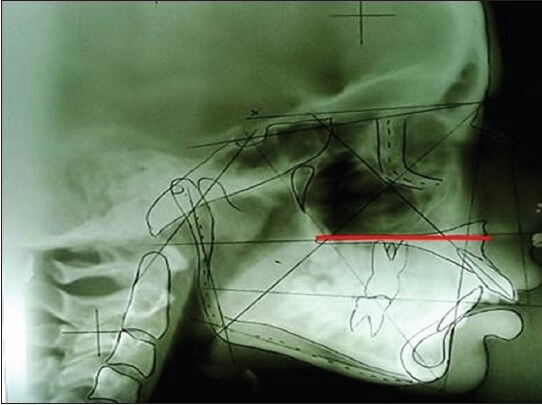
Maxillary extent using Rakosi Jarabak analysis on lateral cephalographs
Figure 2.

Mesiodistal crown widths of the maxillary anterior permanent six teeth using vernier calliper
In order to define sexual dimorphism and the morphometric criteria for mandibular canines, the intercanine distance, right and left mandibular canine width were observed on study casts as shown in Figures 3 and 4 of same 60 study subjects (M:F::30:30) selected above.
Figure 3.

Intercanine distance using vernier calliper
Figure 4.

Right and left mandibular canine width using vernier caliper
Further the mandibular canine index was calculated as:

The mandibular canine index was calculated and the readings obtained were subjected to statistical analysis to derive conclusions and sexual dimorphism in right and left mandibular canines.
Comparisons were made between the measurements recorded with respect to the gender using statistical mean, standard deviation, range and Student's t-test. Combined data, male and female data were analyzed separately for linear regression of height to the parameters recorded.
Results
The descriptive statistics for the measurements of the four parameters was recorded in the study subjects.[1] Table 1 shows that the sample of 60 subjects presented with a mean height of 1616.17 mm with a standard deviation of 64.87. The mean circumference of the skull is 534.55 mm with a standard deviation of 18.52. The MD width of upper anterior is 50.13 mm with a standard deviation of 6.694. The distance from anterior nasal spine to posterior nasal spine is with a mean value of 54.92 mm and standard deviation of 6.95. The inferential statistics of the parameters were recorded [Table 2]. The ANOVA of linear regression analysis of height and its predictors (i.e., three parameters) was found to be statistically significant with the P value of 0.001.
Table 1.
Descriptive statistics

Table 2.
The inferential statistics of the parameters recorded using ANOVA linear regression analysis

On linear regression analysis, the regression equations were derived [Table 3]. Maxillary extent was found to be statistically significant predictor of height with P = 0.027 and the circumference of the skull as a highly significant predictor with P = 0.004.
Table 3.
On linear regression analysis the regression equations were derived as the constants listed in the table

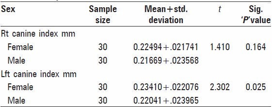
The inferential comparative statistics of canine index using Karl Pearson's coefficient correlation [Table 4] shows the mean value, which evidenced that the left canine index was higher in female than male having less standard deviation of 0.022. Graph 1 depicts left canine to show statistically significant sexual dimorphism. Karl Pearson's coefficient correlation established that the left canine index for female was statistically significant by t-test with P = 0.025.
Table 4.
Group statistics
Graph 1.

Depicting left canine to show statistically significant sexual dimorphism
Discussion
Routine identification tasks are a simple one-to-one matching process. This is not the case in disasters. Mass fatality incidences represent a big challenge in the identification of deceased victims. In such circumstances it necessitates putting a hierarchy system of forensic science.[10] Various methods are used to establish the identity of unknown human remains.[11] The reliability of each method varies. Estimation of stature, as part of the identification process has a long history in physical anthropology. A drawback to physical anthropology techniques is limited applicability to fragmentary remains.[12] When the body has been mutilated, it is common to have the extremities or head amputated from the trunk. An estimate must then be made based on the known relationship of the remains to stature.[3]
In the present study, estimation and correlation of height and gender from odontometry and anthropometric data of the skull was done. In cases, where identification has to be performed based on skeletal remains, the most common stature estimates are derived from long bones.[13] The literature, however, is lacking in the derivation of height from anthropometry of the skull along with odontometric parameter. Osteometry seems to be the preferred technique in the identification process with several advantages as the anatomical landmarks are standard, well defined and easy to locate.
The estimation of living stature from long bones is based upon the principle that the long bones correlate positively with the stature. Since this is true, parts of each bone should also be related to stature even though they may not correlate as highly.[13] The above principle was found to hold good in the present study. We found highest correlation when individual measurements of combined data were plotted against height. Furthermore, adding the odontometric and craniometric parameters elevated the correlation with the stature estimation.
The study of the tooth is a non-destructive and simple process which can be applied to both living and deceased persons, in contrast to other time consuming, expensive, less reliable and destructive methods, which may not be acceptable for ethical, religious, cultural or scientific reasons.[14] Further, procedures such as digitization of lateral cephalograph analysis avoid the bias inherent in observer subjectivity and improve reliability, accuracy and precision.[15]
Any tooth can be used to assess age. Canine teeth are a good candidate for sex determination because they are often present in old age. They are less likely than other anterior teeth to suffer wear as a result of age or any particular work.[16]
Based on the above background, this study was conducted to develop a method for stature and gender determination and their correlation using odontometry and skull anthropometry based on the relationship between various morphological variables of teeth and skull measurements using advanced and more reliable procedures.
Introna et al. in 1993 discussed correlations between cranial diameter and height in the same age group as in the present study, but other parameters were not undertaken.[17]
Chiba and Terazawa in 1998 conducted head and skull measurements for height estimation but did not include gender determination, which is a preliminary unit of an investigation procedure.[18] Kalia et al. in 2010 estimated the height of a person from teeth and skull but again lacked the critical gender determination in individual recognition.[19]
The present study provides a statistically valid technique to achieve accurate clues of stature as well as conspicuous need of gender determination from an individual's fragmentary remains. In the present study, it was observed that circumference of skull and maxillary extent was statistically significant in correlation to height. While in contrast to past studies, mesiodistal dimension of anteriors was found to be statistically insignificant.
In contrast to rare past studies, the present study takes into account “maxillary extent”, which is never taken into account as one of the parameter in stature determination using Rakosi Jarabak analysis. Rakosi Jarabak analysis is considered to be a crucial and precise analysis of skull dimensions in lateral cephalometry. On analysis, the three parameters i.e. anterior nasal spine- posterior nasal spine, ANS-PNS (maxillary extent), mesiodistal dimension of anteriors, circumference of skull corresponded with the variation in height by a highly significant correlation.
In respect to gender determination, Garn and Lewis (1967), Lysell and Myrberg (1982) concluded that the mandibular canine demonstrated the greatest sexual dimorphism amongst all teeth. Lew and Keng in 1991 showed same results in their study on ethnic Chinese population. Hashim and Murshid in 1993 studied the age group of 13-20 years found that only the canines in both jaws exhibited a significant sexual difference.[20,21,22,23]
Nair et al. (1999) concluded that the left mandibular canine with 7.7% followed by the right mandibular canine with 6.2% shows the maximum sexual dimorphism. Further, when these parameters were measured intraorally, they were found to be statistically insignificant.[24] Omar, Azab in 2009 demonstrated that maxillary canine showed greater sexual dimorphism than mandibular canine in variable age groups. This was contradictory to the present study as they did not take into consideration the age dependant wearing away of the tooth. Gorea, Sharma in 2010 observed the effectiveness of intercanine distance in their study for predicting the sex. Their study considered intercanine distance in contrast to the present study and found that intercanine distance was greater in males for both the jaws. They also lacked in even distribution of patients in each age group.
In the present study for gender determination, the left mandibular canine index was found to exhibit greater sexual dimorphism with a highly statistically significant P value of 0.025, however, the right mandibular canine index was found to be statistically insignificant.
Conclusion
In the emerging field of forensic odontology, skull anthropometry including maxillary extent, mesiodistal dimension of anteriors and circumference of skull are beneficial for stature estimation. In concern with gender determination, which is a preliminary investigation in genetic, legal and forensic odontology, paleontology, left mandibular canine index exhibits a strong sexual dimorphism.
Further research is warranted with larger samples in the direction of definite improvement in the accuracy of stature estimation from odontometry. We recommend studies on cadavers and skulls without soft tissue covering as well as assessment of the effect of aging on the regression coefficients in stature estimation.
Footnotes
Source of Support: Nil
Conflict of Interest: None declared
References
- 1.Ratnakar P, Singaraju GS. Methods of identification in forensic dentistry. Annals and Essences of Dentistry. 2010;2(1):26–8. [Google Scholar]
- 2.Jason DR, Taylor K. Estimation of stature from the length of the cervical, thoracic, and lumbar segments of the spine in American whites and blacks. J Forensic Sci. 1995;40:59–62. [PubMed] [Google Scholar]
- 3.Burns K. Forensic Anthropology Training Manual. Upper Saddle River, New Jersey: Prentice-Hall; 2007. [Google Scholar]
- 4.Iscan MY. Forensic anthropology of sex and body size. Forensic Science International. 2005;147(2):107–12. doi: 10.1016/j.forsciint.2004.09.076. [DOI] [PubMed] [Google Scholar]
- 5.González-Colmenares G, Botella-López MC, Moreno-Rueda G, Fernández-Cardenete JR. Age estimation by a dental method: A comparison of Lamendin's and Prince and Ubelaker's technique. J Forensic Sci. 2007;52:1156–60. doi: 10.1111/j.1556-4029.2007.00508.x. [DOI] [PubMed] [Google Scholar]
- 6.Sopher IM. The dentist, the forensic pathologist, and the identification of human remains. J Am Dent Assoc. 1972;85:1324–9. doi: 10.14219/jada.archive.1972.0536. [DOI] [PubMed] [Google Scholar]
- 7.Stedman TL. Stedman's Medical Dictionary. Baltimore: Lippincott Williams and Wilkins; 2000. p. 211. [Google Scholar]
- 8.Marathiotou LL, Papadopoulos MA. Assessment of cephalometric analyses used for craniofacial complex evaluation: Part I-Sagittal maxillary position. Hell Orthod Rev. 2005:8. [Google Scholar]
- 9.Evereklioglu C, Doganay S, Er H, Gunduz A, Tercan M, Balat A, et al. Craniofacial anthropometry in a Turkish population. Cleft Palate Craniofac J. 2002;39:208–18. doi: 10.1597/1545-1569_2002_039_0208_caiatp_2.0.co_2. [DOI] [PubMed] [Google Scholar]
- 10.Al-Amad SH. Forensic odontology. Smile Dent J. 2009;4(1):22–4. [Google Scholar]
- 11.Brickley M, McKinley JI. Guidelines to the Standards for Recording Human Remains. Highfield, Southampton, BABAO. 2004 [Google Scholar]
- 12.Holland TD. Estimation of adult stature from fragmentary tibias. J Forensic Sci. 1992;37:1223–9. [PubMed] [Google Scholar]
- 13.Simmons T, Jantz RL, Bass WM. Stature estimation from fragmentary femora: A revision of the Steele method. J Forensic Sci. 1990;35:628–36. [PubMed] [Google Scholar]
- 14.Kvaal SI, Kolltveit KM, Thomsen IO, Solheim T. Age estimation of adults from dental radiographs. Forensic Sci Int. 1995;74:175–85. doi: 10.1016/0379-0738(95)01760-g. [DOI] [PubMed] [Google Scholar]
- 15.Cameriere R, Ferrante L, Cingolani M. Variations in pulp/tooth area ratio as an indicator of age: A preliminary study. J Forensic Sci. 2004;49:317–9. [PubMed] [Google Scholar]
- 16.Cameriere R, Ferrante L, Belcastro MG, Bonfiglioli B, Rastelli E, Cingolani M. Age estimation by pulp/tooth ratio in canines by peri-apical X-rays. J Forensic Sci. 2007;52:166–70. doi: 10.1111/j.1556-4029.2006.00336.x. [DOI] [PubMed] [Google Scholar]
- 17.Introna F, Jr, Di Vella G, Petrachi S. Determination of height in life using multiple regression of skull parameters. Boll Soc Ital Biol Sper. 1993;69:153–60. [PubMed] [Google Scholar]
- 18.Chiba M, Terazawa K. Estimation of stature from somatometry of skull. Forensic Sci Int. 1998;97:87–92. doi: 10.1016/s0379-0738(98)00145-5. [DOI] [PubMed] [Google Scholar]
- 19.Kalia S, Shetty SK, Patil K, Mahima VG. Stature estimation using odontometry and skull anthropometry. Indian J Dent Res. 2008;19:150–4. doi: 10.4103/0970-9290.40471. [DOI] [PubMed] [Google Scholar]
- 20.Garn SM, Lewis AB, Kerewsky RS. Buccolingual size asymmetry and its developmental meaning. Angle Orthod. 1967;37:186–93. doi: 10.1043/0003-3219(1967)037<0186:BSAAID>2.0.CO;2. [DOI] [PubMed] [Google Scholar]
- 21.Lysell L, Myrberg N. Mesiodistal tooth size in the deciduous and permanent dentitions. Eur J Orthod. 1982;4:113–22. doi: 10.1093/ejo/4.2.113. [DOI] [PubMed] [Google Scholar]
- 22.Lew KK, Keng SB. Anterior crown dimensions and relationship in an ethnic Chinese population with normal occlusions. Aust Orthod J. 1991;12:105–9. [PubMed] [Google Scholar]
- 23.Hashim HA, Murshid ZA. Mesiodistal tooth width. A comparison between Saudi males and females. Part 1. Egypt Dent J. 1993;39:343–6. [PubMed] [Google Scholar]
- 24.Nair P, Rao BB, Annigeri RG. A study of tooth size, symmetry and sexual dimorphism. J Forensic Med Toxicol. 1999;16(2):10–3. [Google Scholar]