Abstract
Aims:
To determine and compare the reliability of pulp tissue in determination of sex and to analyze whether caries have any effect on fluorescent body test.
Materials and Methods:
This study was carried on 50 maxillary and mandibular teeth (25 male teeth and 25 female teeth), which were indicated for extraction. The teeth are categorized into 5 groups, 10 each (5 from males and 5 from females) on the basis of caries progression. The pulp cells are stained with quinacrine hydrochloride and observed with fluorescent microscope for fluorescent body. Gender is determined by identification of Y chromosome fluorescence in dental pulp.
Results:
Fluorescent bodies were found to be more in sound teeth in males as the caries increase the mean percentage of fluorescent bodies observed decreases in males. We also observed the fluorescent spots in females, and the value of the spot increases in female as the caries progresses, thereby giving false positive results in females.
Conclusion:
Sex determination by fluorescent staining of the Y chromosome is a reliable technique in teeth with healthy pulps or caries with enamel or up to half way of dentin. Teeth with caries involving pulp cannot be used for sex determination.
Keywords: Fluorescent microscope, forensic odontology, pulp tissue, quinacrine hydrochloride
Introduction
The main attributes of biological identity are sex, age, stature, and ethnic background of the individual, which are also called the ‘Big Four’ in forensic context.[1] Human identification is of paramount importance, for both legal as well as humanitarian purpose.[2] Albeit there have been tremendous advancements in forensic medicine, this remains a practical problem. The use of dental identification has long been considered a reliable method, especially when other methods (fingerprints) fail because body conditions are not available, and over time, forensic odontology has emerged as a complete branch.[2]
Teeth can survive and remain virtually unaffected long after other soft tissue and skeletal tissues have been destroyed.[2] The recognition of teeth as a tissue that withstands great variation in environment (temperature upto 1600° C, humidity, and pH) has lead to its application in personal identification.[3] It has been observed in past time that dental tissues have witnessed its importance worldwide where it has contributed enormously in identification of victims in disaster, 80% of non-Thai in tsunami in Thailand and 20% in 26/11 incidence where 12 coordinated shooting and bombing attacks were carried by terrorists in Mumbai, India's most populous city on 26 November, 2010 were identified using teeth. Other than this, it has been reported that renowned people like Adolf Hitler and Indian Prime Minister Rajiv Gandhi were identified using tooth.[4]
Several parameters have been proposed for sex determination to identify an individual from teeth, but none of them have proved its reliability. Recently, DNA analysis in forensic have gained attraction, but it is expensive and time-consuming. This prompted us to look for other methods of dental identification, which will be cost-effective yet reliable.[1]
Tooth pulp is encased in a hard tissue casting, where it may be protected from detrimental effects of impact, trauma, and heat.[5] So, Casperson et al. developed a technique using pulpal tissue stained with quinacrine mustard, specific for Y chromosome to determine sex of an individual.[6] In literature, there is scarcity of relevant studies that analyzed the effect of caries on viability of pulpal tissue in sex determination. So, keeping in mind this fact, present study was conducted to analyze the effect of caries on pulpal tissue stained with quinacrine hydrochloride with the application of fluorescent microscope.
Materials and Methods
This study was carried out in the Department of Oral Pathology and Microbiology, Swami Devi Dyal Hospital and Dental College Barwala, Panchkula. The study protocol was approved by the Ethical Committee of college before commencement of the procedure.
Fifty maxillary and mandibular permanent teeth that were indicated for extraction for periodontal and endodontic reasons were included in the study. Study sample consisted of 25 male and 25 female teeth, which were further categorized into 5 groups of 10 each (10 from males and 10 from females) based on the extent of caries progression. Group A includes freshly extracted tooth specimens with no caries, group B consisted of tooth specimens with caries in enamel, group C comprised of tooth specimens with caries less than half way of dentin, group D consisted of freshly extracted teeth with caries more than half way of dentin, and group E includes freshly extracted tooth specimen with caries involving pulp.
The teeth were examined radiographically and clinically for analyzing the various stages of caries progression [Figure 1].
Figure 1.
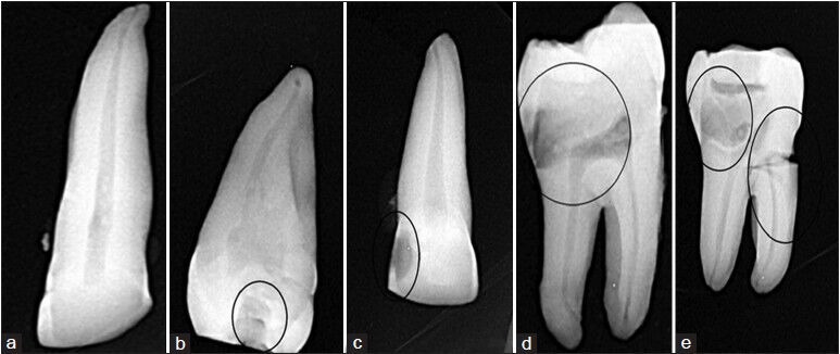
Intra-oral periapical radiographs representing progression of caries. (a) Sound teeth, (b) enamel, (c) less than half way of dentin, (d) more than half way of dentin, (e) pulp
Sectioning of teeth
Modeling wax was folded and made into a block. The tooth was embedded on the modeling block. To remove the pulp, the crown was separated longitudinally by using a carborundum disc at 30,000 rpm. Similarly, the root was split for pulp removal. The pulp tissue was transferred to a conical tube containing normal saline. It was adequately washed in normal saline to remove any calcified bone or dentine particles.
The pulp tissue was then transferred to the dry and clean conical centrifuge tubes containing 5 ml of fixative (3 Methanol:1 Glacial acetic acid) and left as such for about half an hour to 2 hours for the fixation of the pulp cells. It was then crushed with the glass rod sufficiently to isolate the pulp cells. A suspension thus obtained was centrifuged for 10 minutes at 1000 rpm. The supernatant was discarded, leaving behind the pellet in the centrifuge tube. Five ml of fresh fixative was then added to re-suspend the pellet, and the process was repeated thrice till a clear suspension of the pulp cells was obtained.
Thin smears were prepared on chilled microscope slides of 1 mm thickness by the air-drying method i.e., by dropping 2-3 drops of the above suspension on the slide from a distance of inches to get a homogenous population of cells. The cells were stained with 5% Quinacrine hydrochloride to stain the Y chromosome. The slide was mounted in buffer of pH 5.5 and was observed under LEICA DMR fluorescent microscope (Oil immersion in dark field at ×40) by BV exciting method (emitting a blue-violet color, mainly at 4.047A0 and 4.038A0). Only those cells which contained the characteristic Y chromatin i.e., a brightly fluorescent spot attached to the nuclear membrane were counted as positive cells while those which did not show any such fluorescent spot were labeled as negative.
Results obtained were subjected to statistical analysis by using independent t-test using SPSS (version 9.0) software.
Results
This study was done to evaluate the use of the fluorescent body test as a diagnostic test for sex determination. Table 1 showed sensitivity, specificity, positive predictive value, negative predictive value, and efficiency of each of the five groups. The values obtained were highest for sound teeth with caries in enamel and value decreases with the progression of the caries. The median was used as the cut-off value. Table 2 depicted that fluorescent bodies were found to be more in sound teeth in males as the caries increase, the mean percentage of fluorescent bodies observed decreases in males. We also observed the fluorescent spots in females, and the value of the spot increases in female as the caries progresses, thereby giving false positive results in females [Figure 3]. The P value were found to be significant for sound teeth and teeth with caries in enamel and involving less than half the way of dentin, whereas it was non-significant for teeth involved with further caries involvement.
Table 1.
Diagnostic utility of fluorescent body in identifying gender

Table 2.
Percentage of fluorescent bodies (in %) in different groups
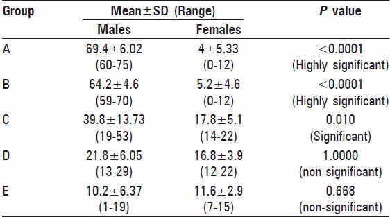
Figure 3.
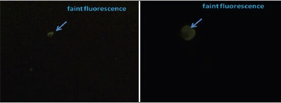
Fluorescent photomicrograph representing faint fluorescence in females
Discussion
Despite the fact that tremendous research work is carried out to determine a reliable method, but none have proven its efficacy. The most reliable means of identification include fingerprints, dental comparisons, and biologic methods such as DNA profiling. In some cases, however, fingerprints are not available from the deceased, or ante-mortem prints cannot be obtained.[5] Sex determination from skull is highly unreliable although number of workers have described differences in the size of male and female teeth, such measurements seem unlikely to be used to determine the sex of an individual.[7] So, a technique has been developed, which may offer a solution whenever soft tissue remains can be recovered.
Casperson et al.[6] showed that chromosome stained with quinacrine mustard fluoresced differentially along their length when viewed in ultraviolet light and the human Y chromosome fluoresced more brightly than other chromosomes. He suggested that alkylating agents like quinacrine acts on the DNA portion rich in guanine and accumulate there. This technique has been used in forensic science for sex determination from dried blood stains, saliva, and hair.[8] Thus, it might be applicable to sex determination from tooth pulp some time after death, because the teeth are a stable part of the skeleton and the pulp tissue is well-protected.
Seno and Ishizu carried out the detection of Y chromosome in the nuclei of dental pulp. They found that f- body positive rate in the nucleated cells of male dental pulp was over 30%, even with the male teeth as old as 5 months, after the extraction. With female teeth, typical f-body cannot be detected, and f-body like spot has been observed in 0.4% of cells, indicating that there can be no error in the identification of male tooth from that of female one, even such an f-body like spot is taken as an f-body itself.[8]
Present study revealed that in males, 60-75% fluorescent y bodies [Figure 2] were observed in sound teeth (mean 69.4% bodies), 59-70% bodies in tooth with caries in enamel (mean 64.2% bodies), 19-53% in teeth with caries in half way of dentin (mean 39.8% bodies), teeth with caries involving more than half way of dentin and involving pulp showed 13-29%, 1-19% (mean 21.8%, 6.37% bodies) off bodies respectively as shown in Table 2. Seno and Ishizu showed a range of 56-72% fluorescent y bodies in freshly extracted sound teeth, which are in accordance to sound teeth in this study.[8]
Figure 2.
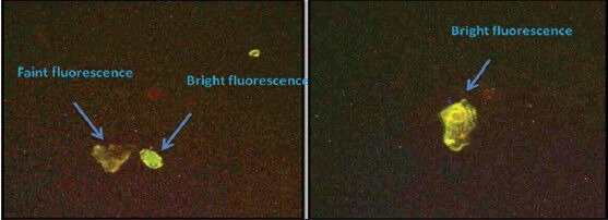
Fluorescent photomicrograph showing bright fluorescent cell in males representing Y chromosome
Pseudo Y body in the present study [Figure 4] in females were found to be in the range of 0-12% (mean 5.2%), 0-12% (mean 5.2%), 14-22% (mean 17.8)%, 12-22 (mean 16.8%), and 7-15% (mean 11.6%) for sound teeth, teeth with caries in enamel, caries in half way of dentin, caries involving more than half way of dentin caries, and caries involving pulp, respectively, as shown in Table 2.
Figure 4.
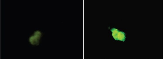
Fluorescent photomicrograph representing artifacts or pseudo f-bodies in females
Discrepancy between various studies could be attributed to random inclusion of carious and non-carious teeth.[5] So, the present study considered the effect of caries on fluorescent body test as literature (when searched via PUBMED and MEDLINE) revealed lack of studies analyzing this fact.
From present study, it can be inferred that as the caries progresses from enamel to dentin and further to pulp tissue, the sensitivity and specificity of determining Y chromosome decreases [Table 1]. When the caries have involved only enamel or half the way of dentin, it has shown significant difference in fluorescent body test determining males and females, unlike when it involved more than half way of dentin and pulp where the difference is non-significant [Table 2]. The decrease in ‘Y’ body could be attributed to the fact that cariogenic bacteria alter the microenvironment of pulp, resulting in alteration in fluorescent staining. As the caries progresses, the inflammatory processes in the pulp are initiated, as the inflammation progresses the internal architecture of the cell is lost, and is also characterized by necrotic poorly stained cellular debris.[9] Many workers attributed the appearance of fluorescent bodies in females to artifacts, whereas others ascribed it to the presence of fluorescent debris. It was necessary to smear pulp cells thinly to prevent masking the fluorescent Y chromosomes by fluorescent debris. Y chromosomes were usually readily visible in thin smears.
Conclusion
Sex determination by fluorescent staining of the Y chromosome is a reliable technique in teeth with healthy pulp tissue or caries with enamel or upto half way of dentin. Teeth with caries involving pulp cannot be used for sex determination.
Footnotes
Source of Support: Nil
Conflict of Interest: None declared
References
- 1.Venkatesh G, Shrivastava R, Mutalik S. Determination of sex in South Indians and immigrant Tibetans from cephalometric analysis and discriminant functions. Forensic Sci Int. 2010;197:122e1–e6. doi: 10.1016/j.forsciint.2009.12.052. [DOI] [PubMed] [Google Scholar]
- 2.Fixott RH. Forensic odontology. Dent Clin North Am. 2001;45:217–27. [PubMed] [Google Scholar]
- 3.Das N, Gorea RK, Gargi J, Singh JR. Sex determination from pulpal tissue. J Indian Acad Forensic Med. 2004;26:122–5. [Google Scholar]
- 4.Mubeen, Pati AR. Forensic odontology: A myriad of possibility. J Indo-Pacific Acad Foren Odont. 2010;1:11–6. [Google Scholar]
- 5.Veeraraghavan G, Lingappa A, Shankara SP, Gowda PM, Sebastian BT, Mujib A. Determination of sex from tooth pulp tissue. Libyan J Med. 2010;5:5084. doi: 10.3402/ljm.v5i0.5084. [DOI] [PMC free article] [PubMed] [Google Scholar]
- 6.Caspersson T. Analysis of human interphase chromosome set by aid of DNA binding fluorescent agents. Expt Cell Res. 1970;62:490–2. doi: 10.1016/0014-4827(70)90586-0. [DOI] [PubMed] [Google Scholar]
- 7.Whittaker DK, Llewelyn DR. Sex determination from necrotic pulp tissue. Br Dent J. 1975;139:403–5. doi: 10.1038/sj.bdj.4803645. [DOI] [PubMed] [Google Scholar]
- 8.Seno M, Ishizu H. Sex identification of human tooth. Int J Forensic Dent. 1973;1:8–11. [PubMed] [Google Scholar]
- 9.Koss LG, Melamed MR. General Cytology. 5th ed. Pennyslvania, USA: Lippincot Williams and Wilkin; 2006. Koss' diagnostic cytology and its histopathologic bases; p. 62. [Google Scholar]


