



Abstract
Purpose
To develop targeted molecular imaging probes for the non-invasive detection of breast cancer lymph node metastasis.
Methods
Six cell surface or secreted markers were identified by expression profiling and from the literature as being highly expressed in breast cancer lymph node metastases. Two of these markers were cell surface carbonic anhydrase isozymes (CAIX and/or CAXII) and were validated for protein expression by immunohistochemistry (IHC) of patient tissue samples on a breast cancer tissue microarray containing 47 normal breast tissue samples, 42 ductal carcinoma in situ, 43 invasive ductal carcinomas without metastasis, 46 invasive ductal carcinomas with metastasis and 49 lymph node macrometastases of breast carcinoma. Targeted probes were developed by conjugation of CAIX and CAXII specific monoclonal antibodies (mAbs) to a near-infrared fluorescent dye.
Results
Together, these two markers were expressed in 100% of the lymph node metastases surveyed. Selectivity of the imaging probes were confirmed by intravenous injection into nude mice bearing mammary fat pad tumors of marker expressing cells, and non-expressing cells or by pre-injection of unlabeled antibody. Imaging of LN metastases showed that peritumorally-injected probes detected nodes harboring metastatic tumor cells. As few as 1,000 cells were detected, as determined by implanting, under ultrasound guidance, a range in number of CAIX and CAXII expressing cells into the axillary LNs.
Conclusion
These imaging probes have potential for non-invasive staging of breast cancer in the clinic and elimination of unneeded surgery, which is costly and associated with morbidities.
Keywords: breast cancer, imaging, lymph node metastases, carbonic anhydrase IX, carbonic anhydrase XII, targeted agent
Introduction
Determining the presence or absence of axillary lymph nodal metastasis is critical to the pathologic staging, prognostication and guidance of treatment in patients with breast cancer (1, 2). The sentinel lymph node (SLN) is the axillary node that first receives drainage from the breast parenchyma in the area of the primary tumor and, therefore has the highest probability of containing metastatic cells. As the standard of care for staging breast cancer, SLNs are identified by peritumoral injection of technetium 99m sulfur colloid or blue dye, followed by SLN biopsy for pathological examination (3-5). If SLNs are negative, then complete axillary lymph node dissection can be avoided (6). However, SLN biopsy is an invasive surgical procedure, requiring a multidisciplinary team with specialized imaging and surgical equipment (6-9), and may have postoperative complications, such as lymphedema, seroma formation, sensory nerve injury, and limitation in range of motion (10). The majority of breast cancer patients (74%) who undergo SLN biopsy are pathologically negative (11). Moreover, biopsies fail to identify axillary disease in 5-10% of patients (8, 9). Therefore, a non-invasive method for a more accurate assessment of lymph node involvement with improved sensitivity and specificity and the elimination of unnecessary surgeries is warranted.
Recently, several groups have developed methods for SLN mapping using different contrast agents, including nanoparticles (12-16). However, a limitation of these approaches is the lack of targeting to tumor metstasis biomarkers. Instead, such agents distribute non-specifically across SLNs providing only an anatomic and non-functional map (17, 18).
Carbonic anydrase isoforms 9 and 12, CAIX and CAXII, are transmembrane proteins that catalyze the reversible dehydration of bicarbonate, and are hypothesized to be involved in intracellular pH regulation (19). Transcription of CA9 can be initiated by hypoxia inducible factor (HIF) binding to a hypoxia response element (HRE) in the promoter region of the gene (20), but the underlying molecular mechanism of CA12 induction by hypoxia remains unknown (21). Expression of CAIX is associated with tumor cell hypoxia in a variety of human tumors including breast (22-27), while it is relatively absent in the corresponding unaffected tissues (28). CAIX is also expressed in normoxia in tumors that constitutively express HIF-1-alpha, which can be stabilized by numerous other factors besides hypoxia (29). CAXII is co-expressed with CAIX in several tumor tissues and is expressed in a number of cancer types, including breast cancer (30, 31). Therefore CAIX and CAXII have been proposed as reliable intrinsic markers for targeting of therapeutic and/or imaging agents and as potential biomarkers of treatment response (28).
In the present study, by gene expression profiling of DNA microarray data from patient tissue samples and literature reports, we have identified six targets (gene names: CA9, CA12, CEACAM6, CXCL9, CXCL10, and MMP9) that are broadly expressed in breast cancer lymph node metastases, and are not expressed in lymph or surrounding tissues. These data were prioritized according to expression level, coverage and availability of known ligands. CAIX and CAXII proteins were selected and validated to be highly and broadly expressed in breast cancer lymph node metastases by immunohistochemistry (IHC). Targeted imaging agents were developed using mAbs specific for binding CAIX and CAXII conjugated to a near-infrared (NIR) fluorescent dye, (termed CA9Ab-680 and CA12Ab-680). After peritumoral injection, these agents were shown to transit through the mammary fat pad and to be specifically retained in target-expressing breast cancer metastases of the axillary lymph node (ALN), and cleared from non-cancerous nodes. Using these antibody-based molecular imaging agents, we have demonstrated the non-invasive detection of breast cancer lymph node metastases with high sensitivity in a mouse model. This technology offers a potentially useful tool for detection of low numbers of tumor cells for the purposes of breast cancer staging.
Methods
Cell Culture
Breast cancer cell lines that express luciferase (Luc), MDA-mb-231/Luc (32), MDA-mb-231/CA12/Luc and ZR-75.1/Luc were grown in RPMI 1640, and MCF-7/Luc were grown in DMEM/F12 containing 10% fetal bovine serum (Life Technologies, Gaithersburg, MD, USA), 0.03% L-glutamine, 100 units/mL penicillin, 100 μg/mL streptomycin in 5% CO2 at 37°C. The cell line was obtained from American Type Culture Collection (ATCC), expanded for two passages, and cryopreserved. All experiments were performed with cells of passage number less than 25. Cell line morphology was monitored by microscopy throughout the study. All lines tested negative for mycoplasma.
Generation of stably transfected MDA-mb-231 cells bearing the CA12 gene
pCMV6-XL5 containing human carbonic anhydrase 12 was purchased (Origene, Rockville, MD, USA) and subcloned into pCDNA3.1-Zeo (Invitrogen, Carlsbad, CA, USA) using the NotI and XbaI restriction sites and competent Escherchia coli DH5α.
The optimal Zeocin (Invitrogen) concentration was determined and used for transfection of 5 μg of pCDNA3.1-Zeo/CA12 into MDA-mb-231/Luc cells. Massive cell death was observed after ∼5 days and resistant colonies appeared after 2 weeks. Large colonies were selected and the clone with the highest CA12 expression was determined by qRT-PCR (33). RNA was extracted using a kit (Qiagen, Valencia, CA, USA). CA12 specific primer sets were designed using Gene Runner Software for Windows version 3.05: forward, 5′- CTGGCATCATGTATTTAGGGGC-3′ and reverse, 5′-GAGTTGCGCCTGTCAGAAAC-3′. β-actin was used for normalization. A clone with the highest expression was selected and maintained in medium containing 300 μg/ml of G418 and 800 ug/ml of zeocin.
Immunocytochemistry (ICC) and fluorescence microscopy
To verify CAXII expression, two sets of MDA-mb-231 (as a negative control) and MDA-mb-231/CAXII cells (each 1 × 10⁴) were plated onto glass coverslips on the bottom of culture wells and incubated for 16 h. Cells were fixed with a 1:1 mixture of cold methanol and acetone, air dried for 20 min, blocked with 3% BSA and 0.2% saponin in PBS for 1 hour at room temperature and incubated with 1:50 CAXII antibody (Sigma-Aldrich, St. Louis, MO, USA) for 1 h. Plates were washed (3 times, 5 min each) with PBS containing 0.2% saponin, and incubated with 1:2000 secondary antibody (Alexa-Fluor 488 goat anti-mouse IgG, Invitrogen). After three washes, coverslips were mounted using mounting medium with DAPI, 4, 6-diamidino-2-phenylindole (Vector Laboratories, Inc., Burlingame, CA, USA). Samples were viewed using an automated Zeiss Observer Z.1 inverted microscope with 40× /1.3NA oil immersion objectives through narrow bandpass DAPI, FITC /A488 Chroma filter cubes, Nomarski Differential Interference Contrast polarizing, and analyzing prisms. Images were produced using the AxioCam MRm CCD camera and Axiovision version 4.6 software suite (Carl Zeiss Inc., Germany).
DNA microarray analysis
A list of 3,800 cell surface genes was compiled from Gene Bank and used to filter Affymetrix expression microarray data from 304 breast cancer samples of which 37 were noted as being from LN positive patients; and from normal tissues including 116 normal breast, 23 lymph node, 4 spleen, 5 lung, 4 heart, 8 kidney and 4 liver samples as described before (34).
Immunohistochemistry (IHC) of tissue mircroarray (TMA)
A TMA containing formalin-fixed and paraffin-embedded (FFPE) human breast tissue specimens was constructed. The TMA contains 47 normal breast tissue, 42 ductal carcinoma in situ, 43 invasive ductal carcinomas without metastasis, 46 invasive ductal carcinomas with metastasis and 49 lymph node macrometastases of breast cancer. Cores were selected from viable tumor regions and did not contain necrosis. The procedure of the TMA construction was reported previously (35). A 1:500 dilution of anti-CAXII antibody (Prestige Antibodies Powered by Atlas Antibodies, Sigma-Aldrich) and a 1:500 dilution of anti-CAIX rabbit polyclonal antibody (abcam, Boston, MA, USA) were used primary antibodies. To determine marker expression in unaffected lymph node of breast cancer patients, 20 existing samples in paraffin embedded blocks were identified, sectioned, stained as described above.
IHC analysis was performed by digitally scanning slides and scoring by three independent reviewers. Positive and negative controls were used. Kidney tumor and normal kidney was used as a positive control for CAIX and CAXII, respectively. For the negative control, an adjacent section of the same tissue was stained without application of primary antibody and any stain pattern observed was considered as non-specific binding of the secondary. Two types of scoring were used. First, two reviewers scored the samples as positive or negative and consensus was reached regarding the scoring of each patient. A positive score represented darker staining compared to the negative control and cytoplasmic and/or membrane staining of greater than 5% of the region of interest. Heterogeneity of expression (staining) within a single TMA core was documented when observed. A second method was used by a third reviewer to determine the degree of positivity. Scores ranged from 0 to 9 and were derived from the product of staining intensity (0-3+) × the percentage of positive tumor stained (on a scale of 0-3). A zero score was considered negative, scores ranging from 1-3 was weak positive, and scores ≥4 were moderate to strong positive.
Conjugation of antibodies to dye and fluorescence microscopy studies
Ten μg anti-human CAIX monoclonal antibody (mAb) (Clone 303123, R&D systems, Minneapolis, MN, USA) was incubated with 10 μg VivoTag-S 680 (VisEn Medical, Bedford, MA, USA) at room temperature for 1 hour. The immunogen for this antibody is rhCA9; accession # NP_001207; aa 59 – 414, which corresponds to the N-terminus and the extracellular domain. The conjugate was purified as described previously (34). The conjugate was termed CA9Ab-680.
The same procedure was used for conjugation of CAXII mAb (Clone 315602, R&D system) to the dye. The immunogen for this antibody is rhCA12; accession # NP_001209; aa 25 – 291, which corresponds to the N-terminus and the extracellular domain. The conjugate was termed CA12Ab-680.
To verify that CA9Ab-680 and CA12Ab-680 retained binding specificity, 1 × 10⁴ MDA-mb-231 cells (constitutively expressing CAIX and non-expressing CAXII) and the same number of MDA-mb-231/CAXII cells (engineered to express CAXII) were seeded and incubated for 16 hours, then incubated with 0.5 μg/μl of the imaging probe (CA9Ab-680 or CA12Ab-680) and 5.0 μg/mL of wheat germ agglutinin (WGA), Oregon Green 488 conjugate (Invitrogen) at 4°C for 10 min, washed 3 times with PBS and fixed as described above.
Micrographs were acquired at 200 Hz using a Leica DMI6000 inverted microscope and TCS SP5 tandem confocal scanner, through a 63X/1.40NA Plan Apochromat oil immersion objective lens (Leica Microsystems, Germany) with triple photomultiplier tube detectors. Lasers, 405 diode (DAPI/ Lysotracker Blue), 488 tunable argon (Green dye) and 543 diode (Rhodamine), were applied to excite the samples and a tunable emission filter was used to eliminate crosstalk between fluorochromes. LAS AF software version 2.1.0 (Leica Microsystems, Germany) was used to acquire and save the images using no compression of the original files.
Tumor xenograft studies
All procedures were carried out in compliance with the Guide for the Care and Use of Laboratory Animal Resources (1996), National Research Council, and approved by the Institutional Animal Care and Use Committee, University of South Florida.
Selectivity, pharmacodynamics, biodistribution and ex vivo studies
To study selectivity of the CA9Ab-680 imaging probe, female nu/nu mice 6-8 weeks old (Harlan Sprague Dawley, Inc., Indianapolis, IN) were implanted with 5 × 10⁶ MDA-mb-231 (CAIX expressing) cells in the right mammary fat pad (MFP). For CA12Ab-680, MDA-mb-231/CAXII and MDA-mb-231 (non-CAXII expressing) cells were implanted in the right and left MFP respectively. Tumor volume was determined with calipers using the formula: volume = (length × width2)/2. Once tumors reached 500-800 mm3, 50 μg CA9Ab-680 in 100 μL sterile saline, was injected into the tail vein. In vivo fluorescence images were acquired using an IVIS 200 small animal imaging system (Caliper LifeSciences, Hopkinton, MA, USA) using a 615-665 nm excitation filter and a 695-770 nm emission filter and the images analyzed as described before (34). Pharmacodynamics, biodistribution and ex vivo studies were performed as described previously (34).
Metastases to ALNs of mouse model
For the CA9Ab-680 study, 5 × 10⁶ MDA-mb-231/Luc cells were implanted into the right MFP of 6-8 weeks old female nu/nu mice. Eight to nine weeks later, bioluminescence imaging was used to follow metastasis formation in the ALN. Animals were anesthetized and 300 μl of D-luciferin potassium salt (GoldBio, St. Louis, MO, USA) was introduced via intraperitoneal (i.p.) injection. Five minutes after the injection, a bioluminescence image was acquired using standard bioluminescence settings on the IVIS 200. The same protocol was used for CA12Ab-680, except that MDA-mb-231/CA12/Luc cells were used.
To determine agent sensitivity, precise numbers of cells with different levels of CAIX and CAXII expression were injected into ALNs using ultrasound image guidance as previously described (34). To measure the level of CA9 and CA12 expression in the breast cancer cell lines, qRT-PCR was done as described above. CA9 specific primer sets were designed using Gene Runner Software for Windows version 3.05: forward, 5′-TTGAATGGGCGAGTGATTGAG -3′ and reverse, 5′-AGGGCTAGGATGTCACCAG -3′.
Four h after injection of cells, animals were bioluminescence imaged using the IVIS-200. Twenty-four hours after injection of cells, 30 μg of agent was injected into the mammary fat pad proximal to axillary nodes, and fluorescence images acquired.
Statistics
Data are represented as mean ± s.d. and Student's t-test was used to determine significance.
Results
Microarray and TMA studies on patient tissue samples
To identify cell-surface markers that are highly and broadly expressed in breast cancer lymph node metastases, expression profiling was performed using DNA microarray data from breast tumor patient samples including ALN positive and normal tissue samples (see Material and Methods). Five genes, CA12, CEACAM6, CXCL9, CXCL10 and MMP9, were identified with high expression in breast cancer including lymph node positive tumors, but low expression in the normal tissues (Fig.1A and supplementary Fig. S1). The identified genes had greater differential expression than general epithelial markers such as E-cadherin and MUC-1 (data not shown). Since CXCL9, CXCL10 and MMP9 products are secreted, and CEACAM6 had significant expression in the lung, we chose to begin by validating CAXII.
Figure 1.

Expression of CA9 and CA12 mRNA in patient and unaffected tissue samples. (A) DNA microarray expression profile of CA9 and CA12 in breast tumor, lymph node positive and corresponding normal samples. Data are represented as mean ± s.d. Note the Log 10 scale. Asterisks indicate a significant difference between breast tumor and normal breast as well as diseased lymph nodes and normal lymph nodes with p values inset in the graphs. (B) CAIX and CAXII protein staining patterns in normal breast, lymph node metastases and adjacent normal tissue. For representative images of CAIX (a) and CAXII (b), staining is localized to the normal breast epithelium. Positive membrane staining in lymph node tumor metastasis (black arrows) and adjacent unaffected lymph nodes for CAIX (c) and CAXII (d) (scale = 25 μm).
CAIX was also selected based on literature reports that expression is observed in a variety of cancer types including breast cancer (22-27), but is relatively absent from corresponding normal tissues (28). CAIX expression is correlated with high tumor grade and increased tumor size in breast cancer (23, 26). Based on DNA microarray data, CA9 and CA12 mRNA expression is significantly higher in breast tumors relative to normal breast, p<0.03 and p<0.0001 respectively (Fig. 1A). However, we did observe CAIX and CAXII protein expression in normal breast epithelia (Fig. 1B & Table 1), which was predominantly cytoplasmic with some membrane staining.
Table 1.
Expression of CAIX and CAXII in normal breast and breast cancer patients.
| Tissue Type* | CAIX† | CAXII† | CAIX and CAXII‡ | |||
|---|---|---|---|---|---|---|
|
| ||||||
| % total positivity | ≥4 score | Total positivity | ≥4 score | Total positivity | ≥4 score | |
|
| ||||||
| Normal breast (n=29) | 74 (n=23) | 0 | 100 (n=28) | 10 | 100 (n=22) | 10% |
| Unaffected LN (n=20) | 55& (n=20) | 0 | 0# (n=20) | 0 | 55&# (n=20) | 0% |
| DCIS (n=34) | 71 (n=28) | 4 | 81 (n= 32) | 16 | 96 (n=27) | 19% |
| IDC without Mets (n=47) | 56 (n=43) | 9.5 | 79 (n=47) | 36 | 98 (n=43) | 40% |
| IDC with Mets (n=46) | 57 (n=46) | 4.5 | 80 (n=46) | 13 | 89 (n=46) | 19% |
| LN Macro Mets (n=49) | 71 (n=49) | 13 | 76 (n=49) | 19 | 100 (n=49) | 35% |
Values (n) represent the number of samples scored for one or the other marker.
Values (n) represent the number scored for a given marker.
Values (n) represent the number of overlapping samples scored for both markers.
Weak staining relative to positive tumor cells.
Some small aggregates of positive histiocytes.
Note: As sections are removed from a TMA, some samples are lost before others. Therefore the numbers used for scoring each marker may differ from the total number originally included on the TMA.
CAXII is coexpressed with CAIX in several tumor tissues and highly expressed in a number of cancer types, including breast cancer (30, 31). Also, CAIX and CAXII were intriguing because of the potential for development of probes targeting the carbonic anhydrase catalytic site using sulfonamide-based inhibitors as imaging probes, which would target both of them.
For validation of protein expression as cell-surface markers for breast cancer LN metastasis, IHC of CAIX and CAXII was performed on a TMA containing normal breast, different types of breast cancer and lymph node macrometastases. From 49 lymph node positive samples included in the TMA, 55 % (n=27), 41% (n=20) and 18% (n=9) were ER+, HER-2+ and triple negative respectively. As shown in Table 1, 74 % and 100% of normal breast tissue samples scored positive for CAIX and CAXII expression in the ductal epithelium, respectively. However, CAIX was not strongly expressed (score ≥4) in any of the normal breast samples, and CAXII was strongly expressed in only 10% of the normal breast samples. These markers were not present in normal breast stroma (Fig. 1B, see panels a and b). CAIX and CAXII staining was distributed in the cell membranes of tumor tissues (Fig. 1B, panels c and d are representative staining of lymph node metastasis samples). Lymph node metastases samples were positive for CAIX (71%) and CAXII (76%), both markers were expressed in 44% of the samples, and all of the positive lymph nodes were found to express either CAIX or CAXII. Heterogeneity of expression was found in 29% and 35% of the tumor samples for CAIX and CAXII, respectively, i.e. different areas of the same tumor on a histological section having different expression patterns for a given marker.
Since CA9 mRNA expression was observed in unaffected lymph nodes by DNA microarray (Fig. 1 A), unaffected lymph node samples from 20 patients with primary breast tumors were IHC stained to determine protein expression (Table 1 and Fig. 1B). Some histiocyte staining was observed for CAXII. Aggregates of histiocytes are smaller than pathologically relevant lesions and have fewer cells than the detection limits of our agents.
Cell models and in vitro studies
To evaluate the selectivity and sensitivity of the imaging probes in vivo as well as in vitro, we needed to identify or engineer breast tumor cells that constitutively express CAIX and CAXII at varying levels. MDA-mb-231 breast cancer cells constitutively express CAIX (22, 29, 36), but do not express the CA12 gene (and presumably the protein) as determined by qRT-PCR. Therefore, MDA-mb-231/Luc cells were engineered to stably express CAXII as confirmed by qRT-PCR and ICC (Supplementary Fig. S2). To identify additional breast cancer cell lines with varying expression of CA9 and CA12, qRT-PCR was performed. Please see supplementary Fig. S3 for the level of expression.
To develop targeted imaging probes for detection of markers expressed on tumor cells in ALN, highly specific monoclonal antibodies were conjugated to near-infrared (NIR) fluorescent dye. To verify the selectivity of the CAIX antibody-dye conjugate (CA9Ab-680) and cell-surface expression of CAIX, CAIX constitutively expressing MDA-mb-231 cells and MCF-7 cells which do not express CAIX in normoxic conditions as a negative control (29), were incubated with CA9Ab-680 at 4°C. The agent was observed at the cell surface co-localized with agglutinin dye (Fig. 2A). For CA12Ab-680, ICC was performed using MDA-mb-231/CAXII cells and the parental cells as a negative control, and the same procedure as described for CAIX. CA12Ab-680 bound only to the cell-surface of expressing cells (Fig. 2B). Hence, the conjugated agents retained specificity for CAIX and CAXII proteins.
Figure 2.
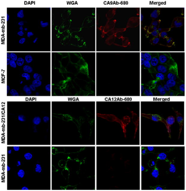
Expression of CAIX and CAXII in MDA-mb-231 cells, and verification of specific binding of CA9Ab-680 and CAX12Ab-680. Confocal micrographs of cells incubated with the nuclear marker DAPI (blue), the plasma- and cytoplasmic-membrane marker, WGA (green) and CA9 and CA12Ab-680 (red). (A) CA9Ab-680 (red) stained MDA-mb-231 cells that constitutively express CAIX, but not MCF-7 cells which do not express CAIX in normoxic conditions. (B) CA12Ab-680 (red) stained MDA-mb-231 cells engineered to express CAXII (up). CA12Ab-680 stained parental MDA-mb-231 cells that do not express CAXII. Note that CA12Ab-680 does not stain these cells (bottom). Please note that merged images show colocalization of agent (red) with membrane marker (green) indicating accumulation of agent on the cell-surface.
In vivo and ex vivo selectivity studies
To study the selectivity of the imaging probes in vivo, orthotopic mammary fat pad (MFP) xenograft models were developed. For CA9Ab-680, MDA-mb-231 cells were used to form positive (+) tumors in nude mice. Since CAIX expression can be induced under hypoxic conditions, all tumors may express CAIX, including small metastases which may not have an established vasculature. Therefore, a blocking experiment was used to determine specificity, where unlabeled CAIX mAb was added prior to CA9Ab-680. For CA12Ab-680, MDA-mb-231/CAXII cells were used to generate the positive (+) tumor in the right MFP, and parental MDA-mb-231 cells were used for the negative (-) left MFP tumor.
After tumor growth to suitable size, agent was intravenously (i.v.) injected into tumor bearing nude mice. CA9Ab-680 was retained in the CAIX positive MDA-mb-231 tumors (n=3 mice) 24 hours after injection (Fig. 3A, left panel), and after blocking with unlabeled CAIX mAb, the amount of probe retained in the tumors (n=3) was decreased significantly by 1.7±0.14 (p<0.02) times relative to the unblocked tumor. (Fig. 3A, middle panel). Following injection with CA12Ab-680 probe, the MDA-mb-231/CAXII tumors (right MFP) also retained high levels of the CA12Ab-680 probe compared to the CAXII negative xenografts (left MFP) (Fig. 3A, right panel). Fluorescence in the CAXII positive tumor was 7.0 ± 1.0 s.d. (n=3, p<0.001) fold greater compared to the negative tumor. These results demonstrate the in vivo targeting specificity of the molecular probes.
Figure 3.
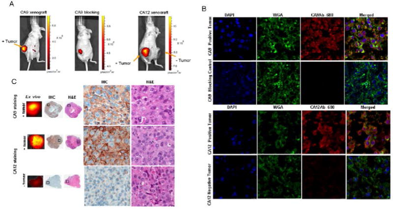
In vivo and ex vivo selectivity of CA9Ab-680 and CA12Ab-680. (A) In vivo fluorescence image of mouse bearing xenograft tumors, 24 hours post intravenous injection of agents: MDA-mb-231 cells that constitutively express CAIX were used to form the positive tumor (left panel). A blocking experiment was performed using pre-injection of 150 μg unlabeled CAIX mAb, followed by injection of 50 μg CA9Ab-680 to determine specificity of the CAIX specific probe (middle panel). The image was acquired 24 hours after injection with labeled probe. MDA-mb-231/CAXII cells were used to form the positive (+) tumor and parental cells were used as the negative (-) tumor (right panel). Tumors are indicated by orange arrows. The agents have cleared from the entire mouse, including the negative tumor at this time-point. (B) Confocal microscopy of negative and positive tumor sections stained with CA9 and CA12Ab-680 (red), membrane marker (green) and nuclear (blue) counterstain. Note the negative tumor is CAXII negative and was stained with CA12Ab-680. (C) Ex vivo images of tumors with corresponding histology (CAIX and CAXII IHC staining) of positive and negative tumors.
For further confirmation of the probes selectivities, an ex vivo analysis was performed. Sections of flash-frozen tumors were stained with the probes, nuclear stain, cell-membrane stain, and were imaged using confocal microscopy (Fig. 3B). CAIX staining was observed in the positive tumor, and was reduced by blocking with unlabled mAb. CAXII staining was only observed in tumors from the positively expressing cell line but not in the negative line (Fig. 3B). Ex vivo images of the corresponding center sections of the tumors confirmed the in vivo results and allowed comparison of probe signal relative to target expression in adjacent sections by IHC and histology (Fig. 3C).
Pharmacokinetics and biodistribution studies
To evaluate the pharmacodynamics of probe uptake and clearance in the tumors, CA9Ab-680 and CA12Ab-680 were intravenously (i.v.) injected into nude mice (n=3 for each probe) and images acquired at intervals from 5 minutes to 8 days post injection (Fig. 4A). Fluorescence signal in positive tumors increased to a maximum at 24 hours following injection of both probes, and signal slowly cleared until approximately 7 days post-injection. Fluorescence signal in CAXII negative tumors increased slightly for about 24 hours and slowly cleared over the following 5 days.
Figure 4.

Pharmacodynamics and biodistribution of CA9Ab-680 and CA12Ab-680. (A) Pharmacodynamics of agents in positive and negative tumors. Note that the peak signal in the positive CAIX and CAXII tumors is 24-hours post-injection, and that the agents are nearly cleared after 8 days. Data represent mean ± s.d. (B) Biodistribution of agents in the CAIX and CAXII tumors and organs, 24 hours and 48 hours post-injection shows predominant positive tumor localization and clearing from the organs by 48 hours post-injection. The values were normalized as percentage of the highest signal.
For biodistribution studies, mice bearing tumors (n=3) were injected with probe, and tissue distribution of fluorescence signal determined after removing tumors and organs at 24-hours and 48-hours post-administration (Fig. 4B). Twenty four hours post-injection, CA9Ab-680 and CA12Ab-680 retained at relatively high levels in the positive tumor relative to a corresponding negative tumor and other organs. However, the CAIX probe was elevated in liver, kidney and stomach and the CAXII probe was elevated in the liver and stomach. After 48 hours, the probe had nearly cleared from tissues except for positive tumors.
Detection of malignant cells in ALNs using CA9Ab-680 and CA12Ab-680 imaging probes
To investigate whether the CAIX and CAXII molecular imaging probes can be delivered through the lymphatics and are selectively retained in positive ALNs, the MDA-mb- 231/Luc (Fig. 5A) as well as MDA-mb-231/CA12/Luc (Fig. 5C) spontaneous metastasis model was used. After injecting cells in the MFP, ALN metastases were observed after 6-8 weeks by bioluminescence imaging (Fig. 5A,C). Probes were then injected peritumorally into the MFP and observed to traverse through the MFP into the lymph node within 4 hours. At 24 h post-injection, a strong fluorescence signal was obtained from the area of the ALN corresponding to a metastasis positive for the marker (Fig. 5B,D), and the probes had mostly cleared from the MFP. A specific and durable fluorescence signal was observed in target positive metastases out to at least 48 hours post-injection, long after completely clearing from the MFP. Probes were not retained in ALN metastases that did not express the target marker.
Figure 5.

Representative images of spontaneous metastases into axillary lymph node of MDA-mb-231/Luc (A,B) and MDA-mb-231/Luc/CA12 (C,D) cells and the ability of the agents to detect the tumor cells in ALN. (A,C) Bioluminescence images of luciferase activity in the primary tumor (yellow arrow) and axillary lymph node (red arrow) of MDA-mb-231/Luc (A) and MDA-mb-231/Luc/CA12 (C) xenografts. Fluorescence surface radiance from primary tumor, injection site and axillary lymph node 24 hours post peritumoral injection of CA9Ab-680 (B) and CA12Ab-680 (D).
Agent selectivity and sensitivity for positive lymph nodes was also measured using an orthotopic model of lymph node metastasis. A range in number (500 to 1 million) of different luciferase expressing breast cancer cell lines with varying levels of CAIX and CAXII expression (Supplementary Fig. S3) were directly injected into the ALN of nude mice (n=3 for each cell number group) using ultrasound image guidance (Supplementary Fig. S4). MDA-mb-231, ZR-75.1 and MCF-7 cell lines were used as high-, medium- and low-CAIX constitutively expressing cells, respectively. MDA-mb-231 cells with engineered expression, and MCF-7 and ZR-75.1 cells with constitutive expression, were used as high-, medium- and low-CAXII expressing cells, respectively.
Successful cell implantation was determined by bioluminescence imaging four hours after implantation (Supplementary Figs. S5A,C). Twenty four hours after cell implantation, CA9Ab-680 and CA12Ab-680 were delivered by MFP injection and fluorescence images were acquired 24 hours after injection (Supplementary Figs. S5B,D). Bioluminescence and fluorescence signals were quantified by drawing a region-of-interest (ROI) surrounding the tumor cells in the ALN. As expected, signal intensities for both bioluminescence and fluorescence decreased with cell number (Fig. 6). With fluorescence, the CA12Ab-680 probe detected as few as 500 high, 1000 medium and 5000 low CAXII expressing cells. CA9Ab-680 detected as few as 1000 high, 5000 medium, and did not detect the low mRNA expressing MCF-7 cells (Fig. 6B and Supplementary Fig. S6 and S5B,D). However, MCF-7 cells do not appear to express CAIX protein as determined by ICC (Fig. 2). Hence, the high range of detection was ∼500 cells and the low range of detection was ∼ 5000 cells for these probes (Supplementary Fig. S6). When CA12Ab-680 was injected into the MFP of animals that were sham injected into the axillary lymph node with matrigel and PBS, probe was not retained at the 24 hour time point (Supplementary Figs. S5B and S5D).
Figure 6.
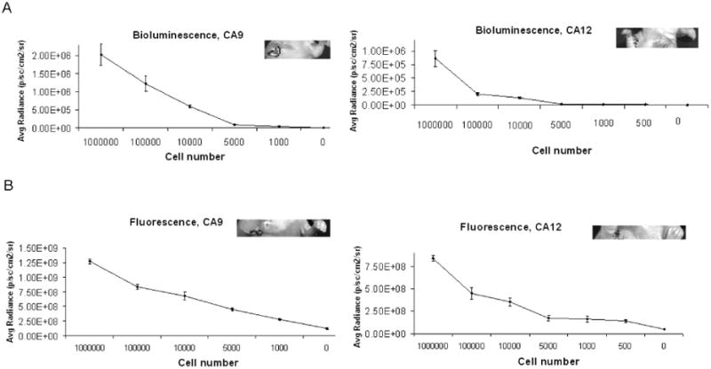
Sensitivity of CA9Ab-680 and CA12Ab-680 for detection of tumor cells in ALN. A range of positive cells (MDA-mb-231 for CAIX and MDA-mb-231 expressing CAXII for CAXII) were injected into ALN using ultrasound image guidance. (A) Bioluminescence activity quantified for a range of injected cell numbers. Insets show signal for mice injected with the lowest cell number. (B) Agent associated fluorescence for a range of cell numbers, 24 hours post-injection. Insets show fluorescence for mice injected with minimum detectable cells by agents (1000 cells for CA9Ab-680 and 500 cells for CA12Ab-680). All data represent mean ± s.d. of pixel values within the ROIs.
Discussion
Optical molecular imaging is a powerful technique for the noninvasive visualization of specific biomarkers in real-time (37). Monoclonal antibodies (mAbs) specific to membrane antigens on cancer cells, have been used for tumor targeting applications and have potential for the selective delivery of therapeutic and imaging agents.
A number of non-invasive imaging modalities for SLN evaluation have been investigated, including single photon emission computed tomography (SPECT), multiphoton microscopy, magnetic resonance imaging (MRI), optical lymphography with indocyanine green dye or multicolor quantum dots, and photoacoustic tomography (PAT) using nanoparticle-based contrast agents (12-16, 38, 39). A limitation of these approaches is the lack of targeting to bona fide tumor metastasis biomarkers. Untargeted probes distribute randomly across the SLNs with only transient and non-specific visualization of the lymphatic system (17, 18). Hence, these methods only provide anatomic maps, and do not detect tumor cells present in lymph nodes. Currently, there is no reliable method to non-invasively identify breast cancer involvement in the ALN. Therefore, there is a need to develop a non-invasive, non-ionizing and accurate detection method for the staging of breast cancer. Using mouse models of breast cancer metastasis and novel mAb-based molecular imaging agents (CA9Ab-680 and CA12Ab-680), we have developed a non-invasive targeted method for detection of ALN metastasis via fluorescence imaging.
Very recently, we reported the development of a mammaglobin-A specific mAb conjugated to a near-infrared (NIR) fluorescent dye (termed MamAb-680), to detect ALN metastases in mouse models (34). Based on our IHC study, mammaglobin-A is expressed in only 45 % of human breast cancer ALN metastases. Therefore, an aim of the current study was to identify a combination of cell-surface markers that cover 100% of breast cancer ALN metastases. By gene-expression profiling, CA12 was identified as a highly and broadly expressed marker in patient samples of lymph node metastasis. Protein expression was subsequently validated using IHC of a TMA containing ALN biopsy samples. CAXII protein is expressed in 75.5% of ALN metastases surveyed and is not expressed in unaffected lymphocytes. However, small aggregates of histiocytes were observed to express CAXII in some unaffected ALN. Others have reported CAXII as a breast cancer marker (31, 40, 41).
CAIX is a cell-surface marker expressed in malignant and invasive breast cancer (23, 42). Although, CAIX was highly expressed in only a small fraction of the breast cancer and lymph node positive samples by DNA microarray (Fig. 1 A), it was scored positive in 71% of ALN metastasis samples on our TMA, which is a direct measure of protein expression but does include weak positives. CAIX expression has been reported in 12.5% of breast cancers, where perinecrotic HIF-1α overexpression is highly associated with CAIX overexpression (43, 44), which is in agreement with our IHC scoring if we consider only moderate to strong expression (score ≥ 4). Weak CAIX expression (score <3) was observed in lymphocytes in a fraction of adjacent and unaffected ALNs, however, at least 13% of the ALN metastases had stronger positivity (Fig. 1B). Based on expression versus non-expression, the combination of CAIX and CAXII covers 100% of patients with ALN metastases represented in our TMA. However, one of these markers is strongly expressed in only 35% of the ALN metastasis samples. Regardless, we have shown that CA9Ab-680 and CA12Ab-680 imaging probes can detect cells with low-levels of marker expression.
Co-expression of these two markers in breast cancer was previously reported (30). We observed co-expression in 44% of samples. Hence, expression of these markers is not interdependent. Notably, these markers are not expressed in unaffected lymph nodes or in organs typically involved in clearance or toxicity, except for CAXII, which is highly expressed in the kidney. This should not be a serious problem for agents administered peritumorally that clear through the lymphatics, as macrophages will likely ingest them preventing circulation and clearance through the kidneys.
CAIX and CAXII are both integral plasma membrane proteins with large extracellular components that are accessible for binding of targeted imaging probes. According to our IHC results, both markers are expressed in normal breast tissue, however, their distribution is limited to the epithelia, which is not accessible to large (e.g. mAb) probes injected into the parenchyma. Furthermore, a large fraction of the expression in normal breast epithelia appeared to be cytoplasmic.
Expression of both CAIX and CAXII can be induced by hypoxia (20). Chiche et al. have reported that both CAIX and CAXII are functionally involved in tumor growth, possibly by regulating intracellular pH in the context of tumor acidosis (45). However, CAIX is known to be constitutively expressed in advanced breast cancers and metastases by a hypoxia independent mechanism (29). Several studies have shown that CAIX expression is associated with a negative prognosis, including resistance to chemo- and radiation-therapy in breast cancer (22, 23, 26) and is associated with increased recurrence and reduced survival (24, 46). In contrast, CAXII has been associated with a good prognosis in invasive breast cancer (40).
Another CAIX-targeted molecular imaging agent has been developed using the chimeric anti-CAIX antibody cG250 radiolabeled with 124I. This probe has completed phase I testing and is currently under a multi-center clinical assessment as a PET diagnostic agent for clear-cell renal carcinoma (47). Hence, there is potential to adapt 124I-cG250 imaging for the detection of CAIX expressing breast cancer ALN metastases. The radioactive half-life of 124I is 4.18 days.
Peak tumor accumulation of both CA9Ab-680 and CA12Ab-680 probes occurred 24 hours post i.v. injection. However, positive lymph nodes were detected by both probes as early as 4 hours post administration. Since these antibody-based agents are relatively large compared to synthetic peptide-based ligands, the time required for ALN uptake and clearance could be improved by the development of a smaller ligands or peptidabodies specific for the surface epitopes or for the carbonic anhydrase catalytic site. However, Carlin et al., have reported that smaller fragments of cG250 antibody are unlikely to have any additional benefits for the imaging of tumor CAIX with 124I-cG250 because of observed lower absolute uptake into areas of high CAIX expression, possibly due to decreased circulation time of the smaller fragment-based probes (48). However, for the purpose of imaging metastases, decreased size will likely increase transit time through the MFP lymphatics to the ALN providing improved timing of optimal imaging after administration of probe.
An orthotopic xenograft model for lymph node metastasis was used to determine the sensitivity of detection, by implanting precise numbers of cells with varying expression levels of CAIX and CAXII based on qRT-PCR. As few as 500 (high CAXII expression) and 5000 (low CAXII expression) cells were quantitatively detected in the ALN using the CAXII specific probe, whereas 1000 (high CAIX expression) and 5000 (low CAIX expression) cells were the limits of detection for the CAIX probe. Metastatic disease is classified as macrometastasis (> 2mm), micrometastasis (between 0.2 and 2 mm) or individual tumor cells or groups of cells (ITCs, <0.2 mm) (49). The smallest micrometastasis will have >1000 cells which is below the limit of detection for the low marker expressing cells, but is above the detection limit for higher expressing markers. Hence, for this approach to work, a combination of markers will need to be identified where at least one will be expressed at high enough levels to allow detection. Also, the lower limit of detection may be further optimized to allow for detection of lower numbers of cells.
In the future, this method may be translated for use in the clinic through use of a fluorescent dye with longer excitation and emission wavelengths for decreased tissue absorbance and by using fluorescence molecular tomography (FMT) (50), or photoacoustic tomography. This method may also be compatible with other more established imaging modalities such as detection of positron or single photon emissions, or MRI. To add a therapeutic component, cancer targeted agents useful for PAT may also be used for photothermal ablation of cells bound by probe (51). Since CAIX and XII are known to promote tumor growth (45), therapeutic capability could be added by developing theragnostic agents capable of detection by noninvasive imaging and of treatment by inhibition of carbonic anhydrase activity. To accomplish this, inhibitors specific for the CAIX and CAXII isoforms, need to be developed (52). Alternatively, inhibitor agents may be developed that do not cross the cell membrane, limiting the antagonist effects to carbonic anhydrases with extracellular active sites (53). Fluorescently labeled and membrane-impermeable CA inhibitors have been reported (54).
Invisible NIR fluorescent light can provide high resolution and sensitivity for real-time intraoperative image-guidance during surgery (55). Troyan et al., have reported image-guided sentinel lymph node surgery using NIR dye in a patient with breast cancer. Our fluorescent targeted agents can offer the additional opportunity to intraoperatively guide resection to remove involved nodes and leave uninvolved nodes (50, 55).
In conclusion, we have shown that either CAIX or CAXII are expressed in 100% of the breast cancer lymph node metastasis samples surveyed in this study. It is not yet known if these two markers will be sufficient for coverage of all clinical LN metastases, or if additional markers will be needed. We continue to study additional markers, including mammaglobin-A (34) and the other markers identified by expression profiling in this study. Also, low expression of CAIX in some unaffected lymphocytes and expression of CAIX and CAXII in small aggregates of histiocytes in unaffected ALN could lead to some amount of false positives. However, some level of false positives may be acceptable by this method, as currently 74% of SLN biopsies are negative. CAIX and CAXII targeted molecular imaging probes were developed for non-invasive in vivo imaging and detection of breast cancer metastases in ALNs using small animal models. These imaging probes detected tumor cells in ALNs with high sensitivity. This targeted imaging strategy has potential for future translation into the clinic for ALN assessment and intraoperative surgical guidance as well as monitoring alteration in CAIX/CAXII expression as an indicator of treatment response. In the future, the agents may be improved by development of small targeting peptides and agents with theragnostic capability.
Supplementary Material
Statement of Translational Relevance.
Determining the presence of lymph node (LN) metastasis is critical for breast cancer staging. Sentinel lymph node (SLN) biopsy is the standard of care, where the SLN is identified by injection of tracer, surgically removed and examined pathologically. However, SLN biopsy is associated with morbidities.
Herein, we have developed targeted fluorescent molecular imaging probes for the non-invasive detection of breast cancer LN metastasis. We report target expression (CAIX or CAXII) in 100% of axillary lymph node samples surveyed and the sensitive detection of lymph node metastases in animal models using these probes. In patients, this approach could greatly decrease or even eliminate unnecessary surgery, significantly reducing associated morbidities in breast cancer patients. This has potential for great impact since nearly all women that present with a primary breast tumor undergo SLN biopsy for staging and it has been reported that the majority (74%) are pathologically negative.
Acknowledgments
The authors wish to acknowledge the following shared resources at the H. Lee Moffitt Cancer Center & Research Institute for their contributions to this work: Analytic Microscopy, Tissue Core, Microarray Core, Comparative Biomedicine, and Small Animal Modeling and Imaging.
Grant Support: University of Florida/Moffitt Cancer Center Collaborative Initiative Grant, UF 69-15540-01-01, University of Florida, Gainesville, FL.
Footnotes
Disclosure of Potential Conflicts of Interest: No potential conflicts of interest were disclosed.
References
- 1.Stacker SA, Achen MG, Jussila L, Baldwin ME, Alitalo K. Lymphangiogenesis and cancer metastasis. Nat Rev Cancer. 2002;2:573–83. doi: 10.1038/nrc863. [DOI] [PubMed] [Google Scholar]
- 2.Tafreshi NK, Kumar V, Morse DL, Gatenby RA. Molecular and functional imaging of breast cancer. Cancer Control. 2010;17:143–55. doi: 10.1177/107327481001700302. [DOI] [PubMed] [Google Scholar]
- 3.Albertini JJ, Lyman GH, Cox C, Yeatman T, Balducci L, Ku N, et al. Lymphatic mapping and sentinel node biopsy in the patient with breast cancer. JAMA. 1996;276:1818–22. [PubMed] [Google Scholar]
- 4.Giuliano AE, Kirgan DM, Guenther JM, Morton DL. Lymphatic Mapping and Sentinel Lymphadenectomy for Breast-Cancer. Annals of Surgery. 1994;220:391–401. doi: 10.1097/00000658-199409000-00015. [DOI] [PMC free article] [PubMed] [Google Scholar]
- 5.Krag DN, Weaver DL, Alex JC, Fairbank JT. Surgical resection and radiolocalization of the sentinel lymph node in breast cancer using a gamma probe. Surg Oncol. 1993;2:335–9. doi: 10.1016/0960-7404(93)90064-6. discussion 40. [DOI] [PubMed] [Google Scholar]
- 6.Douglas-Jones AG, Woods V. Molecular assessment of sentinel lymph node in breast cancer management. Histopathology. 2009;55:107–13. doi: 10.1111/j.1365-2559.2008.03218.x. [DOI] [PubMed] [Google Scholar]
- 7.Krag D, Weaver D, Ashikaga T, Moffat F, Klimberg VS, Shriver C, et al. The sentinel node in breast cancer--a multicenter validation study. N Engl J Med. 1998;339:941–6. doi: 10.1056/NEJM199810013391401. [DOI] [PubMed] [Google Scholar]
- 8.McMasters KM, Tuttle TM, Carlson DJ, Brown CM, Noyes RD, Glaser RL, et al. Sentinel lymph node biopsy for breast cancer: a suitable alternative to routine axillary dissection in multi-institutional practice when optimal technique is used. J Clin Oncol. 2000;18:2560–6. doi: 10.1200/JCO.2000.18.13.2560. [DOI] [PubMed] [Google Scholar]
- 9.Ung OA. Australasian experience and trials in sentinel lymph node biopsy: the RACS SNAC trial. Asian J Surg. 2004;27:284–90. doi: 10.1016/S1015-9584(09)60052-X. [DOI] [PubMed] [Google Scholar]
- 10.Purushotham AD, Upponi S, Klevesath MB, Bobrow L, Millar K, Myles JP, et al. Morbidity after sentinel lymph node biopsy in primary breast cancer: results from a randomized controlled trial. J Clin Oncol. 2005;23:4312–21. doi: 10.1200/JCO.2005.03.228. [DOI] [PubMed] [Google Scholar]
- 11.Krag DN, Anderson SJ, Julian TB, Brown AM, Harlow SP, Ashikaga T, et al. Technical outcomes of sentinel-lymph-node resection and conventional axillary-lymph-node dissection in patients with clinically node-negative breast cancer: results from the NSABP B-32 randomised phase III trial. Lancet Oncol. 2007;8:881–8. doi: 10.1016/S1470-2045(07)70278-4. [DOI] [PubMed] [Google Scholar]
- 12.Kim JW, Galanzha EI, Shashkov EV, Moon HM, Zharov VP. Golden carbon nanotubes as multimodal photoacoustic and photothermal high-contrast molecular agents. Nat Nanotechnol. 2009;4:688–94. doi: 10.1038/nnano.2009.231. [DOI] [PMC free article] [PubMed] [Google Scholar]
- 13.Kobayashi H, Ogawa M, Kosaka N, Choyke PL, Urano Y. Multicolor imaging of lymphatic function with two nanomaterials: quantum dot-labeled cancer cells and dendrimer-based optical agents. Nanomedicine (Lond) 2009;4:411–9. doi: 10.2217/nnm.09.15. [DOI] [PMC free article] [PubMed] [Google Scholar]
- 14.Ogata F, Azuma R, Kikuchi M, Koshima I, Morimoto Y. Novel lymphography using indocyanine green dye for near-infrared fluorescence labeling. Ann Plast Surg. 2007;58:652–5. doi: 10.1097/01.sap.0000250896.42800.a2. [DOI] [PubMed] [Google Scholar]
- 15.Pan D, Pramanik M, Senpan A, Ghosh S, Wickline SA, Wang LV, et al. Near infrared photoacoustic detection of sentinel lymph nodes with gold nanobeacons. Biomaterials. 2010;31:4088–93. doi: 10.1016/j.biomaterials.2010.01.136. [DOI] [PMC free article] [PubMed] [Google Scholar]
- 16.Song KH, Kim C, Cobley CM, Xia Y, Wang LV. Near-infrared gold nanocages as a new class of tracers for photoacoustic sentinel lymph node mapping on a rat model. Nano Lett. 2009;9:183–8. doi: 10.1021/nl802746w. [DOI] [PMC free article] [PubMed] [Google Scholar]
- 17.Galanzha EI, Kokoska MS, Shashkov EV, Kim JW, Tuchin VV, Zharov VP. In vivo fiber-based multicolor photoacoustic detection and photothermal purging of metastasis in sentinel lymph nodes targeted by nanoparticles. J Biophotonics. 2009;2:528–39. doi: 10.1002/jbio.200910046. [DOI] [PMC free article] [PubMed] [Google Scholar]
- 18.McElroy M, Hayashi K, Garmy-Susini B, Kaushal S, Varner JA, Moossa AR, et al. Fluorescent LYVE-1 antibody to image dynamically lymphatic trafficking of cancer cells in vivo. J Surg Res. 2009;151:68–73. doi: 10.1016/j.jss.2007.12.769. [DOI] [PMC free article] [PubMed] [Google Scholar]
- 19.Swietach P, Wigfield S, Cobden P, Supuran CT, Harris AL, Vaughan-Jones RD. Tumor-associated carbonic anhydrase 9 spatially coordinates intracellular pH in three-dimensional multicellular growths. J Biol Chem. 2008;283:20473–83. doi: 10.1074/jbc.M801330200. [DOI] [PubMed] [Google Scholar]
- 20.Wykoff CC, Beasley NJ, Watson PH, Turner KJ, Pastorek J, Sibtain A, et al. Hypoxia-inducible expression of tumor-associated carbonic anhydrases. Cancer Res. 2000;60:7075–83. [PubMed] [Google Scholar]
- 21.Pastorekova S, Parkkila S, Pastorek J, Supuran CT. Carbonic anhydrases: current state of the art, therapeutic applications and future prospects. J Enzyme Inhib Med Chem. 2004;19:199–229. doi: 10.1080/14756360410001689540. [DOI] [PubMed] [Google Scholar]
- 22.Brennan DJ, Jirstrom K, Kronblad A, Millikan RC, Landberg G, Duffy MJ, et al. CA IX is an independent prognostic marker in premenopausal breast cancer patients with one to three positive lymph nodes and a putative marker of radiation resistance. Clin Cancer Res. 2006;12:6421–31. doi: 10.1158/1078-0432.CCR-06-0480. [DOI] [PubMed] [Google Scholar]
- 23.Chia SK, Wykoff CC, Watson PH, Han C, Leek RD, Pastorek J, et al. Prognostic significance of a novel hypoxia-regulated marker, carbonic anhydrase IX, in invasive breast carcinoma. J Clin Oncol. 2001;19:3660–8. doi: 10.1200/JCO.2001.19.16.3660. [DOI] [PubMed] [Google Scholar]
- 24.Colpaert CG, Vermeulen PB, Fox SB, Harris AL, Dirix LY, Van Marck EA. The presence of a fibrotic focus in invasive breast carcinoma correlates with the expression of carbonic anhydrase IX and is a marker of hypoxia and poor prognosis. Breast Cancer Res Treat. 2003;81:137–47. doi: 10.1023/A:1025702330207. [DOI] [PubMed] [Google Scholar]
- 25.Hussain SA, Ganesan R, Reynolds G, Gross L, Stevens A, Pastorek J, et al. Hypoxia-regulated carbonic anhydrase IX expression is associated with poor survival in patients with invasive breast cancer. Br J Cancer. 2007;96:104–9. doi: 10.1038/sj.bjc.6603530. [DOI] [PMC free article] [PubMed] [Google Scholar]
- 26.Span PN, Bussink J, Manders P, Beex LV, Sweep CG. Carbonic anhydrase-9 expression levels and prognosis in human breast cancer: association with treatment outcome. Br J Cancer. 2003;89:271–6. doi: 10.1038/sj.bjc.6601122. [DOI] [PMC free article] [PubMed] [Google Scholar]
- 27.Trastour C, Benizri E, Ettore F, Ramaioli A, Chamorey E, Pouyssegur J, et al. HIF-1alpha and CA IX staining in invasive breast carcinomas: prognosis and treatment outcome. Int J Cancer. 2007;120:1451–8. doi: 10.1002/ijc.22436. [DOI] [PubMed] [Google Scholar]
- 28.Supuran CT, Scozzafava A. Carbonic anhydrases as targets for medicinal chemistry. Bioorg Med Chem. 2007;15:4336–50. doi: 10.1016/j.bmc.2007.04.020. [DOI] [PubMed] [Google Scholar]
- 29.Robey IF, Lien AD, Welsh SJ, Baggett BK, Gillies RJ. Hypoxia-inducible factor-1alpha and the glycolytic phenotype in tumors. Neoplasia. 2005;7:324–30. doi: 10.1593/neo.04430. [DOI] [PMC free article] [PubMed] [Google Scholar]
- 30.Goonewardene TI, Sowter HM, Harris AL. Hypoxia-induced pathways in breast cancer. Microsc Res Tech. 2002;59:41–8. doi: 10.1002/jemt.10175. [DOI] [PubMed] [Google Scholar]
- 31.Ivanov S, Liao SY, Ivanova A, Danilkovitch-Miagkova A, Tarasova N, Weirich G, et al. Expression of hypoxia-inducible cell-surface transmembrane carbonic anhydrases in human cancer. Am J Pathol. 2001;158:905–19. doi: 10.1016/S0002-9440(10)64038-2. [DOI] [PMC free article] [PubMed] [Google Scholar]
- 32.Baggett B, Roy R, Momen S, Morgan S, Tisi L, Morse D, et al. Thermostability of firefly luciferases affects efficiency of detection by in vivo bioluminescence. Mol Imaging. 2004;3:324–32. doi: 10.1162/15353500200403178. [DOI] [PubMed] [Google Scholar]
- 33.Morse DL, Carroll D, Weberg L, Borgstrom MC, Ranger-Moore J, Gillies RJ. Determining suitable internal standards for mRNA quantification of increasing cancer progression in human breast cells by real-time reverse transcriptase polymerase chain reaction. Anal Biochem. 2005;342:69–77. doi: 10.1016/j.ab.2005.03.034. [DOI] [PubMed] [Google Scholar]
- 34.Tafreshi NK, Enkemann SA, Bui MM, Lloyd MC, Abrahams D, Huynh AS, et al. A mammaglobin-A targeting agent for noninvasive detection of breast cancer metastasis in lymph nodes. Cancer Res. 2011;71:1050–9. doi: 10.1158/0008-5472.CAN-10-3091. [DOI] [PMC free article] [PubMed] [Google Scholar]
- 35.Schlauder SM, Steffensen TS, Morgan M, Letson DG, Pledger WJ, Ma L, et al. Assessment of muscarinic and nicotinic acetylcholine receptor expression in primitive neuroectodermal tumor/ewing family of tumor and desmoplastic small round cell tumor: an immunohistochemical and Western blot study of tissue microarray and cell lines. Fetal Pediatr Pathol. 2008;27:83–97. doi: 10.1080/15513810802077529. [DOI] [PubMed] [Google Scholar]
- 36.Li Y, Wang H, Oosterwijk E, Tu C, Shiverick KT, Silverman DN, et al. Expression and activity of carbonic anhydrase IX is associated with metabolic dysfunction in MDA-MB-231 breast cancer cells. Cancer Invest. 2009;27:613–23. doi: 10.1080/07357900802653464. [DOI] [PMC free article] [PubMed] [Google Scholar]
- 37.Massoud TF, Gambhir SS. Molecular imaging in living subjects: seeing fundamental biological processes in a new light. Genes Dev. 2003;17:545–80. doi: 10.1101/gad.1047403. [DOI] [PubMed] [Google Scholar]
- 38.Kitai T, Inomoto T, Miwa M, Shikayama T. Fluorescence navigation with indocyanine green for detecting sentinel lymph nodes in breast cancer. Breast Cancer. 2005;12:211–5. doi: 10.2325/jbcs.12.211. [DOI] [PubMed] [Google Scholar]
- 39.Tagaya N, Yamazaki R, Nakagawa A, Abe A, Hamada K, Kubota K, et al. Intraoperative identification of sentinel lymph nodes by near-infrared fluorescence imaging in patients with breast cancer. Am J Surg. 2008;195:850–3. doi: 10.1016/j.amjsurg.2007.02.032. [DOI] [PubMed] [Google Scholar]
- 40.Watson PH, Chia SK, Wykoff CC, Han C, Leek RD, Sly WS, et al. Carbonic anhydrase XII is a marker of good prognosis in invasive breast carcinoma. Br J Cancer. 2003;88:1065–70. doi: 10.1038/sj.bjc.6600796. [DOI] [PMC free article] [PubMed] [Google Scholar]
- 41.Wykoff CC, Beasley N, Watson PH, Campo L, Chia SK, English R, et al. Expression of the hypoxia-inducible and tumor-associated carbonic anhydrases in ductal carcinoma in situ of the breast. Am J Pathol. 2001;158:1011–9. doi: 10.1016/S0002-9440(10)64048-5. [DOI] [PMC free article] [PubMed] [Google Scholar]
- 42.Bartosova M, Parkkila S, Pohlodek K, Karttunen TJ, Galbavy S, Mucha V, et al. Expression of carbonic anhydrase IX in breast is associated with malignant tissues and is related to overexpression of c-erbB2. J Pathol. 2002;197:314–21. doi: 10.1002/path.1120. [DOI] [PubMed] [Google Scholar]
- 43.Vleugel MM, Greijer AE, Shvarts A, van der Groep P, van Berkel M, Aarbodem Y, et al. Differential prognostic impact of hypoxia induced and diffuse HIF-1alpha expression in invasive breast cancer. J Clin Pathol. 2005;58:172–7. doi: 10.1136/jcp.2004.019885. [DOI] [PMC free article] [PubMed] [Google Scholar]
- 44.Vleugel MM, Shvarts D, van der Wall E, van Diest PJ. p300 and p53 levels determine activation of HIF-1 downstream targets in invasive breast cancer. Hum Pathol. 2006;37:1085–92. doi: 10.1016/j.humpath.2006.03.015. [DOI] [PubMed] [Google Scholar]
- 45.Chiche J, Ilc K, Laferriere J, Trottier E, Dayan F, Mazure NM, et al. Hypoxia-inducible carbonic anhydrase IX and XII promote tumor cell growth by counteracting acidosis through the regulation of the intracellular pH. Cancer Res. 2009;69:358–68. doi: 10.1158/0008-5472.CAN-08-2470. [DOI] [PubMed] [Google Scholar]
- 46.Kilbride KE, Lee MC, Nees AV, Cimmino VM, Diehl KM, Sabel MS, et al. Axillary staging prior to neoadjuvant chemotherapy for breast cancer: predictors of recurrence. Ann Surg Oncol. 2008;15:3252–8. doi: 10.1245/s10434-008-0136-3. [DOI] [PubMed] [Google Scholar]
- 47.Divgi CR, Pandit-Taskar N, Jungbluth AA, Reuter VE, Gonen M, Ruan S, et al. Preoperative characterisation of clear-cell renal carcinoma using iodine-124-labelled antibody chimeric G250 (124I-cG250) and PET in patients with renal masses: a phase I trial. Lancet Oncol. 2007;8:304–10. doi: 10.1016/S1470-2045(07)70044-X. [DOI] [PubMed] [Google Scholar]
- 48.Carlin S, Khan N, Ku T, Longo VA, Larson SM, Smith-Jones PM. Molecular targeting of carbonic anhydrase IX in mice with hypoxic HT29 colorectal tumor xenografts. PLoS One. 2010;5:e10857. doi: 10.1371/journal.pone.0010857. [DOI] [PMC free article] [PubMed] [Google Scholar]
- 49.Green FL. AJCC cancer staging handbook-TNM classification of malignant tumors. New York: Springer; 2002. [Google Scholar]
- 50.Sevick-Muraca EM, Sharma R, Rasmussen JC, Marshall MV, Wendt JA, Pham HQ, et al. Imaging of lymph flow in breast cancer patients after microdose administration of a near-infrared fluorophore: feasibility study. Radiology. 2008;246:734–41. doi: 10.1148/radiol.2463070962. [DOI] [PMC free article] [PubMed] [Google Scholar]
- 51.Sharma P, Brown SC, Bengtsson N, Zhang Q, Walter GA, Grobmyer SR, et al. Gold-Speckled Multimodal Nanoparticles for Noninvasive Bioimaging. Chem Mater. 2008;20:6087–94. doi: 10.1021/cm801020s. [DOI] [PMC free article] [PubMed] [Google Scholar]
- 52.Supuran CT, Scozzafava A. Carbonic anhydrase inhibitors: aromatic sulfonamides and disulfonamides act as efficient tumor growth inhibitors. J Enzyme Inhib. 2000;15:597–610. doi: 10.3109/14756360009040713. [DOI] [PubMed] [Google Scholar]
- 53.Thiry A, Supuran CT, Masereel B, Dogne JM. Recent developments of carbonic anhydrase inhibitors as potential anticancer drugs. J Med Chem. 2008;51:3051–6. doi: 10.1021/jm701526d. [DOI] [PubMed] [Google Scholar]
- 54.Dubois L, Douma K, Supuran CT, Chiu RK, van Zandvoort MA, Pastorekova S, et al. Imaging the hypoxia surrogate marker CA IX requires expression and catalytic activity for binding fluorescent sulfonamide inhibitors. Radiother Oncol. 2007;83:367–73. doi: 10.1016/j.radonc.2007.04.018. [DOI] [PubMed] [Google Scholar]
- 55.Troyan SL, Kianzad V, Gibbs-Strauss SL, Gioux S, Matsui A, Oketokoun R, et al. The FLARE intraoperative near-infrared fluorescence imaging system: a first-in-human clinical trial in breast cancer sentinel lymph node mapping. Ann Surg Oncol. 2009;16:2943–52. doi: 10.1245/s10434-009-0594-2. [DOI] [PMC free article] [PubMed] [Google Scholar]
Associated Data
This section collects any data citations, data availability statements, or supplementary materials included in this article.