Abstract
Purpose:
The Farnsworth-Munsell (FM) 100-hue test is well known but is also time consuming, especially its analytical component. To reduce this needless time-waste during precious working hours, a simple modification was devised.
Design:
Prospective, comparative, observational study.
Materials and Methods:
A transparent clear plastic carrier box replaced the opaque one, allowing ready digital photodocumentation of top and bottom without even opening the box, or handling/inverting the caps -200 reportedly normals and 50 known color vision defectives could be easily tested on this modified-FM and results stored, allowing rapid turnover. The captured scores with patient ID were analyzed, at leisure, outside hospital time, saving 45-60 minutes/patient. After recording, the box was promptly handed over to the next subject for rearrangement. Times taken for test/patient were recorded.
Results:
Running time was reduced from 60-75 min to ~15 min/patient with no waste of invaluable lab hours. Turnover time is limited to capturing two photographs (~60 sec). The box is relatively cheap and easy to maintain.
Conclusions:
Our simplified FM 100-hue test allowed rapid assessment of color visions with easy data storage of both top and bottom.
Keywords: Color vision, color vision testing, FM 100-Hue, Ishihara
The Farnsworth-Munsell 100-hue test is a psycho-technical arrangement test designed to test hue discrimination among people with normal color vision and to measure the areas of color confusion in color-defective observers. Clinically, the test has been used in conjunction with the Nagel anomaloscope to classify color vision abnormalities.[1] This test can be applied to patients afflicted with either congenital or acquired color deficiencies.[2]
The FM 100- hue test was initiated and popularized by Farnsworth in the early 1940s – since then, various tests have been developed such as Farnsworth-Munsell Dichotomous D-15 or Panel D-15 test, Lanthony Desaturated D-15 and Adams Desaturated D-15.[3,4,5,6,7]
Administration
The test consists of 85 movable color samples arranged in four opaque boxes of 22 colors in the first box and 21 colors in each of the remaining 3 [no. 85-21, 22-42, 43-63, and 64-84]. The hue samples were designed to represent perceptually equal steps of hue and to form a natural hue circle. The colors are set in plastic caps and subtend 1.5° at 50 cm. They are numbered on the back according to the correct color order of the hue instruction manual and scoring sheets are provided. One box is presented at a time. The examiner prearranges the caps in random order on the upper lid of the box. The observer is instructed to “arrange the caps in order according to color” in the lower tray [with a see-through bottom], where the two fixed caps appear. The box is presented for each eye at a comfortable distance under illuminate C providing at least 270 lux of natural light. The generally recommended time for arranging each panel is 2 minutes. For each panel, the time spent on it is recorded, depending on the first panel, second panel, etc.
Scoring
Errors are made whenever caps are misplaced from the correct order. Error scores are calculated according to the distance between any two caps. Score of a cap is the sum of the differences between the number of that cap and the numbers of the caps adjacent to it on either side, e.g., if cap number 50 is wrongly positioned say between 55 and 56, then the score of this cap is 55-50 + 56 − 50 = 5 + 6 = 11. The score of each cap is plotted on a circular graph provided. The error score is the score with 2 subtracted –i.e., 11 − 2 = 9. [If this cap no. 50 had been correctly positioned, then the score of that cap would have been 50 − 49 + 51 − -50 = 2: and with 2 subtracted, its error score would have been zero]. Sum of the error scores of the entire set of caps goes to make the total error score (TES). By plotting the scores graphically, characteristic patterns are obtained in specific defects.
Manual scoring of error scores and plotting graphs are of course extremely time consuming and very tedious. To overcome this, various computer-based [but expensive] methods have been developed.[8] Our attempt was to save needless time-wastage during precious hospital working hours, and increase maximum patient turnover in the limited time available to the professional specialist.
Materials and Methods
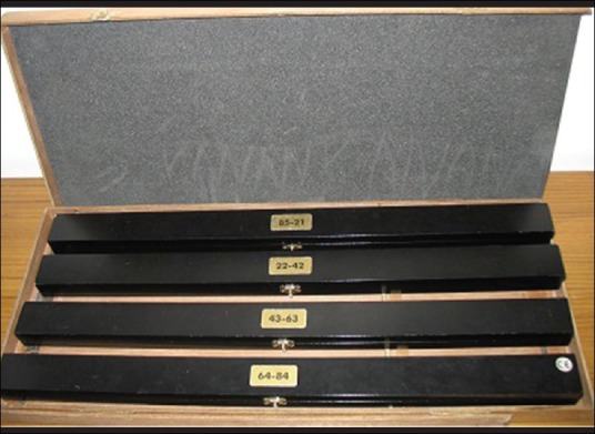
The original FM 100-hue carrier box consists of a wooden cover with 4 elongated boxes inside it. This is an opaque outer cover, so there is obvious difficulty in photo-documentation. One had to manually record the scores of caps on sheets, which hugely prolonged the time spent per patient [Fig. 1].
Figure 1.

The original FM 100-hue elongated boxes with an opaque wooden cover
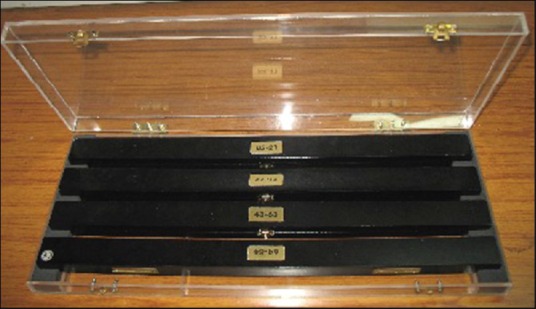
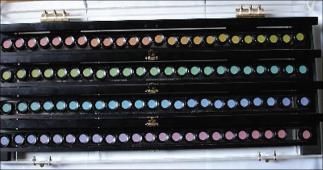
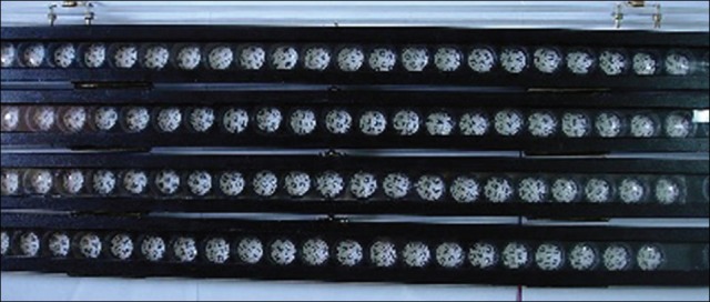
We modified the FM 100-hue procedure by converting the outer opaque box into a transparent clear plastic carrier container that allowed a ready photo-documentation of the ‘arranged’ open trays from the top and bottom without even opening the box or inverting the caps [Fig. 2]. (The inner trays already have a flip-top opaque cover, but a see-through bottom) [Figs. 3 and 4]. We screened 200 reportedly normal and 50 known color defectives using this simply modified FM 100 -hue test, first OD then OS. After capturing the digital images from both the bottom and top of the ‘arranged’ FM 100-hue box with patient ID, the [used] box was promptly handed over to the next subject for rearrangement. Scores were analyzed, at leisure, outside hospital time. Time taken by patient for each eye per test was noted. Written informed consents were taken from all subjects. Our study followed the tenets of the Declaration of Helsinki and was duly approved by our Institutional Ethics Committee.
Figure 2.

Our modified transparent carrier box for the FM-100 hue test
Figure 3.

The caps arranged by a reportedly normal subject, according to hue perceived [photographed through the transparent plastic top cover] - the arrangement is almost normal but slightly defective
Figure 4.

Showing arranged numbers imaged from the bottom through the transparent plastic [along with the Patient ID] - note the slight defects
Results
The immediate test running time for each subject including the technician's measuring and plotting time was reduced from 60-75 min to ~15 min with no waste of invaluable lab hours. Turnover time was limited to only capturing two photographs (~60 secs). The box is relatively cheap and easy to maintain, and also replaces whenever required [Figs. 3 and 4].
These numbers on the caps are normally not visible because of the original opaque cover – evidently, the investigator is standing by to observe that the patient does not turn over the entire box to peep at the numbers of the color buttons.
Conclusion
Nowadays, the Farnsworth Munsell 100-Hue Test often includes Windows based scoring software, which calculates a numerical score and provides a graphic display and printout of the subject's score and status.[2,3,4,5,6,7,8] Our simple modification and adjustment of the original box of the FM 100-hue test alone proved to be not just cost-effective but also permitted rapid assessment of color vision and quick patient turnover with easy data storage, without the need of any expensive scoring software with a requirement of a routine camera, preferably digital, for easy retrieval. It could also be very useful for telescreening and quick comparisons.
Acknowledgement
Mr. J. P Singh, Chief Technical Officer, RPC, AIIMS.
Footnotes
Source of Support: Nil
Conflict of Interest: None declared
References
- 1.Ghose S, Shrey D, Singh JP, Venkatesh P, Dada T, Ravi AK, et al. Hyderabad: Proc 65 th All India Ophthalmol (AIOS) Conference; 2007. Comparative evaluation of Colour Vision (CVn) tests and Colour Perception (CP) in actual work conditions among normals and known Colour Deficients (CDf) p. 328. [Google Scholar]
- 2.Cooper H, Bener A. Application of a laserJet printer to plot the Farnsworth-Munsell 100-Hue colour test. Optom Vis Sci. 1990;67:372–6. doi: 10.1097/00006324-199005000-00013. [DOI] [PubMed] [Google Scholar]
- 3.Farnsworth D. The Farnsworth-Munsell 100 hue and Dichotomous tests for colour vision. J Opt Soc Am. 1943;33:568–78. [Google Scholar]
- 4.Donaldson GB. Instrumentation for the Farnsworth-Munsell 100-Hue test. J Opt Soc Am. 1977;67:248–9. doi: 10.1364/josa.67.000248. [DOI] [PubMed] [Google Scholar]
- 5.Birch J. A method of qualitative scoring of the Farnsworth D-15 panel. Acta Ophthalmol. 1982;60:907–16. doi: 10.1111/j.1755-3768.1982.tb00621.x. [DOI] [PubMed] [Google Scholar]
- 6.Good GW, Schepler A, Nichols JJ. The reliability of the lanthony desaturated D-15 test. Optom Vis Sci. 2005;82:1054–9. doi: 10.1097/01.opx.0000192351.63069.4a. [DOI] [PubMed] [Google Scholar]
- 7.Dain SJ. Clinical colour vision tests. Clin Exp Optom. 2004;87:276–93. doi: 10.1111/j.1444-0938.2004.tb05057.x. [DOI] [PubMed] [Google Scholar]
- 8.Melamud A, Simpson E, Traboulsi EI. Introducing a new computer-based test for the clinical evaluation of color discrimination. Am J Ophthalmol. 2006;142:953–60. doi: 10.1016/j.ajo.2006.07.027. [DOI] [PubMed] [Google Scholar]


