

Abstract
Successful navigation requires interactions among multiple but overlapping neural pathways mediating distinct capabilities, including egocentric (self-oriented, route-based) and allocentric (spatial, map-based) learning. Route-based navigation has been shown to be impaired following acute exposure to the dopaminergic (DA) drugs (+)-methamphetamine and (+)-amphetamine, but not the serotoninergic (5-HT) drugs (±)-3,4-methylenedioxymethamphetamine or (±)-fenfluramine. The dopaminergic-rich neostriatum is involved in both allocentric and egocentric navigation. This experiment tested whether dorsal striatal DA loss using bilateral 6-hydroxydopamine (6-OHDA) injections impaired one or both types of navigation. Two weeks following 6-OHDA injections, rats began testing in the Cincinnati water maze (CWM) followed by the Morris water maze (MWM) for route-based and spatial navigation, respectively. 6-OHDA treatment significantly increased latency and errors in the CWM and path length, latency, and cumulative distance in the MWM with no difference on cued MWM trials. Neostriatal DA levels were reduced by 80% at 2 and 7 weeks post-treatment. In addition, 6-OHDA increased DA turnover and decreased norepinephrine (NE) levels. 6-OHDA injections did not alter monoamine levels in the prefrontal cortex. The data support that neostriatal DA modulates both types of navigation.
Keywords: Dopamine, Route-based learning, Spatial learning, 6-hydroxydopamine, Egocentric, Allocentric, Striatum
Introduction
Impairments in navigational ability are present in numerous human conditions including Huntington’s disease, Alzheimer’s disease, schizophrenia, Parkinson’s disease, stroke, traumatic brain injury, as well as during normal aging (Aguirre and D’Esposito, 1999;Iaria et al., 2009;Laczo et al., 2009;Livingstone and Skelton, 2007;Sanders et al., 2008;Weniger and Irle, 2006). Successful navigation requires complex interactions among multiple distinct, but parallel cognitive processes that can be subdivided into egocentric (self-oriented) and allocentric (spatial, map-based) wayfinding. In the allocentric process, the navigator’s spatial orientation to distal cues in the environment is fluid and represented in a common coordinate map system external to the navigator (Byrne, 1982;Garber, 2000). Spatial learning is frequently studied in rodents using the Morris water maze (MWM), and acquisition of the place navigation task is dependent on the hippocampus (Kesner, 1990;Morris, 1981;Morris et al., 1982;Sutherland et al., 1983), although other regions also influence the process. For example, MWM learning is sensitive to damage to cortical regions including frontal, cingulate, and parietal areas (Galani et al., 2002;Kesner et al., 1989;Whishaw et al., 2001), as well as the neostriatum (Devan and White, 1999;Devan et al., 1999).
Egocentric wayfinding is subdivided into path integration and route-based navigation. For path integration, the navigator can return to a starting point through vector addition of the route segments taken on an outbound journey using cues of direction, speed, and distance to determine a direct path home without having to retrace steps (Etienne et al., 1996). Route-based navigation is a self-oriented representation of space that is connected by “nodes” or choice points representing successive navigational decision points (Aguirre and D’Esposito, 1999;Byrne, 1982). The neostriatum has been implicated in egocentric learning pathways (Cook and Kesner, 1988;Packard, 2009;Potegal, 1971). Striatal head direction cells are thought to signal context-dependent directional information as opposed to orientating relative to a visual cue (Mizumori et al., 2009;Ragozzino et al., 2001;Taube, 1998). Dorsolateral striatal lesions in rodents impair egocentric adjacent-arm radial arm maze (RAM) performance and right-left discrimination tasks, with no effect on allocentric 8-arm RAM performance, motivation, or motor ability (Cook and Kesner, 1988).
The Cincinnati water maze (CWM) is a 9-unit multiple-T swimming maze that when run under infrared conditions eliminates spatial cues, thus leaving only self-movement cues to make it a route-based learning task (Vorhees, 1987;Vorhees et al., 1991). CWM deficits are observed under infrared conditions following exposure to drugs that reduce the levels of neostriatal dopamine (DA) (i.e., (+)-methamphetamine and (+)-amphetamine), but not to drugs that primarily reduce the levels of forebrain serotonin (5-HT) (i.e., (±)-3,4-methylenedioxymethampetamine (MDMA) or (±)-fenfluramine) (Herring et al., 2008;Herring et al., 2010;Vorhees et al., 2010a). These data suggest that route-based navigation may be predominately mediated by dopaminergic neurons in the neostriatum. However, route-based navigation may also be affected by pathways outside the neostriatum, as the effects of the aforementioned drugs are not regionally-specific.
While striatal DA reductions have previously been shown to impair spatial learning (De Leonibus et al., 2007a;Whishaw and Dunnett, 1985a;Lindner et al., 1999;Mura and Feldon, 2003), this appears to depend on the magnitude of DA loss (Da Cunha C. et al., 2003;Miyoshi et al., 2002;Hagan et al., 1983). To test the role of neostriatal DA reduction on route-based and spatial learning, rats were injected with 6-hydroxydopamine (6-OHDA) into the dorsal striatum and tested in both the CWM and MWM tasks.
Methods
Animals
Adult male Sprague-Dawley CD IGS rats (300-325 g) were purchased from Charles River Laboratories, Raleigh, NC. Animals were pair-housed in polycarbonate cages (46 × 24 × 20 cm) containing woodchip bedding for at least a 2-week acclimation period prior to surgery. Animals had free access to food and water and were housed in an environmentally controlled vivarium (21 ± 1°C), and were on a 14 h light-dark cycle (lights on at 600 h). Body weights were taken prior to surgery and weekly thereafter. Some animals were provided wet food if they failed to restart eating spontaneously pelleted rat chow. All procedures were in compliance with the Institutional Animal Care and Use Committee and the vivarium is fully accredited by the Association for the Assessment and Accreditation of Laboratory Animal Care.
Surgery (day 0)
Rats were anesthetized with isoflurane (IsoThesia; Butler Animal Health Supply, Dublin OH), with continuous administration via a nose cone throughout surgery. Animals were then placed in a motorized, computer-controlled stereotaxic apparatus (StereoDrive, Stoelting Co., Wood Dale, IL). Animals for behavioral testing were given bilateral injections of 6-hydroxydopamine hydrobromide (6-OHDA; Sigma, St. Louis, MO) in the dorsal striatum. A volume of 2 μl of a 1.25 μg /μl in 0.2% ascorbic acid saline solution (each animal received 10 μg 6-OHDA total, 5 μg on each side) was injected at each site automatically (Quintessential Stereotaxic Injector, Stoelting Co., Wood Dale, IL) at a rate of 0.2 μl/min using a 26 gauge 10 μl Hamilton Gastight syringe (Reno, NV). The syringe was left in place for 5 min following completion of each injection to maximize absorption. Coordinates were based on the Paxinos and Watson brain atlas (Paxinos et al., 1985) (from bregma AP: +1.6 mm; ML: ± 2.4 mm; DV: -4.2 mm; AP: +0.2 mm; ML: ± 2.6 mm; DV: -7.0 mm). Control animals (SHAM) received the same amount of vehicle using the same procedure. Following surgery, animals were given 0.1 ml buprenorphine hydrochloride to minimize pain, and placed in a new cage singly. Animals were allowed to recover for 2 weeks before the beginning of testing.
To determine striatal monoamine alterations 2 weeks after surgery, a separate group of animals was given unilateral 6-OHDA lesions (2 μl/injection site, total 5 μg of 6-OHDA given), along with contralateral vehicle injections which were used for comparison. All other surgery conditions were identical to those described above.
Behavioral Testing
Straight Channel (day 13)
One day prior to CWM testing, animals were tested for swimming in a 244 cm long × 15 cm wide × 51 cm high water filled (38 cm deep) straight channel for 4 consecutive trials with a maximum time limit of 2 min/trial (Herring et al., 2008;Vorhees et al., 2008)). Straight channel swimming served three functions: (a) swimming acclimation, (b) to teach that escape was possible by climbing on the submerged platform at the opposite end of the channel, and (c) to determine if all animals had comparable swimming ability.
Cincinnati water maze (days 14-28)
The CWM is a nine-unit multiple T maze placed in water (21 ± 1 °C) as described previously (Vorhees, 1987;Vorhees et al., 2008;Vorhees et al., 1991). Animals had to locate a submerged escape platform; the room was dark in order to eliminate visual cues with infrared lighting for the camera. Two trials/day (5 min limit/trial) were given. If an animal failed to find the escape within 5 min on trial-1 of each day, they were given not less than 5 min of rest before trial-2. If they found the escape on trial-1 in less than 5 min, trial-2 was given immediately. Animals reaching the time limit were removed and not guided to the goal. Latency to escape and number of errors (defined as head and shoulder entry in a stem or arm of a T that was not on the path to the goal) were recorded. To correct for animals that stopped searching for the escape, animals failing to find the platform within 5 min were given an error score equal to the highest number of errors made by the animal that did find the escape and had the most errors in under 5 min + 1. Data for the CWM were analyzed in 2-day (4 trials) blocks similar to the 4-trial blocks used to analyze MWM data.
Morris water maze hidden platform (days 29-35)
MWM hidden platform testing began the day following CWM completion. Animals were placed in a 210 cm diameter tank of water (21 ± 1 °C) and were required to find a submerged platform (10 cm diameter) in a stationary position with pseudo-randomized, balanced cardinal and ordinal start positions. For 6 days, rats were given 4 trials/day with a 2 min trial limit and an ITI of 15 s (on the platform). If a rat failed to find the platform within the time limit, it was placed on the platform. On the 7th day, a 30 s probe trial was from a novel start position with the platform removed. Data were collected using video tracking software (AnyMaze, Stoelting Co., Wood Dale, IL).
Morris water maze cued (days 36-37)
Cued MWM testing began the day following the hidden platform phase for 2 days. Curtains were closed around the tank to minimize distal cues, and a yellow plastic ball was attached to the top of a brass rod mounted in the center of the submerged platform (10 cm diameter) to mark its location. On each day, animals were given 4 trials with the locations of the platform and starting positions randomized (2 min trial limit with ITI of 15 s on the platform + 15-20 s to reposition the platform). Latency was manually recorded.
Tissue Collection
Tissue collection took place following the completion of behavioral testing for animals that received bilateral 6-OHDA lesions, or 14 days following unilateral 6-OHDA lesions for those not tested. Animals were brought to an adjacent suite and decapitated. Brains were removed and dissected and the neostriatum, hippocampi, and prefrontal cortex (PFC) were frozen for later monoamine assay as previously described (Williams et al., 2007).
Monoamine assays
Monoamines were assayed via high performance liquid chromatography with electrochemical detection (HPLC-ECD). Frozen tissues were weighed, thawed, and sonicated in appropriate volumes of 0.1 N perchloric acid (Fisher Scientific, Pittsburgh, PA). Samples were centrifuged for 14 min at 13,000 RCF at 4°C. The supernatant sample was transferred to a new vial for injection onto a Supelco Supelcosil™ LC-18 column (150 × 4.6 mm, 3 μm; Sigma-Aldrich Co., St. Louis, MO). The HPLC system consisted of a Waters 717plus autosampler (Waters Corp., Milford, MA), ESA 584 pump, and Coulochem III electrochemical detector. The potential settings were -150 mV for E1 and +250 mV for E2, with a guard cell potential set at +350 mV. MD-TM mobile phase (ESA Inc.) was used and consisted of 75 mM sodium dihydrogen phosphate (monohydrate), 1.7 mM 1-octanesulfonic acid sodium salt, 100 μl/l triethylamine, 25 μM EDTA, and 10% acetonitrile, with a final pH of 3.0. The pump flow rate was set at 0.7 ml/min, and the samples were run at 28°C. Standards for DA, 3, 4-dihydroxyphenylacetic acid (DOPAC), homovanillic acid (HVA), norepinephrine (NE), 5-HT, and 5-hydroxyindoleacetic acid (5-HIAA) (all obtained from Sigma-Aldrich Co., St. Louis, MO) were prepared in 0.1 N perchloric acid. All neurotransmitters were run on a single chromatogram.
Statistical Analysis
Data were analyzed using mixed linear ANOVA models (SAS Proc Mixed, SAS Institute 9.2, Cary, NC). The covariance matrix for each dataset was checked using best fit statistics. In most cases, the best fit was to the autoregressive-1 covariance structure. Kenward-Rodger adjusted degrees of freedom were used. Measures taken repetitively on the same animal, such as week, day, or block, were within-subject factors. For the MWM, an analysis of covariance (ANCOVA) using swim speed as a covariate was also performed to account for lesion-induce motor differences. Significant interactions were analyzed using simple-effect slice ANOVAs at each level of the repeated measure factor. Biochemical data were analyzed using two-tailed t-tests. Significance was set at p ≤ 0.05 and trends at p ≤ 0.10. Data are presented as least squared (LS) mean ± LS SEM.
Results
Body Weights
No differences between body weights were observed on the day of surgery (Fig. 1, week 0). At 5 and 6 weeks post-surgery, lesioned animals weighed significantly less than their SHAM counterparts [lesion x week interaction: 5 weeks: F(1,30.5) = 4.43, p < 0.05; 6 weeks: F(1,34.7) = 5.41, p < 0.05]. However, the main effect of lesion was not significant.
Figure 1. Body Weights.
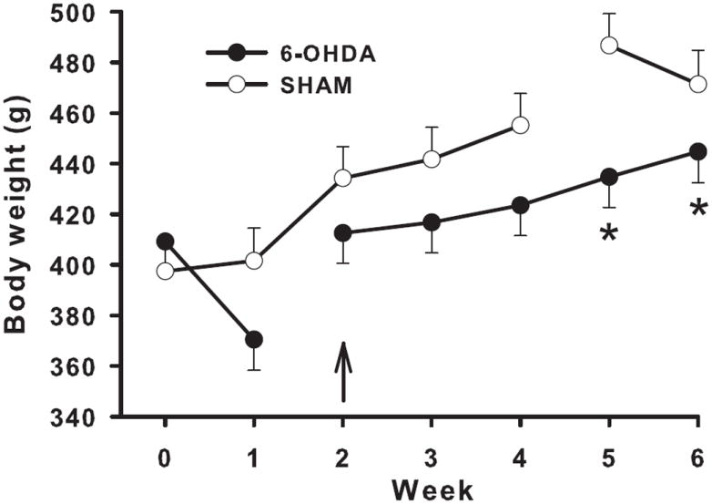
No initial weight difference was observed between groups prior to surgery. While there was no significant overall effect of 6-OHDA on body weight, animals that received striatal 6-OHDA injections weighed less at 5 and 6 weeks post-surgery compared with Sham controls. Arrow denotes start of behavioral testing. N = 13/6-OHDA; 12/Sham. * p < 0.05.
Straight Channel
No difference in time to swim the straight channel was observed across trials between 6-OHDA-lesioned and SHAM control animals (LS mean ± LSSEM across trials: 6-OHDA: 15.67 ± 1.85 s; SHAM: 13.87 ± 2.00 s).
Cincinnati water maze
6-OHDA-treated animals had significantly increased latencies to find the platform compared with SHAM animals (F(1,26.5) = 6.09, p < 0.01; Fig 2A) with significantly longer latencies observed from block-5 through block-9 (treatment x block, F(8,146) = 2.77, p < 0.01). 6-OHDA-treated animals committed significantly more errors overall compared with SHAM controls (F(1, 24.1) = 5.01, p < 0.05; Fig 2B) with significantly more errors observed during blocks 5-7 and block 9 (treatment x block effect: F(8,145) = 2.68, p < 0.01).
Figure 2. Cincinnati water maze.
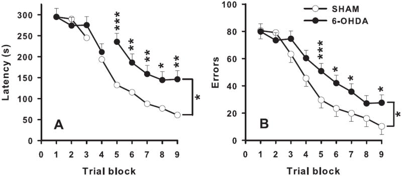
Throughout testing, striatal 6-OHDA injections increased latency to find the submerged platform (A), as well as number of errors made during the trial (B), compared with Shams. N = 13/6-OHDA; 12/Sham. * p < 0.05, ** p < 0.01, *** p < 0.001.
Because of the difficulty of finding the escape under infrared lighting, 100% of animals had one or more trials in which they reached the 5-min time limit. Most 5-min trials occurred on early test days (mostly days 1-3) then declined rapidly thereafter. For SHAM, 40.5% of trials reached the time limit whereas for 6-OHDA animals, 57.3% reached the time limit (p < 0.05), providing further evidence that lesioned rats had greater difficulty learning the task than SHAM.
Morris water maze
All animals learned to find the hidden platform during testing, however latency to find the platform (F(1,23.3) = 13.03, p < 0.001; Fig 3A), path length (F(1,23.3) = 5.55, p < 0.05; Fig 3B), and cumulative distance (F(1,23.4) = 14.67, p < 0.001; not shown) were significantly increased in 6-OHDA-treated animals compared with SHAM controls. There was no treatment interaction with day. 6-OHDA-treated animals had reduced speed compared with SHAM animals (F(1,23) = 8.71, p < 0.01; 6-OHDA: 0.23 ± 0.01 m/s, SHAM: 0.24 ± 0.01 m/s). However, ANCOVA with swim speed as the covariate showed that speed did not account for the increase in latency (F(1,22.2) = 5.79, p < 0.05), path length (F(1,22.2) = 7.53, p < 0.01), or cumulative distance (F(1,22.3) = 6.33, p < 0.01) of 6-OHDA-treated animals compared with SHAM animals.
Figure 3. Morris water maze.
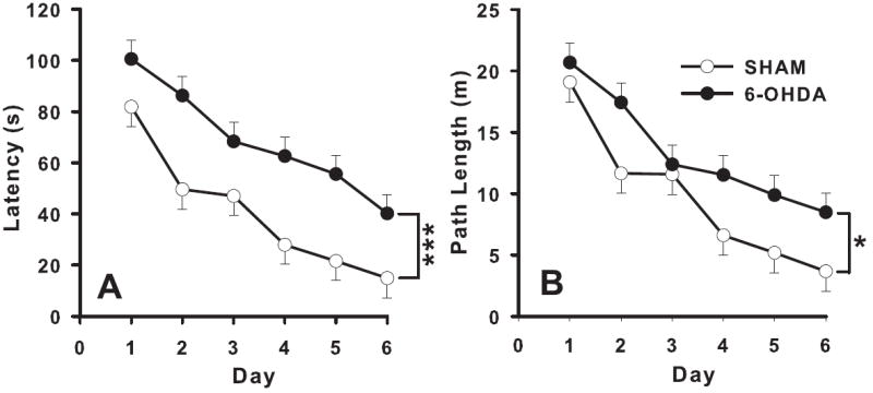
Throughout testing, striatal 6-OHDA injections increased latency to find the submerged platform (A), as well as path length (B), compared with Shams. N = 13/6-OHDA; 12/Sham. * p < 0.05, *** p < 0.001.
During the probe trial, 6-OHDA-treated animals showed a decreased percentage of time in the target quadrant compared with SHAM animals (t(23) = 1.84, p < 0.05; 6-OHDA: 31.79% ± 4.4%, SHAM: 43.63% ± 4.6%) and had greater average distance from the platform site compared with SHAM animals (t(23) = 1.89, p < 0.05; 6-OHDA: 0.76 ± 0.05 m, SHAM: 0.61 ± 0.06 m). ANCOVA with swim speed as the covariate did not alter these results (percent time in target quadrant: t(22) = 1.79, p < 0.05; average distance: t(22) = 1.83, p < 0.05, respectively).
For cued platform trials, there were no significant differences between 6-OHDA-treated and SHAM-treated animals for latency to find the platform (data not shown) further supporting the notion that performance factors cannot account for the spatial learning and retention deficits observed in the 6-OHDA lesioned animals.
Monoamine Assessment
In the neostriatum at 2 weeks, DA concentrations on the 6-OHDA-injected side were decreased by 80% compared with the vehicle-injected side (t(10) = 9.55, p < 0.001) and were decreased bilaterally in 6-OHDA-lesioned-behaviorally tested animals at 7 weeks compared with SHAM-treated animals (t(23) = 8.85, p < 0.001; Fig 4A). DOPAC levels were also decreased at both time points (2 weeks: t(10) = 3.65, p < 0.01; 7 weeks: t(23) = 6.16, p < 0.001; Table 1). 6-OHDA also decreased striatal HVA levels (2 weeks: t(10) = 4.58, p < 0.001; 7 weeks: t(23) = 7.48, p < 0.001; Table 1) and increased DOPAC/DA ratios [Table 1; 2 weeks t(10) = -4.25, p < 0.01; 7 weeks: t(23) = -5.36, p < 0.001] compared with the SHAM-treated animals. Similar patterns were found for both intracellular (DOPAC/DA ratio) and extracellular (HVA/DA ratio) DA ratios. A trend was observed in 5-HT reductions at both time points (2 weeks: t(10) = 1.92, p = 0.08; 7 weeks: t(23) = 1.84, p = 0.08; Fig 4B) in 6-OHDA-injected striata compared with the appropriate controls. No differences were observed between treatments for 5-HIAA levels at either time point. Levels of NE were decreased in 6-OHDA-injected animals 7 weeks post-surgery (t(23) = 2.26, p < 0.05; Fig 4C), but not in 6-OHDA-injected striata 2 weeks post-surgery compared with vehicle-injected striata.
Figure 4. Striatal monoamine levels.
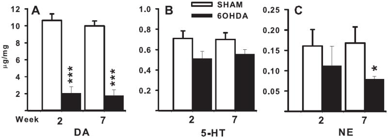
Striatal 6-OHDA injections decreased striatal DA (A), 5-HT (B), and NE (C) levels at both 2 and 7 weeks post-surgery. 2 weeks: N = 6/6-OHDA, 6/Sham. 7 weeks: N = 13/6-OHDA, 12/Sham. * p < 0.05, *** p < 0.001.
Table 1.
Neostriatal DA metabolite levels and utilization at 2 and 7 weeks post-treatment (pg/mg tissue)
| DOPAC | HVA | (DOPAC+HVA) ÷ DA | ||||
|---|---|---|---|---|---|---|
| 2 wks | 7 wks | 2 wks | 7 wks | 2 wks | 7 wks | |
| Sham | 1618.9±176.0 | 1435.0±151.2 | 875.8±73.9 | 711.1±48.9 | 0.23±0.01 | 0.21±0.01 |
| 6-OHDA | 580.3±223.3** | 345.6±97.2*** | 307.7±99.6*** | 178.5±51.4*** | 0.47±0.05** | 0.43±0.04** |
| % -Δ | 35.8% | 24.0% | 35.1% | 25.1% | 48.9% | 48.8% |
Hippocampus
Monoamine levels in the hippocampus and PFC were only collected at the 7 week time point. NE levels were decreased in the 6-OHDA-treated animals compared with SHAM-treated animals (t(23) = 10.02, p < 0.001; 6-OHDA: 125.9 ± 17.3 pg/mg, SHAM: 349.8 ± 13.8 pg/mg). No differences were observed between treatments for 5-HT, 5-HIAA, or the 5-HT utilization ratio (5-HIAA/5-HT).
Prefrontal Cortex
Monoamine levels for the PFC at the 7-week time point were not different between treatment groups for NE, 5-HT, 5-HIAA levels, or the 5-HIAA/5-HT ratio.
Discussion
Both spatial learning in the MWM and route-based learning in the CWM were impaired following 80% reductions of neostriatal DA via bilateral injections of 6-OHDA. These deficits were present independently of motivational factors (no differences in straight channel or visible platform MWM escape times), or motor deficits (slightly slower swim speeds in the MWM that did not significantly affect efficient platform finding parameters). The observed reductions in DA metabolites (DOPAC and HVA) are consistent with previous reports following bilateral 6-OHDA neostriatal injection (Aguiar et al., 2008;Chen et al., 2007;Henze et al., 2005;Tadaiesky et al., 2008). Monoamines in the PFC were unaffected by 6-OHDA striatal treatment, and only NE levels were altered in the hippocampus. As hippocampal NE levels do not play a significant role in spatial navigation (Hagan et al., 1983;Thomas and Palmiter, 1997) and route-based navigation is thought to be independent of hippocampal function (Devan et al., 1999;Devan and White, 1999;McDonald and White, 1993;McDonald and White, 1994), the deficits seen herein are most likely a result of the neostriatal DA reductions.
6-OHDA treatment resulted in an impairment of route-based navigation. This finding is consistent with data that drugs that target DA systems and produce decreases in DA levels affect CWM performance, while drugs that preferentially act on 5-HT do not (Herring et al., 2008;Herring et al., 2010;Vorhees et al., 2010b). However, drugs such as methamphetamine affect DA, 5-HT, and glutamate making it unclear which mechanism contributes most to the effect of the drug on route-based navigation. However, 6-OHDA is specific; therefore, this is the first study to show that deficits in the CWM may be attributed to DA depletion in the dorsal striatum.
Striatal DA has been shown to be involved in procedural learning of another kind: cued MWM deficits after intrastriatal 6-OHDA (Tadaiesky et al., 2008) or substantia nigra pars compacta 6-OHDA or 1-methyl-4-phenyl-1,2,3,6-tetrahydropyridine (MPTP) injections (Ferro et al., 2005;Miyoshi et al., 2002;Whishaw and Dunnett, 1985b). We did not observe such a deficit in this study, and this may be the result of testing differences. Here, the cued task followed spatial acquisition and conducting spatial learning prior to procedural learning has been shown to eliminate deficits in cued learning following intranigral MPTP injections (Da Cunha et al., 2007); therefore, test order may account for this apparent inconsistency. Cued vs. hidden platform MWM testing was not counterbalanced here in order to keep the methods between this study and our previous study consistent. Furthermore, while test order shows practice effects, order does not affect spatial learning per se. There are examples where drugs that induce sensorimotor interference impair MWM performance and these effects can be attenuated by giving practice trials (cued or nonspatial) prior to spatial trials (Saucier et al., 1996), but there is no evidence for such effects in the present context with 6-OHDA lesions.
In addition, only large nigrostriatal DA reductions result in spatial navigation deficits following 6-OHDA or MPTP- injections (Whishaw and Dunnett, 1985b). We and others have observed MWM spatial learning acquisition deficits when DA levels are depleted by 60% or more (De Leonibus et al., 2007b;Lindner et al., 1999;Mura and Feldon, 2003;Whishaw and Dunnett, 1985b), but smaller reductions do not produce this effect (Da Cunha C. et al., 2003;Miyoshi et al., 2002).
Multiple lines of evidence indicate that motoric effects are unlikely to account for the learning and memory impairments observed in the 6-OHDA lesioned rats. First, the 6-OHDA group showed no change in straight channel swimming times, a task in which there is essentially no learning required. Accordingly, this task assesses a relatively direct measure of swim speed that reflects motor ability and motivation to escape from the water. The results show that 6-OHDA rats swim a straight corridor as fast as sham controls. Second, the 6-OHDA group showed increased CMW errors, a measure not influences by swim speed or motor coordination. Third, the 6-OHDA group showed no deficits in the MWM on measures immune from performance factors, included path length and cumulative distance. Fourth, the 6-OHDA group, while they swam slower in the MWM on hidden platform acquisition trials, this did not affect learning indices based on ANCOVA results using swim speed as a covariate for each dependent measure (latency, path length, and cumulative distance) and showed no change in the finding of impaired spatial learning in the lesioned group. Fifth, a similar covariate analysis with swim speed during the probe trial confirmed that this did not alter the finding that the 6-OHDA group was impaired during the transfer trial requiring the rats to relocate the spot where the platform had formerly been. Sixth, the cued trials showed no reduction in ability to reach the platform compared with sham control group, providing no evidence that 6-OHDA impaired these animals’ ability to see and swim directly the platform even though it was moved unpredictably on every trial. Overall, the small swim speed reduction found during the MWM hidden platform trials but not on no other indices of swimming provides convergent evidence that the allocentric and egocentric navigation deficits seen in the 6-OHDA group are upon learning and memory processes.
In summary, we show here that both route-based and spatial navigation are substantially determined by dorsal striatal DA. This provides further evidence for the role of neostriatal DA on these forms of navigation, and is the first study to explore this in route-based egocentric navigation in the CWM. Future research may benefit from a dose-response investigation of DA reduction on performance to elucidate the threshold of DA for both types of navigational processes and determine if there is a differential sensitivity for the effect of DA on these two mazes. In addition, selective subregional lesions within the neostriatum have differential effects on spatial and non-spatial learning as it has been shown that dorsolateral striatum is implicated in egocentric tasks, whereas the dorsomedial striatum is implicated more heavily in spatial acquisition (Devan and White, 1999;Devan et al., 1999;Divac et al., 1967).
Egocentric and allocentic learning investigated after striatal 6-OHDA lesions.
Egocentric learning in the Cincinnati water maze was compromised
Spatial learning in the Morris water maze was detrimentally affected.
The results suggest dopamine modulates egocentric and allocentric learning.
Acknowledgments
We thank Dr. Stephen Benoit for advice on an earlier draft of this manuscript and to Mary Moran for statistical assistance. This research was supported by NIH project grants ES015689 (MTW) and DA021394 (CVV) and training grant ES07051 for trainee support to (AAB, DLG, & TLS).
Footnotes
Publisher's Disclaimer: This is a PDF file of an unedited manuscript that has been accepted for publication. As a service to our customers we are providing this early version of the manuscript. The manuscript will undergo copyediting, typesetting, and review of the resulting proof before it is published in its final citable form. Please note that during the production process errors may be discovered which could affect the content, and all legal disclaimers that apply to the journal pertain.
Reference List
- Aguiar LM, Macedo DS, Vasconcelos SM, Oliveira AA, de Sousa FC, Viana GS. CSC, an adenosine A(2A) receptor antagonist and MAO B inhibitor, reverses behavior, monoamine neurotransmission, and amino acid alterations in the 6-OHDA-lesioned rats. Brain Res. 2008;1191:192–199. doi: 10.1016/j.brainres.2007.11.051. [DOI] [PubMed] [Google Scholar]
- Aguirre GK, D’Esposito M. Topographical disorientation: a synthesis and taxonomy. Brain. 1999;122(Pt 9):1613–1628. doi: 10.1093/brain/122.9.1613. [DOI] [PubMed] [Google Scholar]
- Byrne RW. Normality and pathology in cognitive function. London: Academic; 1982. Geographical knowledge and orientation; pp. 239–264. [Google Scholar]
- Chen H, Jing FC, Li CL, Tu PF, Zheng QS, Wang ZH. Echinacoside prevents the striatal extracellular levels of monoamine neurotransmitters from diminution in 6-hydroxydopamine lesion rats. J Ethnopharmacol. 2007;114:285–289. doi: 10.1016/j.jep.2007.07.035. [DOI] [PubMed] [Google Scholar]
- Cook D, Kesner RP. Caudate nucleus and memory for egocentric localization. Behav Neural Biol. 1988;49:332–343. doi: 10.1016/s0163-1047(88)90338-x. [DOI] [PubMed] [Google Scholar]
- Da Cunha C, Wietzikoski S, Wietzikoski EC, Miyoshi E, Ferro MM, Anselmo-Franci JA, Canteras NS. Evidence for the substantia nigra pars compacta as an essential component of a memory system independent of the hippocampal memory system. Neurobiol Learn Mem. 2003;79:236–242. doi: 10.1016/s1074-7427(03)00008-x. [DOI] [PubMed] [Google Scholar]
- Da Cunha C, Wietzikoski S, Wietzikoski EC, Silva MH, Chandler J, Jr, Ferro MM, Andreatini R, Canteras NS. Pre-training to find a hidden platform in the Morris water maze can compensate for a deficit to find a cued platform in a rat model of Parkinson’s disease. Neurobiol Learn Mem. 2007;87:451–463. doi: 10.1016/j.nlm.2006.12.004. [DOI] [PubMed] [Google Scholar]
- De Leonibus E, Pascucci T, Lopez S, Oliverio A, Amalric M, Mele A. Spatial deficits in a mouse model of Parkinson disease. Psychopharmacology (Berl) 2007a;194:517–525. doi: 10.1007/s00213-007-0862-4. [DOI] [PubMed] [Google Scholar]
- De Leonibus E, Pascucci T, Lopez S, Oliverio A, Amalric M, Mele A. Spatial deficits in a mouse model of Parkinson disease. Psychopharmacology (Berl) 2007b;194:517–525. doi: 10.1007/s00213-007-0862-4. [DOI] [PubMed] [Google Scholar]
- Devan BD, McDonald RJ, White NM. Effects of medial and lateral caudate-putamen lesions on place- and cue-guided behaviors in the water maze: relation to thigmotaxis. Behav Brain Res. 1999;100:5–14. doi: 10.1016/s0166-4328(98)00107-7. [DOI] [PubMed] [Google Scholar]
- Devan BD, White NM. Parallel information processing in the dorsal striatum: relation to hippocampal function. J Neurosci. 1999;19:2789–2798. doi: 10.1523/JNEUROSCI.19-07-02789.1999. [DOI] [PMC free article] [PubMed] [Google Scholar]
- Divac I, Rosvold HE, Szwarcbart MK. Behavioral effects of selective ablation of the caudate nucleus. J Comp Physiol Psychol. 1967;63:184–190. doi: 10.1037/h0024348. [DOI] [PubMed] [Google Scholar]
- Etienne AS, Maurer R, Seguinot V. Path integration in mammals and its interaction with visual landmarks. J Exp Biol. 1996;199:201–209. doi: 10.1242/jeb.199.1.201. [DOI] [PubMed] [Google Scholar]
- Ferro MM, Bellissimo MI, Anselmo-Franci JA, Angellucci ME, Canteras NS, Da CC. Comparison of bilaterally 6- J Neurosci Methods. 2005;148:78–87. doi: 10.1016/j.jneumeth.2005.04.005. [DOI] [PubMed] [Google Scholar]
- Galani R, Obis S, Coutureau E, Jarrard L, Cassel JC. A comparison of the effects of fimbria-fornix, hippocampal, or entorhinal cortex lesions on spatial reference and working memory in rats: short versus long postsurgical recovery period. Neurobiol Learn Mem. 2002;77:1–16. doi: 10.1006/nlme.2000.3998. [DOI] [PubMed] [Google Scholar]
- Garber PA. Evidence for the use of spatial, temporal, and social information by some primate foragers. In: Boinski SB, Garber PA, editors. On the move: how and why animals travel in groups. Chicago: University of Chicago Press; 2000. pp. 261–298. [Google Scholar]
- Hagan JJ, Alpert JE, Morris RG, Iversen SD. The effects of central catecholamine depletions on spatial learning in rats. Behav Brain Res. 1983;9:83–104. doi: 10.1016/0166-4328(83)90015-3. [DOI] [PubMed] [Google Scholar]
- Henze C, Earl C, Sautter J, Schmidt N, Themann C, Hartmann A, Oertel WH. Reactive oxidative and nitrogen species in the nigrostriatal system following striatal 6-hydroxydopamine lesion in rats. Brain Res. 2005;1052:97–104. doi: 10.1016/j.brainres.2005.06.020. [DOI] [PubMed] [Google Scholar]
- Herring NR, Gudelsky GA, Vorhees CV, Williams MT. (+)-Methamphetamine-induced monoamine reductions and impaired egocentric learning in adrenalectomized rats is independent of hyperthermia. Synapse. 2010;64:773–785. doi: 10.1002/syn.20784. [DOI] [PMC free article] [PubMed] [Google Scholar]
- Herring NR, Schaefer TL, Gudelsky GA, Vorhees CV, Williams MT. Effect of +-methamphetamine on path integration learning, novel object recognition, and neurotoxicity in rats. Psychopharmacology (Berl) 2008;199:637–650. doi: 10.1007/s00213-008-1183-y. [DOI] [PMC free article] [PubMed] [Google Scholar]
- Iaria G, Palermo L, Committeri G, Barton JJ. Age differences in the formation and use of cognitive maps. Behav Brain Res. 2009;196:187–191. doi: 10.1016/j.bbr.2008.08.040. [DOI] [PubMed] [Google Scholar]
- Kesner RP. Memory for frequency in rats: role of the hippocampus and medial prefrontal cortex. Behav Neural Biol. 1990;53:402–410. doi: 10.1016/0163-1047(90)90282-b. [DOI] [PubMed] [Google Scholar]
- Kesner RP, Farnsworth G, DiMattia BV. Double dissociation of egocentric and allocentric space following medial prefrontal and parietal cortex lesions in the rat. Behav Neurosci. 1989;103:956–961. doi: 10.1037//0735-7044.103.5.956. [DOI] [PubMed] [Google Scholar]
- Laczo J, Vlcek K, Vyhnalek M, Vajnerova O, Ort M, Holmerova I, Tolar M, Andel R, Bojar M, Hort J. Spatial navigation testing discriminates two types of amnestic mild cognitive impairment. Behav Brain Res. 2009;202:252–259. doi: 10.1016/j.bbr.2009.03.041. [DOI] [PubMed] [Google Scholar]
- Lindner MD, Cain CK, Plone MA, Frydel BR, Blaney TJ, Emerich DF, Hoane MR. Incomplete nigrostriatal dopaminergic cell loss and partial reductions in striatal dopamine produce akinesia, rigidity, tremor and cognitive deficits in middle-aged rats. Behav Brain Res. 1999;102:1–16. doi: 10.1016/s0166-4328(98)00160-0. [DOI] [PubMed] [Google Scholar]
- Livingstone SA, Skelton RW. Virtual environment navigation tasks and the assessment of cognitive deficits in individuals with brain injury. Behav Brain Res. 2007;185:21–31. doi: 10.1016/j.bbr.2007.07.015. [DOI] [PubMed] [Google Scholar]
- McDonald RJ, White NM. A triple dissociation of memory systems: hippocampus, amygdala, and dorsal striatum. Behav Neurosci. 1993;107:3–22. doi: 10.1037//0735-7044.107.1.3. [DOI] [PubMed] [Google Scholar]
- McDonald RJ, White NM. Parallel information processing in the water maze: evidence for independent memory systems involving dorsal striatum and hippocampus. Behav Neural Biol. 1994;61:260–270. doi: 10.1016/s0163-1047(05)80009-3. [DOI] [PubMed] [Google Scholar]
- Miyoshi E, Wietzikoski S, Camplessei M, Silveira R, Takahashi RN, Da CC. Impaired learning in a spatial working memory version and in a cued version of the water maze in rats with MPTP-induced mesencephalic dopaminergic lesions. Brain Res Bull. 2002;58:41–47. doi: 10.1016/s0361-9230(02)00754-2. [DOI] [PubMed] [Google Scholar]
- Mizumori SJ, Puryear CB, Martig AK. Basal ganglia contributions to adaptive navigation. Behav Brain Res. 2009;199:32–42. doi: 10.1016/j.bbr.2008.11.014. [DOI] [PubMed] [Google Scholar]
- Morris RG. Spatial Localization Does Not Require the Presence of Local Cues. Learning and Motivation. 1981;12:239–260. [Google Scholar]
- Morris RG, Garrud P, Rawlins JN, O’Keefe J. Place navigation impaired in rats with hippocampal lesions. Nature. 1982;297:681–683. doi: 10.1038/297681a0. [DOI] [PubMed] [Google Scholar]
- Mura A, Feldon J. Spatial learning in rats is impaired after degeneration of the nigrostriatal dopaminergic system. Mov Disord. 2003;18:860–871. doi: 10.1002/mds.10472. [DOI] [PubMed] [Google Scholar]
- Packard MG. Exhumed from thought: basal ganglia and response learning in the plus-maze. Behav Brain Res. 2009;199:24–31. doi: 10.1016/j.bbr.2008.12.013. [DOI] [PubMed] [Google Scholar]
- Paxinos G, Watson C, Pennisi M, Topple A. Bregma, lambda and the interaural midpoint in stereotaxic surgery with rats of different sex, strain and weight. J Neurosci Methods. 1985;13:139–143. doi: 10.1016/0165-0270(85)90026-3. [DOI] [PubMed] [Google Scholar]
- Potegal M. A note on spatial-motor deficits in patients with Huntington’s disease: a test of a hypothesis. Neuropsychologia. 1971;9:233–235. doi: 10.1016/0028-3932(71)90047-9. [DOI] [PubMed] [Google Scholar]
- Ragozzino KE, Leutgeb S, Mizumori SJ. Dorsal striatal head direction and hippocampal place representations during spatial navigation. Exp Brain Res. 2001;139:372–376. doi: 10.1007/s002210100795. [DOI] [PubMed] [Google Scholar]
- Sanders AE, Holtzer R, Lipton RB, Hall C, Verghese J. Egocentric and exocentric navigation skills in older adults. J Gerontol A Biol Sci Med Sci. 2008;63:1356–1363. doi: 10.1093/gerona/63.12.1356. [DOI] [PMC free article] [PubMed] [Google Scholar]
- Saucier D, Hargreaves EL, Boon F, Vanderwolf CH, Cain DP. Detailed behavioral analysis of water maze acquisition under systemic NMDA or muscarinic antagonism: nonspatial pretraining eliminates spatial learning deficits. Behav Neurosci. 1996;110:103–116. [PubMed] [Google Scholar]
- Sutherland RJ, Whishaw IQ, Kolb B. A behavioural analysis of spatial localization following electrolytic, kainate- or colchicine-induced damage to the hippocampal formation in the rat. Behav Brain Res. 1983;7:133–153. doi: 10.1016/0166-4328(83)90188-2. [DOI] [PubMed] [Google Scholar]
- Tadaiesky MT, Dombrowski PA, Figueiredo CP, Cargnin-Ferreira E, Da CC, Takahashi RN. Emotional, cognitive and neurochemical alterations in a premotor stage model of Parkinson’s disease. Neuroscience. 2008;156:830–840. doi: 10.1016/j.neuroscience.2008.08.035. [DOI] [PubMed] [Google Scholar]
- Taube JS. Head direction cells and the neurophysiological basis for a sense of direction. Prog Neurobiol. 1998;55:225–256. doi: 10.1016/s0301-0082(98)00004-5. [DOI] [PubMed] [Google Scholar]
- Thomas SA, Palmiter RD. Disruption of the dopamine beta-hydroxylase gene in mice suggests roles for norepinephrine in motor function, learning, and memory. Behav Neurosci. 1997;111:579–589. doi: 10.1037//0735-7044.111.3.579. [DOI] [PubMed] [Google Scholar]
- Vorhees CV. Maze learning in rats: a comparison of performance in two water mazes in progeny prenatally exposed to different doses of phenytoin. Neurotoxicol Teratol. 1987;9:235–241. doi: 10.1016/0892-0362(87)90008-0. [DOI] [PubMed] [Google Scholar]
- Vorhees CV, He E, Skelton MR, Graham DL, Schaefer TL, Grace CE, Braun AA, Amos-Kroohs R, Williams MT. Comparison of (+)-methamphetamine, +/--Methylenedioxymethamphetamine, (+)-amphetamine and +/--fenfluramine in rats on egocentric learning in the Cincinnati water maze. Synapse. 2010a doi: 10.1002/syn.20854. [DOI] [PMC free article] [PubMed] [Google Scholar]
- Vorhees CV, He E, Skelton MR, Graham DL, Schaefer TL, Grace CE, Braun AA, Amos-Kroohs R, Williams MT. Comparison of (+)-methamphetamine, +/--Methylenedioxymethamphetamine, (+)-amphetamine and +/--fenfluramine in rats on egocentric learning in the Cincinnati water maze. Synapse. 2010b doi: 10.1002/syn.20854. [DOI] [PMC free article] [PubMed] [Google Scholar]
- Vorhees CV, Herring NR, Schaefer TL, Grace CE, Skelton MR, Johnson HL, Williams MT. Effects of neonatal (+)-methamphetamine on path integration and spatial learning in rats: effects of dose and rearing conditions. Int J Dev Neurosci. 2008;26:599–610. doi: 10.1016/j.ijdevneu.2008.04.002. [DOI] [PMC free article] [PubMed] [Google Scholar]
- Vorhees CV, Weisenburger WP, Acuff-Smith KD, Minck DR. An analysis of factors influencing complex water maze learning in rats: effects of task complexity, path order and escape assistance on performance following prenatal exposure to phenytoin. Neurotoxicol Teratol. 1991;13:213–222. doi: 10.1016/0892-0362(91)90013-m. [DOI] [PubMed] [Google Scholar]
- Weniger G, Irle E. Posterior parahippocampal gyrus lesions in the human impair egocentric learning in a virtual environment. Eur J Neurosci. 2006;24:2406–2414. doi: 10.1111/j.1460-9568.2006.05108.x. [DOI] [PubMed] [Google Scholar]
- Whishaw IQ, Dunnett SB. Dopamine depletion, stimulation or blockade in the rat disrupts spatial navigation and locomotion dependent upon beacon or distal cues. Behav Brain Res. 1985a;18:11–29. doi: 10.1016/0166-4328(85)90165-2. [DOI] [PubMed] [Google Scholar]
- Whishaw IQ, Dunnett SB. Dopamine depletion, stimulation or blockade in the rat disrupts spatial navigation and locomotion dependent upon beacon or distal cues. Behav Brain Res. 1985b;18:11–29. doi: 10.1016/0166-4328(85)90165-2. [DOI] [PubMed] [Google Scholar]
- Whishaw IQ, Maaswinkel H, Gonzalez CL, Kolb B. Deficits in allothetic and idiothetic spatial behavior in rats with posterior cingulate cortex lesions. Behav Brain Res. 2001;118:67–76. doi: 10.1016/s0166-4328(00)00312-0. [DOI] [PubMed] [Google Scholar]
- Williams MT, Herring NR, Schaefer TL, Skelton MR, Campbell NG, Lipton JW, McCrea AE, Vorhees CV. Alterations in body temperature, corticosterone, and behavior following the administration of 5-methoxy-diisopropyltryptamine (‘foxy’) to adult rats: a new drug of abuse. Neuropsychopharmacology. 2007;32:1404–1420. doi: 10.1038/sj.npp.1301232. [DOI] [PMC free article] [PubMed] [Google Scholar]