Case Summary
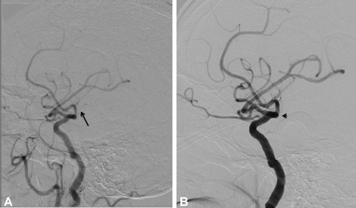
A 72-year-old male with hypertension and an old-left-middle-cerebral-artery (MCA) stroke presented with symptoms of left-sided hemiparesis, left facial drop, left-sided neglect syndrome, and left-partial homonymous hemianopia. Symptoms were suggestive of a right MCA infarct; however, advanced neuroimaging was inconclusive. Conventional diagnostic angiogram of the right internal carotid artery (ICA) was suggestive of absent right anterior choroidal artery (AChA) compared with a previous angiogram. The AChA supplies structures such as the posterior limb of the internal capsule causing contralateral hemiparesis.1 Hemisensory loss is due to damaged ventral posterolateral nucleus of the thalamus.1 Homonymous hemianopia is secondary to damaged lateral geniculate body.1 Features distinguishing AChA from larger arterial pathology such as superficial MCA involves the absence of aphasia, depressed level of consciousness, or headache.1 Anatomically the AChA originates from the posterior wall of the ICA, 2–5 mm distal to the posterior communicating artery and 2–5 mm proximal to the carotid bifurcation.2 Visualization may be challenging owing to its small diameter or secondary to the MCA branches obscuring it.2 A thorough knowledge about cerebral vascular anatomy including the small penetrators such as the AChA is important to diagnose such rare types of stroke. In the end, our case demonstrates the utility of a diagnostic cerebral angiogram to confirm the involvement of small vessels such as the AChA by providing optimal visualization.
Figure 1. Cerebral angiogram. (A) Previous angiogram showing the presence of atherosclerotic right AChA (black arrow). (B) Current angiogram with absent right AChA (black arrowhead).
Figure 2. Diffusion weighted MRI of the brain. (A) Showing hyperintense signal of the lateral geniculate body (white arrowhead). (B) Reveals hyperintense signal of the internal capsule (arrow).
References
- Bruno A, Graff-Radford NR, Biller J, Adams HP, Jr, et al. Anterior choroidal artery territory infarction: a small vessel disease. Stroke. 1989;20:616–9. doi: 10.1161/01.str.20.5.616. [DOI] [PubMed] [Google Scholar]
- Takahashi S, Suga T, Kawata Y, Sakamoto K. Anterior choroidal artery: angiographic analysis of variations and anomalies. AJNR. 1990;11:719–29. [PMC free article] [PubMed] [Google Scholar]


