Abstract
Skin-sparing mastectomy with sentinel lymph node biopsy (SLNB) and synchronous breast reconstruction are widely used in breast cancer surgery nowadays. Difficulties in feeling confident in this technique and postoperative surgical complications are the major obstacles against the widespread usage of this technique. Compared with the other surgical techniques, the complications are hard to treat. Cutaneous necrosis because of methylene blue used for sentinel lymph node mapping in patients who underwent skin-sparing mastectomy and SLNB is already reported in the literature. We present here two cases with cutaneous necrosis because of isosulphane blue injection after skin-sparing mastectomy and SLNB as a rare complication of dye injection.
Keywords: cutaneous necrosis, isosulphane blue, sentinel lymph node mapping
Introduction
Skin-sparing mastectomy is a surgical technique of removing breast tissue while sparing breast skin. It is nowadays accepted as the most appropriate technique for patients, esthetically and physiologically.1–3 The most important problem with this technique is the local control of the disease in breast cancer patients. Recent studies showed that there are no significant differences between skin-sparing mastectomy and other surgical techniques for local relapse and overall survival in breast cancer patients, and it is accepted as a safe technique in breast cancer surgery.1–3 Difficulties to feel confident in this technique and postoperative surgical complications are the major obstacles against the widespread use of this technique. After the removal of breast tissue, blood circulation of the breast skin is only supported by skin vessels and it is important for reconstruction of the breast. Although the incidence of ischemic complications such as flap necrosis can vary between different studies, but nowadays average complication rates are acceptable.1–3 As a current approach for determining axillary lymph node metastasis and avoiding complications of unnecessary axillary dissection in early breast cancer, sentinel lymph node biopsy (SLNB) is accepted as the golden standard in breast surgery.4 The first lymph node mapping with blue dye in breast cancer was performed by Giuliano et al, and they used isosulphane blue in their study.5 Injection of the coloring substance can be performed by multiple approaches such as subdermal, subareolar, and peritumoral injection. Although blue dye for SLNB is usually used in 1% concentration, it can be possible to get acceptable results with lower concentrations.6
Cutaneous necrosis because of methylene blue used for sentinel lymph node mapping in patients who underwent skin-sparing mastectomy and sentinel lymph node biopsy is already reported in the literature.6–9 We present here two cases with cutaneous necrosis because of isosulphane blue injection after skin-sparing mastectomy and SLNB as a rare complication of dye injection.
Case Report
The first patient was a 61-year-old postmenopausal woman who was admitted to hospital with right retroareolar palpable mammarian mass. In physical examination, a retroareolar solid mass of approximately 3 cm diameter was palpated in the right breast. There was no inflammation or retraction on breast skin and no palpable lymph nodes in axilla. Ultrasound scan demonstrated a solid, irregular mass of 3 cm diameter in the right retroareolar area. In mammography scan, the lesion was categorized as BIRADS 4. Invasive ductal cancer was revealed by an ultrasound-guided tru-cut biopsy, and the patient underwent skin-sparing mastectomy and SLNB after this diagnosis. Injection of isosulphane blue was performed using subareolar and subdermal approaches. Because of positive sentinel lymph node biopsy in frozen section examination, skin-sparing mastectomy, level 2 axillary dissection, and simultaneous breast reconstruction with implantation were performed. The patient was discharged at the postoperative second day without complications.
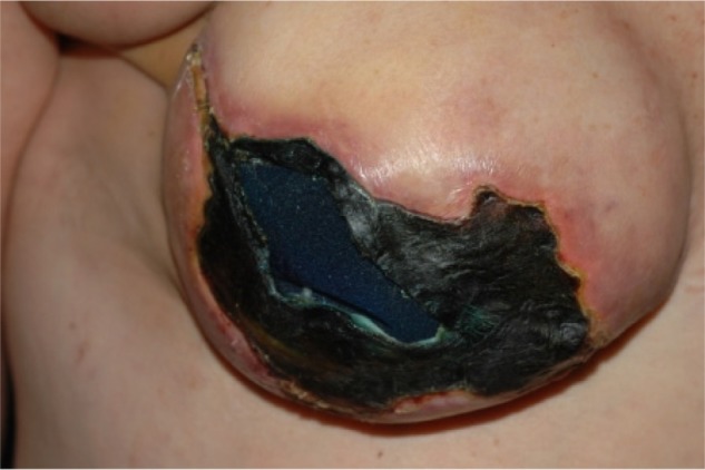
The patient was admitted to a hospital at the postoperative fourth day with skin necrosis (Fig. 1).
Figure 1.

Skin necrosis and underlying breast implant at the postoperative fourth day.
The second patient was a 57-year-old postmenopausal woman who was admitted to a hospital with palpable mass in the superior outer quadrant of the right breast. In physical examination, solid mass of approximately 2 cm diameter palpated in the superior outer quadrant of the right breast. There were no inflammation and retraction on breast skin and no palpable lymph nodes in axilla. Ultrasound scan demonstrated a solid, irregular mass of 2 cm diameter in the superior outer quadrant of the right breast. In mammography scan, the lesion was categorized as BIRADS 4. Invasive ductal cancer was revealed with an ultrasound guided tru-cut biopsy, and the patient underwent surgery with this diagnosis. Isosulphane blue was injected using subareolar and subdermal approaches. Because of positive sentinel lymph node biopsy in frozen section examination, skin-sparing mastectomy, level 2 axillary dissection, and simultaneous breast reconstruction with implantation were performed. The patient was discharged at the postoperative second day without complications.
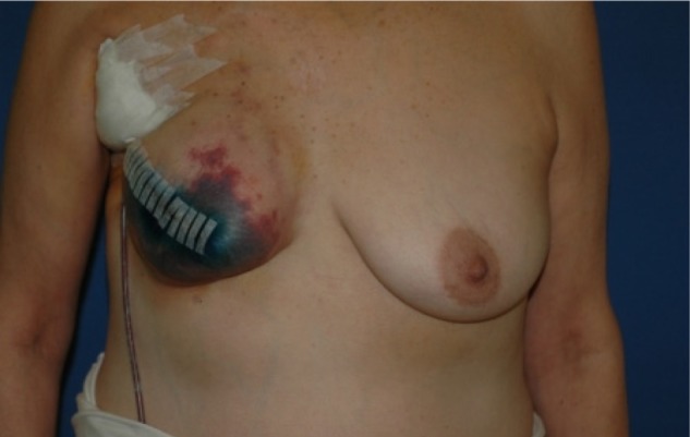
The patient was admitted to the hospital at the postoperative fourth day with skin necrosis (Fig. 2).
Figure 2.

Skin necrosis at the postoperative fourth day.
The patient was treated with a conservative approach, and there were no need for surgical interventions.
Discussion
Determining axillary lymph node metastasis is very important for the postoperative following period and adjuvant therapies in patients with breast cancer which treated with surgery.10 Because of the limitations and complications as a result of axillary dissection, it is important to determine patients who really needed axillary dissection. Although sentinel lymph node is usually detected in level one of the axillary lymph node, it can be detected in level two in approximately 18–23% of cases.11
While the possibility of allergic reaction because of isosulphane blue was reported as 2–3% in the literature, it was lower in percentage with methylene blue without decreasing the success of the procedure.6–9
Skin necrosis as a result of dermal and subareolar injection of methylene blue was reported in some cases, and because of this some authors recommended to perform methylene blue injection deeper into the breast parenchyma.6,9 But it must be considered that the success rate will decrease with this approach depending on the decrease of the absorption of blue dye because of the narrowing of the lymph ducts in the breast parenchyma and the pressure of the mass; hence, it is thought that subareolar injection ensures the optimal success rate.6,12
The incidence of skin necrosis in patients who undergo skin-sparing mastectomy without use of isosulphane blue dye varies from 0.2 to 20%.1–3
Although there is no direct evidence that the colorant dye injection caused skin necrosis, absence of predisposing factors such as smoking, increased body mass index, preoperative radiotherapy, thin skin flaps, and low ischemic complication rates associated with skin-sparing and nipple-sparing mastectomies led to suspicion of colorant dye as a possible cause of skin necrosis. Our patient’s body mass indexes were 28 and 26, respectively, and they were nonsmokers. The thickness of skin flaps was 1 cm, and they did not receive preoperative radiotherapy. Because of positive sentinel lymph nodes, nipple–areolar complex was not spared.
Moreover, isosulphane blue dye is commonly used in melanoma surgery and there really has been scarcity of descriptions of this complication with those surgeries.
In conclusion, besides predisposing factors of ischemic complications, it is clear that a lower risk of cutaneous necrosis and anaphylactic complications because of dye injection of both methylene blue and isosulphane blue exists in SLNB procedure. Because of this, it can be recommended to use radionuclide SLNB instead of blue dye or use the blue dye in lower concentrations to avoid these complications.
Footnotes
Author Contributions
BK drafted the manuscript while HYB and UO helped in the case summary. BK and HYB are surgeons who operated the patient. AD and OK amended the English and OK proof read the manuscript. All authors reviewed and approved the final manuscript.
ACADEMIC EDITOR: Athavale Nandkishor, Associate Editor
FUNDING: Authors disclose no funding sources.
COMPETING INTERESTS: Authors disclose no potential conflicts of interest.
This paper was subject to independent, expert peer review by a minimum of two blind peer reviewers. All editorial decisions were made by the independent academic editor. All authors have provided signed confirmation of their compliance with ethical and legal obligations including (but not limited to) use of any copyrighted material, compliance with ICMJE authorship and competing interests disclosure guidelines and, where applicable, compliance with legal and ethical guidelines on human and animal research participants.
REFERENCES
- 1.Kasem A, Mokbel K. Evolving role of skin sparing mastectomy. World J Clin Oncol. 2014;5(2):33–5. doi: 10.5306/wjco.v5.i2.33. [DOI] [PMC free article] [PubMed] [Google Scholar]
- 2.Colwell AS, Tessler O, Lin AM, et al. Breast reconstruction following nipple-sparing mastectomy: predictors of complications, reconstruction outcomes, and 5-year trends. Plast Reconstr Surg. 2014;133(3):496–506. doi: 10.1097/01.prs.0000438056.67375.75. [DOI] [PubMed] [Google Scholar]
- 3.Stanec Z, Zic R, Budi S, et al. Skin and nipple-areola complex sparing mastectomy in breast cancer patients: 15-year experience. Ann Plast Surg. 2012 doi: 10.1097/SAP.0b013e31827a30e6. [DOI] [PubMed] [Google Scholar]
- 4.Singh-Ranger G, Mokbel K. The sentinel node biopsy is a new standard of care for patients with early breast cancer. Int J Fertil Womens Med. 2004;49:225–7. [PubMed] [Google Scholar]
- 5.Giuliano AE, Kirgan DM, Guenther JM, Morton DL. Lymphatic mapping and sentinel lymphadenectomy for breast cancer. Ann Surg. 1994;220(3):391–8. doi: 10.1097/00000658-199409000-00015. [DOI] [PMC free article] [PubMed] [Google Scholar]
- 6.Zakaria S, Hoskin TL, Degnim AC. Safety and technical success of methylene blue dye for lymphatic mapping in breast cancer. Am J Surg. 2008;196(2):228–33. doi: 10.1016/j.amjsurg.2007.08.060. [DOI] [PubMed] [Google Scholar]
- 7.Thevarajah S, Huston TL, Simmons RM. A comparison of the adverse reactions associated with isosulfan blue versus methylene blue dye in sentinel lymph node biopsy for breast cancer. Am J Surg. 2005;189:236–9. doi: 10.1016/j.amjsurg.2004.06.042. [DOI] [PubMed] [Google Scholar]
- 8.Salhab M, Al Sarakbi W, Mokbel K. Skin and fat necrosis of the breast following methylene blue dye injection for sentinel node biopsy in a patient with breast cancer. Int Semin Surg Oncol. 2005;2:26. doi: 10.1186/1477-7800-2-26. [DOI] [PMC free article] [PubMed] [Google Scholar]
- 9.Stradling B, Aranha G, Gabram S. Adverse skin lesions after methylene blue injections for sentinel lymph node localization. Am J Surg. 2002;184:350–2. doi: 10.1016/s0002-9610(02)00945-5. [DOI] [PubMed] [Google Scholar]
- 10.Chua B, Olivotto IA, Donald JC, et al. Outcome of sentinel node biopsy for breast cancer in British Colombia, 1996 to 2001. Am J Surg. 2003;185:118–26. doi: 10.1016/s0002-9610(02)01215-1. [DOI] [PubMed] [Google Scholar]
- 11.Roumen RMH, Valkenborg JGM, Geuskens LM. Lymphosintigraphy and feasibility of sentinel node biopsy in 83 patients with primary breast cancer. Eur J Surg Oncol. 1997;23:495–502. doi: 10.1016/s0748-7983(97)92885-7. [DOI] [PubMed] [Google Scholar]
- 12.Kern KA. Sentinel lymph node mapping in breast cancer using subareolar injection of blue dye. J Am Coll Surg. 1999;189:539–45. doi: 10.1016/s1072-7515(99)00200-8. [DOI] [PubMed] [Google Scholar]


