Abstract
Purpose
Patients with strabismus often complain of difficulty navigating through visually stimulating environments without clear explanation for this symptom. Binocular summation (BiS), defined as the superiority of binocular over monocular viewing on visual threshold tasks, is decreased in conditions that cause large interocular differences in visual acuity, but is not well studied in strabismic populations without amblyopia. The authors hypothesized that strabismus may lead to decreased BiS for tasks related to discrimination within increased background complexity. The goal of this study was to test the extent of BiS in patients with strabismus during discrimination of a luminance target disk embedded in visual noise.
Methods
Participants included 10 exotropic, 10 esotropic, and 13 age-matched control patients. Performance of a task detecting a luminance-target was measured at 0, 10, and 20 μdeg2 of visual noise for binocular and monocular conditions. BiS was calculated as the ratio of binocular contrast sensitivity to monocular contrast sensitivity for the target embedded in noise.
Results
Patients with strabismus had lower BiS values than controls, with a significant decrease on linear regression in patients with strabismus at 20 μdeg2 of noise (P = .05), with a trend toward significance at 10 μdeg2 of noise (P = .07). Patients with strabismus showed a mean binocular inhibition (summation ratio < 1) at both noise levels.
Conclusions
These findings support the hypothesis that strabismus can lead to decreased BiS and even binocular inhibition. Despite literature showing enhanced BiS in visually demanding situations such as high levels of visual noise or low contrast, BiS was not significantly affected by visual noise in either group.
INTRODUCTION
Binocular summation (BiS) is defined as the superiority of binocular over monocular performance on visual threshold tasks.1 Extensive studies in normal patients have shown approximately a 1.4-fold improvement in performance binocularly compared to monocularly on psychophysical tests at low contrast.2 The amount of BiS is affected by various factors, including target size,3,4 stimulus contrast,5–7 type of task,8,9 and background complexity.10 When these factors increase the difficulty with which an image is seen, monocular vision worsens more than binocular vision and BiS occurs. Therefore, BiS may play a significant role in visual function in daily life, where visual noise, low contrast, and background complexity are pervasive.11
Factors that impair BiS include advanced age12 and interocular differences in visual acuity.1,12–14 Binocular inhibition may occur at the extremes of age and interocular difference in visual acuity, indicating better monocular vision than binocular vision. Strabismus, or binocular misalignment, causes an image to fall extrafoveally in the deviated eye, which may impair BiS by causing an induced interocular difference. Recent work by our group has shown that BiS for low contrast visual acuity is negatively impacted by the presence of strabismus.15
In this study, we aimed to investigate the effect of visual noise, or background complexity, on BiS in patients with strabismus using a target embedded in pixel noise. Anecdotally, many patients in our practice have complained about increased visual difficulty in visually demanding situations (such as grocery stores). Our goal therefore was to further understand the complex binocular deficits of patients with strabismus, hypothesizing that BiS may be lower in patients with strabismus than in normal control patients.
PATIENTS AND METHODS
This study was approved by the University of California, Los Angeles Institutional Review Board and conformed to the requirements of the United States Health Insurance Portability and Accountability Act.
Ten patients with exotropia and ten patients with esotropia were prospectively recruited from a university Pediatric Ophthalmology and Strabismus Clinic in 2012. Patients were included if they were diagnosed as having esotropia or exotropia and did not have a diagnosis of amblyopia or meet any of the exclusion criteria. Exclusion criteria included current amblyopia, age younger than 7 years or older than 65 years, pathologic nystagmus, neurologic disease, or any structural lesion causing an interocular difference exceeding 0.3 logMAR (eg, cataract, macular degeneration). Age-matched non-strabismic control patients were recruited from patients without strabismus, family members of patients, and staff volunteers. For age matching, an age within 5 years of the study subject was required. All patients underwent a screening examination in which their visual acuity was tested using the Early Treatment of Diabetic Retinopathy (ETDRS) protocol with their habitual refractive correction.16 If visual acuity was worse than 0.20 logMAR in either eye, a manifest refraction was performed and the study tests were performed with the best-corrected visual acuity. Next, binocular alignment was measured at distance (5 m) and near (30 cm) using cover/uncover and alternate prism cover testing. Stereoacuity was tested using the Titmus test (StereoOptical, Chicago, IL).
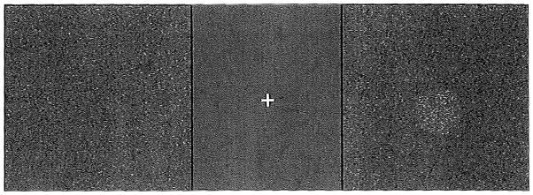
Testing conditions were based in part on previous studies of binocular summation for luminance detection within visual noise.17 The stimulus was generated on a laptop (screen size of 15.4″, spatial resolution of 1,680 x 1,050 pixels, temporal resolution of 60 Hz, background luminance of 163.92 cd/m2) using software created by Pyskinematix (Montreal, Canada) at a viewing distance of 61 cm. Luminance contrast threshold was tested using a circular target randomly positioned in one of two square fields with granular background noise presented side by side (Figure 1). After the stimulus was presented for 1 second, patients were instructed to indicate which field contained the target by pressing one of two buttons. Two feedback tones indicated either a correct or incorrect response to the patient. Contrast threshold was detected at 0, 10, and 20 μdeg2 of noise for binocular and monocular conditions with a 79% correct criterion using a one-up, two-down adaptive staircase method. Each viewing condition was scored by percent contrast (Michelson definition). Amount of BiS was then calculated by dividing the better eye score into the binocular score (binocular/better eye score). Binocular inhibition was determined to have occurred when binocular/better eye score was less than 1.
Figure 1.

Stimulus: a circular target randomly positioned in one of two fields with granular background noise.
Statistical Analysis
The demographic features of control and patients with strabismus were compared using the Student’s t test for continuous variables. BiS ratios were compared using a Wilcoxon test. Because interocular differences is a known covariate that is associated with a decrease in BiS,17 a multiple linear regression model of BiS scores was created with interocular difference and strabismus versus control status as covariates.
RESULTS
Ten exotropic, ten esotropic, and thirteen age-matched control patients were enrolled. The mean age was 25 ± 5 years (range: 8 to 63 years) for the control patients, 29 ± 6 years (range: 10 to 64 years) for the patients with exotropia, and 25 ± 6 years (range: 9 to 64 years) for the patients with esotropia. The mean deviation at near distance was 18.5 ± 4.2 prism diopters (PD) (range: 61 to 45 PD) for the patients with exotropia and 20.1 ± 5.0 PD (range: 6 to 50 PD) for the patients with esotropia. The age of onset ranged from 1 year to 56 years (median: 2 years) for patients with esotropia and from 1 to 14 years (median: 1 year) for patients with exotropia. Stereoacuity in the patients with strabismus ranged from nil to 40 seconds of arc. For the patients with esotropia, 4 patients had nil stereoacuity, 2 patients had 3,000 seconds of arc, 2 patients had 800 seconds or arc, and one subject each had 400 and 200 seconds of arc. For the patients with exotropia, 7 patients had nil stereoacuity, 1 subject had 3,000 seconds or arc, 1 subject had 100 seconds of arc, and 1 subject had 40 seconds of arc.
Mean BiS scores are summarized in Table 1. When the esotropic and patients with exotropia were grouped together, there was a significant decrease in BiS in the strabismus group compared to controls at 10 μdeg2 noise levels (P = .02). For the exotropia group alone there was also a significantly lower amount of BiS for the 10 μdeg2 noise level. In addition, both the esotropia and exotropia groups showed a mean BiS ratio of less than 1, indicating a mean binocular inhibition except for patients with esotropia at the 10 μdeg2 noise level. Conversely, the normal patients all had a mean BiS ratio of 1 or greater, indicating binocular summation.
TABLE 1.
Mean Binocular Summation Scores for Luminance Detection for Strabismic and Control Patients at 0 and 20 μdeg2 Levels if Noisea
| Comparision | 0 μdeg2
|
10 μdeg2
|
20 μdeg2
|
|||
|---|---|---|---|---|---|---|
| Mean ± SD | P | Mean ± SD | P | Mean ± SD | P | |
| ET vs control | 0.94 ± 0.3 vs 1.1 ± 0.3 | .40 | 0.8 ± 0.3 vs 1.0 ± 0.3 | .10 | 1.08 ± 0.5 vs 1.1 ± 0.3 | .50 |
| XT vs control | 0.99 ± 0.3 vs 1.1 ± 0.2 | .30 | 0.7 ± 0.3 vs 1.0 ± 0.3 | .02 | 0.75 ± 0.3 vs 1.1 + 0.4 | .30 |
| Strabismus vs control | 0.96 ± 0.3 vs 1.1 ± 0.3 | .30 | 0.8 ± 0.2 vs 1.1 ± 0.3 | .02 | 1.0 ± 0.4 vs 1.2 ± 0.4 | .80 |
SD = standard deviation; ET = esotropia; XT = exotropia
Binocular summation was calculated as a ratio between the binocular and better eye scores for contrast sensitivity within visual noise.
A multiple linear regression model of BiS scores was created with interocular difference and strabismus versus control status as covariates. The results are summarized in Figure 2 and Table 2. For the linear regression model, strabismus was found to be significantly associated with a decrease in BiS at all three noise levels.
Figure 2.

Linear regression models for binocular summation scores for strabismus (gray line) and control (black line) groups. (A) 0 μdeg2 level of noise. Significant effect of strabismus versus control (P = .05) but not for interocular difference (P = .20). (B) 10 μdeg2 level of noise. Significant effect of interocular difference (P = .04) but not strabismus versus control (P = .07). (C) 20 μdeg2 level of noise. Significant effect of strabismus versus control (P = .05) but not interocular difference (P = .10).
TABLE 2.
Linear Regression Model for Binocular Summation Scores at 0, 10, and 20 μdeg2 Levels of Noisea
| Noise Level | Coefficient | P | R2 |
|---|---|---|---|
| 0 μdeg2 | 0.11 | ||
| Strabismus vs contral | 0.06 | .05 | |
| Interocular difference in contrast sensitivity | −0.07 | .20 | |
| 10 μdeg2 | 0.14 | ||
| Strabismus vs control | 0.10 | .07 | |
| Interocular difference in contrast sensitivity | −0.04 | .04 | |
| 20 μdeg2 | 0.12 | ||
| Strabismus vs control | 0.20 | .05 | |
| Interocular difference in contrast sensitivity | −0.19 | .10 |
Binocular summation was calculated as a ratio between the binocular and better eye scores for contrast sensitivity within visual noise.
DISCUSSION
The results of this study suggest that strabismus reduces BiS scores for detection of a luminance-defined contrast target embedded in visual noise. The mean BiS score of patients with strabismus at 10 and 20 μdeg2 was less than one for all groups, suggesting that binocular inhibition may be occurring in some patients, with their monocular scores being better than their binocular scores for all noise levels (including zero noise).
Although previous studies have shown increased BiS with visually difficult tasks (especially tasks with higher “noise” levels),2–10 background noise did not significantly affect BiS in this study. In addition, BiS in the control patients was lower (approximately 1.1) than the estimated 1.4-fold that has been proposed in previous literature regarding BiS for contrast threshold.2 These findings replicate another recent study evaluating binocular summation for targets within visual noise (in patients without strabismus),10 which found significantly increased BiS in a noisy background compared to a noise-free background, but only at 15° and 18° eccentricities. Similarly to our findings, BiS was minimal (< 1.05) with central vision, regardless of noise level. Given that our target was always presented centrally, the non-significant effect of background noise on BiS and the lower-than-expected BiS scores in our study are consistent with these results.
Patients with strabismus often complain of difficulty navigating through environments that are visually demanding.18 Although our study outcome was characterized by laboratory testing on a computer module, we believe that the measurement of contrast sensitivity within a background of visual noise could be a reasonable surrogate for visually “noisy” situations, such as driving through a snowstorm or looking through a dirty windshield.19 Our findings of decreased BiS suggest that there may be a small degree of binocular inhibition that causes binocular degradation in both visually demanding “noisy” situations and those that are less “noisy” or demanding. In the patients with exotropia, the addition of visual noise further decreased their BiS ratio, which contradicts the results in normal control patients. This may explain why at least this subset of patients finds visually demanding environments more difficult. The results of patients with exotropia are in contrast to those of esotropic and control patients who experienced a small increase in BiS with the addition of visual noise, which is what one would expect from previous studies.10 Although we do not know why patients with exotropia appear to have less BiS in noisy situations than esotropes or control patients, one possible explanation is that the suppression area in patients with exotropia is larger than that of patients with esotropia.
The results of this study must be understood within the context of its limitations. Although our goal was to study the impact that strabismus has on a subject’s ability to distinguish low contrast within visual noise, our testing conditions were somewhat artificial and not necessarily representative of “real life.” In addition, our use of the same age-matched controls for the esotropic and exotropic groups might result in overrepresentation of these patients in the analysis. Finally, our subject numbers are small because this was a pilot study.
Further research is needed in characterizing the functional deficit of BiS in patients with strabismus. Our results support a reduction in BiS in contrast threshold tasks with strabismus. However, the effect of background noise is less clear, although it may contribute to the development of binocular inhibition at high noise levels, especially in patients with exotropia. Other factors on BiS such as task type and motion discrimination should be explored in this patient population.
Footnotes
The authors have no financial or proprietary interest in the materials presented herein.
References
- 1.Blake R, Sloane M, Fox R. Further developments in binocular summation. Percept Psychophys. 1981;30:266–276. doi: 10.3758/bf03214282. [DOI] [PubMed] [Google Scholar]
- 2.Campbell FW, Green DG. Monocular versus binocular visual acuity. Nature. 1965;208:191–192. doi: 10.1038/208191a0. [DOI] [PubMed] [Google Scholar]
- 3.Wood JM, Collins MJ, Carkeet A. Regional variations in binocular summation across the visual field. Ophthalmic Physiol Opt. 1973;34:2841–2848. [PubMed] [Google Scholar]
- 4.Wakayama A, Matsumoto C, Ohmure K, Matsumoto F, Shimomura Y. Properties of receptive field on binocular fusion stimulation in the central visual field. Graefes Clin Exp Ophthalmol. 2002;240:743–747. doi: 10.1007/s00417-002-0526-3. [DOI] [PubMed] [Google Scholar]
- 5.Banton T, Levi DM. Binocular summation in vernier acuity. J Opt Soc Am A Opt Image Sci Vis. 1991;8:673–680. doi: 10.1364/josaa.8.000673. [DOI] [PubMed] [Google Scholar]
- 6.Bearse MA, Jr, Freeman RD. Binocular summation in orientation discrimination depends on stimulus contrast and duration. Vision Res. 1994;34:19–29. doi: 10.1016/0042-6989(94)90253-4. [DOI] [PubMed] [Google Scholar]
- 7.Pardhan S. Binocular recognition summation in the peripheral visual field: contrast and orientation dependence. Vision Res. 2003;43:1249–1255. doi: 10.1016/s0042-6989(03)00093-2. [DOI] [PubMed] [Google Scholar]
- 8.Zlatkova MB, Anderson RS, Ennis FA. Binocular summation for grating detection and resolution in foveal and peripheral vision. Vision Res. 2001;41:3093–3100. doi: 10.1016/s0042-6989(01)00191-2. [DOI] [PubMed] [Google Scholar]
- 9.Wakayama A, Matsumoto C, Shimomura Y. Binocular summation of detection and resolution thresholds in the central visual field using parallel-line targets. Invest Ophthalmol Vis Sci. 2005;46:2810–2815. doi: 10.1167/iovs.04-1421. [DOI] [PubMed] [Google Scholar]
- 10.Wakayama A, Matsumoto C, Ohmure K, Inase M, Shimomura Y. Influence of background complexity on visual sensitivity and binocular summation using patterns with and without noise. Invest Ophthalmol Vis Sci. 2012;53:387–93. doi: 10.1167/iovs.11-8022. [DOI] [PubMed] [Google Scholar]
- 11.Legge GE. Binocular contrast summation: I. Detection and discrimination. Vision Res. 1984;24:373–383. doi: 10.1016/0042-6989(84)90063-4. [DOI] [PubMed] [Google Scholar]
- 12.Gagnon RW, Kline DW. Senescent effects on binocular summation for contrast sensitivity and spatial interval acuity. Curr Eye Res. 2003;27:315–321. doi: 10.1076/ceyr.27.5.315.17225. [DOI] [PubMed] [Google Scholar]
- 13.Jiménez JR, Ponce A, Anera RG. Induced aniseikonia diminishes binocular contrast sensitivity and binocular summation. Optom Vis Sci. 2004;81:559–562. doi: 10.1097/00006324-200407000-00019. [DOI] [PubMed] [Google Scholar]
- 14.Pineles SL, Birch EE, Talman LS, et al. One eye or two: a comparison of binocular and monocular low-contrast acuity testing in multiple sclerosis. Am J Ophthalmol. 2011;152:133–140. doi: 10.1016/j.ajo.2011.01.023. [DOI] [PMC free article] [PubMed] [Google Scholar]
- 15.Pineles SL, Velez FG, Isenberg SJ, et al. Functional burden of strabismus: decreased binocular summation and binocular inhibition. JAMA Ophthalmol. 2013;131:1413–1419. doi: 10.1001/jamaophthalmol.2013.4484. [DOI] [PMC free article] [PubMed] [Google Scholar]
- 16.Early Treatment of Diabetic Retinopathy Study. Early treatment diabetic retinopathy study design and baseline characteristics. ETDRS report number 7. Ophthalmology. 1991;98:741–756. doi: 10.1016/s0161-6420(13)38009-9. [DOI] [PubMed] [Google Scholar]
- 17.Wagner D, Manahilov V, Loffler G, Gordon GE, Dutton GN. Visual noise selectively degrades vision in migraine. Invest Ophthalmol Vis Sci. 2010;51:2294–2299. doi: 10.1167/iovs.09-4318. [DOI] [PubMed] [Google Scholar]
- 18.Otto JM, Bach M, Kommerell G. Advantage of binocularity in the presence of external visual noise. Graefes Arch Clin Exp Ophthalmol. 2010;248:535–541. doi: 10.1007/s00417-010-1304-2. [DOI] [PubMed] [Google Scholar]
- 19.Reiner J. Irritation des binokulaten Sehens durch Wischerspuren an Windschutzscheiben. Klin Monbl Augenheilkd. 1989;194:62–64. doi: 10.1055/s-2008-1046338. [DOI] [PubMed] [Google Scholar]


