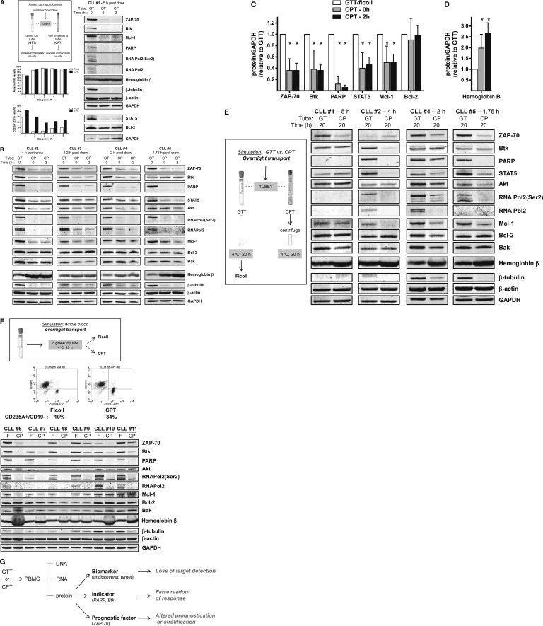
Figure 1.
Blood leukemia cell isolation methods have an impact on intracellular protein integrity. All studies were performed using fresh primary lymphocytes isolated from blood obtained from patients with CLL. Patients gave informed consent to participate in this laboratory protocol, which was approved by the Institutional Review Board of The University of Texas MD Anderson Cancer Center. Informed consent was provided according to the Declaration of Helsinki. (A) Immediate processing of whole blood on-site at the clinical trial location. Blood from the same patient (n = 5) was collected directly into a Vacutainer GTT (#366480; Becton Dickinson, Franklin Lakes, NJ) and a CPT (#362753; Becton Dickinson) simultaneously. Each tube contained sodium heparin and was kept at ambient temperature until processing. Briefly, for the GTT processing using Ficoll, the blood was mixed with 2 volumes of phosphate-buffered saline (PBS) and then overlaid gently onto 10 mL of Ficoll-Hypaque and centrifuged for 20 minutes at 20°C. The PBMCs were isolated, washed twice with PBS, and counted. The cell number was determined by using a Coulter Channelyzer (Coulter Electronics, Hialeah, FL). The CPT was centrifuged at 1800g for 20 minutes at ambient temperature, and then the tube was inverted several times to recover the isolated cells above the polyester gel barrier per the manufacturer’s recommendation. Isolated cells were washed twice with cold PBS and counted. For measurement of apoptosis levels, CLL cells (n = 5) isolated by Ficoll (white bars) or CPT (black bars) were incubated with annexin V, fluorescein isothiocyanate (FITC), and propidium iodide and were analyzed by flow cytometry. Similarly, to measure percentages of erythrocytes and B cells, CLL cells were stained with CD19-phycoerythrin (PE) and CD235A-FITC and analyzed by flow cytometry. Immunoblot analysis was performed as previously described5 by using the following antibodies: Akt (BD Pharmingen); Bak (Millipore); Bcl-2 (Dako, Carpinteria, CA); Btk (Abcam, Cambridge, MA); hemoglobin β, β-actin, and β-tubulin (Sigma); glyceraldehyde-3-phosphate dehydrogenase (GAPDH; Novus Biologicals, Littleton, CO); Mcl-1 and STAT5 (Santa Cruz Biotechnology, Santa Cruz, CA); PARP (Enzo Life Sciences International, Plymouth Meeting, PA); total RNA Pol2 (8WG16) and phospho-RNA Pol2 (Ser2) (Covance, Emeryville, CA); and ZAP-70 (Cell Signaling Technologies, Beverly, MA). Immunoblot analysis of cellular proteins of PBMCs isolated from blood collected in GTT (lane GT) or CPT (lane CP) from patient #1 is shown. Protein levels were quantified relative to GAPDH, then normalized to protein levels in Ficoll-isolated PBMCs from GTT-collected blood (lane GT). (B) Immunoblot analysis of cellular proteins of PBMCs isolated from blood collected in GTTs (lane GT) or CPTs (lane CP) from patients #2 to #5. (C-D) Quantified cellular protein levels in GTT- and CPT-isolated cells. Immunoblots shown in Figure 1A-B were analyzed by using Li-Cor Odyssey software (Li-Cor Inc., Lincoln, NE), and protein levels were normalized to GAPDH loading control. Protein levels in CPT-isolated cells (lanes CP) were expressed relative to levels in GTT-isolated cells (lane GT), and asterisks denote statistical significance (*P < .05) as determined by using GraphPad Prism software (GraphPad Software, Inc. San Diego, CA). (E) Simulation of blood drawn into a GTT or CPT and then shipped overnight to an off-site reference laboratory. Blood was collected as described in (A), the CPT was processed by centrifugation, and the PBMCs were kept in the CPT at 4°C for 20 hours to simulate overnight transport on icepacks. The GTT was kept at 4°C for 20 hours, and then the PBMCs were isolated using the Ficoll method. Immunoblot analysis of cellular proteins of PBMCs isolated from blood collected in GTTs (lane GT) or CPTs (lane CP) from patients #1, #2, #4, and #5 are shown. (F) Isolation of primary leukemia cells drawn into GTTs and then shipped overnight to an off-site reference laboratory. Blood from patients with CLL (n = 6) was collected into GTTs and kept at 4°C for 20 hours to simulate shipment on wet ice to a reference laboratory. Half the sample was then processed using Ficoll and half was transferred into a CPT containing sodium heparin and centrifuged. Shown is the flow cytometry analysis of CLL cells from patient #11 isolated by Ficoll (left) or CPT (right) and stained with CD235A-FITC/CD19-PE. Immunoblot analysis of CLL primary cells from patients #6 to #11 isolated by Ficoll (lane F) or CPT (lane CP) is shown. (G) Implications and consequences of cellular protein loss on CLL biology, diagnostics, and therapeutic research.

