Abstract
UbiA prenyltransferase domain containing 1 (UBIAD1) is a novel vitamin K2 biosynthetic enzyme screened and identified from the human genome database. UBIAD1 has recently been shown to catalyse the biosynthesis of Coenzyme Q10 (CoQ10) in zebrafish and human cells. To investigate the function of UBIAD1 in vivo, we attempted to generate mice lacking Ubiad1, a homolog of human UBIAD1, by gene targeting. Ubiad1-deficient (Ubiad1 −/−) mouse embryos failed to survive beyond embryonic day 7.5, exhibiting small-sized body and gastrulation arrest. Ubiad1 −/− embryonic stem (ES) cells failed to synthesize vitamin K2 but were able to synthesize CoQ9, similar to wild-type ES cells. Ubiad1 +/− mice developed normally, exhibiting normal growth and fertility. Vitamin K2 tissue levels and synthesis activity were approximately half of those in the wild-type, whereas CoQ9 tissue levels and synthesis activity were similar to those in the wild-type. Similarly, UBIAD1 expression and vitamin K2 synthesis activity of mouse embryonic fibroblasts prepared from Ubiad1 +/− E15.5 embryos were approximately half of those in the wild-type, whereas CoQ9 levels and synthesis activity were similar to those in the wild-type. Ubiad1 −/− mouse embryos failed to be rescued, but their embryonic lifespans were extended to term by oral administration of MK-4 or CoQ10 to pregnant Ubiad1 +/− mice. These results suggest that UBIAD1 is responsible for vitamin K2 synthesis but may not be responsible for CoQ9 synthesis in mice. We propose that UBIAD1 plays a pivotal role in embryonic development by synthesizing vitamin K2, but may have additional functions beyond the biosynthesis of vitamin K2.
Introduction
Vitamin K is a cofactor for gamma-glutamyl carboxylase (GGCX), an enzyme that converts specific glutamic acid residues in several substrate proteins involved in blood coagulation and bone metabolism to gamma-carboxyglutamic acid (Gla) residues [1], [2]. To date, 19 Gla-containing proteins have been found in vertebrates. Besides its role as a cofactor for GGCX, vitamin K is involved in the transcriptional regulation of the nuclear receptor SXR/PXR [3]–[5] and regulates PKA signalling in osteoblasts and hepatocellular carcinoma cells [6]. Vitamin K functions as a mitochondrial electron carrier during ATP production by the electron transport chain in Drosophila [7].
There are two naturally occurring forms of vitamin K, phylloquinone (PK) or vitamin K1 and the group of menaquinones (MKs). All forms of vitamin K share a common 2-methyl-1,4-naphthoquinone nucleus, differing from one another in the length and degree of saturation of the aliphatic side chain at the 3 position. PK has a monounsaturated side chain of four isoprenyl residues, and is primarily found in leafy green vegetables. MKs can be classified into 14 types on the basis of the length of their unsaturated side chains. MK-4 or vitamin K2 is predominantly present in poultry products, whereas MK-7–MK-10 are exclusively produced by bacteria and gut microflora in mammals. Menadione (MD) or vitamin K3 is a synthetic compound that lacks a side chain, although it is believed to be biologically active by virtue of its conversion to MK-4 in the body [8].
Interestingly, dietary PK releases MD by the cleavage of the side chain in the intestine, followed by the delivery of MD via the mesenteric lymphatic system and blood circulation to tissues, where it is converted to MK-4 by the prenylating enzyme UBIAD1 and accumulates in the form of MK-4 [9]. UBIAD1 is a recently identified vitamin K2/MK-4 biosynthetic enzyme exhibiting various subcellular localisations including the endothelial reticulum [10], [11], Golgi complex [11], [12] and mitochondria [13] in a variety of tissues and cell types of vertebrates. Whether UBIAD1 has any functions beside the biosynthesis of MK-4 is unknown, but UBIAD1/ubiad1 mutations in zebrafish have been reported to cause cardiac oedema and cranial haemorrhages [12], [14] and UBIAD1/heixuedian (heix) mutations in Drosophila cause defects in mitochondrial ATP production [7]. In humans, mutations in UBIAD1 cause a rare autosomal-dominant eye disease called Schnyder corneal dystrophy (SCD). SCD is characterised by abnormal deposition of cholesterol and phospholipids in the cornea, resulting in progressive corneal opacification and vision loss [15]. UBIAD1 (also known as transitional epithelial response protein 1 (TERE1)) suppressed the proliferation of transitional cell carcinoma cell lines and prostate cancer cell lines [16]–[20]. However, whether UBIAD1 is involved directly in the above biological responses or indirectly through the biosynthesis of MK-4 remains unknown. It has recently been reported that UBIAD1 catalyses the non-mitochondrial biosynthesis of CoQ10 in zebrafish [12]. Coenzyme Q (CoQ) exists in several forms and can be found in microorganisms, plants and mammals, including humans. CoQ6, Q7 and Q8 are found in yeast and bacteria, whereas CoQ9 is found in rats and mice. CoQ10 is prevalent in humans and zebrafish. CoQ is an endogenously synthesized electron carrier that is critical for electron transfer in the mitochondrial membrane for respiratory chain activity, and as a lipid-soluble antioxidant it plays an important role in protecting biological membranes from oxidative damage. The biosynthesis of CoQ in mitochondria has been studied exclusively in bacteria and yeasts. To investigate the functions of UBIAD1 in vivo, we attempted to generate mice completely lacking Ubiad1 by gene targeting. We found that Ubiad1-deficient (Ubiad1 −/−) mice uniformly died between embryonic day (E) 7.5 and E10.5 and that Ubiad1 −/− mouse embryos failed to be rescued, but their embryonic lifespans were extended partially to term by oral administration of MK-4 or CoQ10 to pregnant Ubiad1 +/− mice, indicating that UBIAD1 plays a pivotal role in the embryonic development of mice.
Materials and Methods
Materials
Deuterium-labelled MD (MD-d8) was purchased from C/D/N Isotopes, Inc. 13C-labelled 4-hydroxybenzoate (13C6-4HB) was purchased from Santa Cruz. PK epoxide, MK-4 epoxide, 18O-labelled PK and MK-4 (PK-18O and MK-4-18O), deuterium-labelled PK and PK epoxide (PK-d7 and PK-d7 epoxide), deuterium-labelled MK-4 and MK-4 epoxide (MK-4-d7 and MK-4-d7 epoxide) were synthesized in our laboratory as reported previously [8], [21]. CoQ9 and CoQ10 were donated by Eisai Co. The water-soluble type CoQ10 powder (PureSorb-Q™40. CoQ10 content is 40 w/w% hereafter P40) developed by Nisshin Pharma Inc. (Tokyo, Japan) was used [22].
Ethics statement
All animal experimental protocols were performed in accordance with the Guidelines for Animal Experiments at Kobe Pharmaceutical University and were approved by The Animal Research and Ethics Committee of Kobe Pharmaceutical University, Kobe Japan. All surgery was performed under sodium pentobarbital anesthesia, and all efforts were made to minimize suffering.
Generation of Ubiad1-deficient mice
pPE7neoW-F2LF, which contains a single loxP site, two flippase recombination target (FRT) sites and a neomycin resistance (neoR) cassette, and pMC-DTA, which contains the diphtheria toxin A gene (DTA), a negatively selective marker, were kindly provided by Dr. K. Yusa (Osaka University, Osaka, Japan) [23]. In this study, pPE7neoW-F2LF was digested with EcoRI and HindIII, and an oligonucleotide linker containing NheI, KpnI and loxP sites was inserted. The resulting plasmid pPE7neoWF2LR/loxP was digested with SacI and SalI to generate a loxP-FRT-neoR-FRT-loxP fragment. The DNA fragment was ligated into the SacI and SalI sites of the pMCS-DTA vector to generate pMCS-DTA-cKO. The Ubiad1 exon1 was amplified with an SpeI-anchored sense primer (SpeI_DA_F: CCCTGAAATCCCAGGAGGGCTAAACAG) and a KpnI-anchored antisense primer (KpnI_DA_R: CAAAGACGCCTTACTAAAGTAGGCCACTT) from mouse genomic DNA (Clontech Lab., Inc.), and cloned into NheI–KpnI-digested pMCS-DTA-cKO. The Ubiad1 5′-flanking region was amplified with a SalI-anchored sense primer (SalI_5A_F: GCTCGTAAGCGCTACAACCAATCAG) and a ClaI-anchored antisense primer (ClaI_5A_R: CCCAGTATAACGCAAAGCGACACG) from mouse genomic DNA and cloned into pMCS-DTA-cKO. The Ubiad1 3′-flanking region was amplified with a NotI-anchored sense primer (NotI_3A_F: GACTGGAAATCCAAAATGTGTGTATCG) and a SacII-anchored antisense primer (SacII_3A_R: GGTGTTTCACTGGGGTCTTTCAAACCA) from mouse genomic DNA and cloned into pMC-DTA-cKO. The targeting vector was linearised with SacII and used for electroporation of the RENKA ES cell lines derived from C57BL/6. Homologous recombination at the Ubiad1 locus resulted in replacement of the first exon of Ubiad1 with the neomycin-resistance cassette. Random integration was reduced because of a DTA cassette at the 5′ end of the targeting construct [24]. A total of 5 of 528 neomycin-resistant embryonic stem (ES) clones were correctly targeted (1.67% efficiency), as confirmed by nested PCR and Southern blotting at the 3′-flanking region. Heterozygous ES cell clones were injected into C57BL/6 blastocysts, and two of them formed germline chimaeras that transmitted the targeted allele to their offspring. The resulting male chimeras were mated with C57BL/6 females and their offspring were examined for heterozygosity by Southern blotting and PCR. Heterozygous Ubiad1 +/− mice of a C57BL/6 background identified by PCR were viable and fertile. Genotypes were confirmed by PCR with a sense primer (loxP-F, 5′- CCTTGAATTCTCTTCCTGTCGTCGTCTC-3′) and an antisense primer (GTP-R2, 5′-AGTGTTCATAATCCACTGCCAAACC-3′).
Ubiad1 −/− ES cell derivation and ES cell culture
Cryopreserved two-cell embryos obtained by in vitro fertilisation (IVF) were thawed and cultured to blastocyst stage in potassium simplex optimised medium (KSOM medium) at 37°C under 5% CO2. ES cell lines were established by a method described previously [25]. In brief, blastocysts were transferred into a 10-cm culture dish with feeder cells and cultured in ES cell medium for 8–10 days at 37°C under 5% CO2. When ES cell colonies appeared as cell clumps, each colony was isolated and transferred to a well of a 24-well plate containing feeder cells. The ES cells were subsequently cultured in ES cell medium for 8–10 days. Each ES cell line culture was passaged once before preparing frozen stocks. Genotyping was performed by PCR applied to a DNA template derived from ES cells using the primers described above. ES cells were maintained in Knockout Dulbecco's modified Eagle medium (Gibco BRL) supplemented with 16% knockout serum replacement (KSR; Gibco BRL), 1% non-essential amino acid solution (Invitrogen), 1% glutamine, 0.1 mM beta-mercaptoethanol (Sigma) and LIF solution (ESGRO, 107 units/ml; CHEMICON).
Morphological analysis of Ubiad1 −/− mouse embryos
IVF was performed using unfertilised eggs and sperm prepared from female and male mice carrying a mutation in Ubiad1, according to standard methods [26], [27]. Fertilised eggs were cultured to the two-cell embryo stage in KSOM medium at 37°C under 5% CO2 [28]. The embryos were cryopreserved by a simple verification method [29]. Besides cryopreservation, some of the embryos were cultured to the blastocyst stage in KSOM at 37°C under 5% CO2 to examine their morphology. The cryopreserved two-cell embryos were thawed by the method described previously [28] and washed in KSOM. To evaluate the embryonic development of Ubiad1 −/− mice, 10 viable two-cell embryos recovered from cryopreservation were transferred into an oviduct of a pseudopregnant female mouse 1 day after mating with a vasectomised male mouse [30]. The embryos were collected at E7 and E10 to examine their morphology under a dissection microscope. Images of the embryos were captured with a Nikon DXM1200 camera attached to a Nikon TE2000-U microscope for blastocysts and Pixera Pro 600ES with an OLYMPUS SZX9 dissection microscope for E7 and E10 embryos. Genotypes of individual embryos were identified by PCR applied to a DNA template derived from a yolk sac at E10, a whole embryo at E7 or a blastocyst, using forward (loxP-F) and reverse (GTP-R2) primers.
Mouse embryonic fibroblast isolation
Mouse embryonic fibroblast (MEF) cell cultures were prepared from E15.5 embryos of Ubiad1 +/+ and Ubiad1 +/− mice. The embryos were dissected from the uterus under sterile conditions and washed with PBS. Embryo paws and legs were minced and digested with 0.25% trypsin for 20 minutes at 37°C. Cell suspensions were plated in Dulbecco's modified Eagle medium containing 10% foetal bovine serum and penicillin/streptomycin.
Measurements of MK-4 and MK-4-d7 in tissues of mice administered MD-d8
Ten-week-old male Ubiad1 +/+ and Ubiad +/− mice were orally administered MD-d8 as a single dose at 10 µmol/kg body weight. After 24 hours, mice were sacrificed and tissues were removed and stored at −80°C for analysis. MK-4, MK-4 epoxide, MK-4-d7 and MK-4-d7 epoxide were measured by LC-APCI-MS/MS as reported previously [8], [21].
Measurements of CoQ9, CoQ10, 13C6-CoQ9 and 13C6-CoQ10
Tissues (wet weight, 100–200 mg) were minced and transferred to a brown glass tube with a Teflon-lined screw cap. Next, 0.1 mL of ethanol containing MK-4-18O as internal standard, 1.9 mL of ethanol, 1.0 mL distilled water and 3 mL of hexane followed by thorough mixing on a voltex mixer for 5 minutes. The resulting mixture was centrifuged at 2,500 rpm for 5 minutes at 4°C, and the upper layer was transferred to a small brown glass tube and evaporated to dryness under reduced pressure. The residue was dissolved in 1 mL hexane and evaporated under reduced pressure. This residue was dissolved in 60 µL methanol. An aliquot of this solution was analyzed by APCI3000 LC-MS/MS (Applied Biosystems, Foster City, CA). HPLC analyses were performed on a Shimadzu HPLC system (Shimadzu, Kyoto, Japan) consisting of a binary pump (LC-10AD liquid chromatography), an automatic solvent degasser (DGU-14A degasser) and an autosampler (SIL-10AD autoinjector). Separations were performed using a reversed-phase C18 column (Capcell Pak C18 UG120, 5 µm; 4.6 mm inner diameter×250 mm, Shiseido, Tokyo, Japan) with a solvent system consisting of isocratic solvent A. Solvent A contained methanol∶isopropanol (3∶1, v/v) and was delivered at 1.0 mL/minute. This mobile phase was passed through the column at 1.0 mL/minute. The column was maintained at 35°C by a column oven (CTO-10AC column oven). All MS data were collected in positive ion mode with atmospheric pressure chemical ionisation (APCI). The following settings were used: corona discharge needle voltage, 5.5 kV; vaporizer temperature, 400°C; sheath gas (high-purity nitrogen) pressure, 50 p.s.i. and transfer capillary temperature, 220°C. The electron multiplier voltage was set at 850 eV. Identification and quantification were based on MS/MS using multiple reaction monitoring (MRM) mode. The range for the parent scan was 400–900 atomic mass units. MRM transitions (precursor ion and product ion, m/z) and retention time (minute) for each analyte were as follows: MK-4-18O: precursor ion, 449.3; product ion, 191.2; retention time, 3.6; CoQ9: precursor ion, 796.5; product ion, 197.1; retention time, 8.7; CoQ10: precursor ion, 864.6; product ion, 197.0; retention time, 11.5; 13C6-CoQ9: precursor ion, 802.5; product ion, 203.1; retention time, 8.6 and 13C6-CoQ10: precursor ion, 870.6; product ion, 203.0; retention time, 11.4 [31]–[33]. Calibration, using internal standardisation, was performed by linear regression using five different concentrations of 100, 200, 400, 800 and 1,600 ng/mL.
Conversion of MD-d8 to MK-4-d7 and 13C6-4HB to 13C6-CoQ9 in ES cells
ES cells described above were maintained in Knockout Dulbecco's modified Eagle medium (Gibco BRL.) supplemented with 16% KSR (Gibco BRL), 1% non-essential amino acid solution (Invitrogen), 1% glutamine, 0.1 mM β-mercaptoethanol (Sigma) and LIF solution (ESGRO, 107 units/ml; CHEMICON). ES cells were cultured on MEF in 6-well tissue culture plates (2×10⁵ cells/well) for 3 days and treated with culture medium containing MD-d8 (10−6 M) and 13C6-4HB for 24 hours. ES cells were trypsinised, washed with ES medium and cultured on gelatin-coated plates for 40 minutes. Floated ES cells were collected and washed with cold PBS(−) twice and then stored at −30°C. After warming to room temperature, cells were lysed in 1 mL of PBS(−). Cell lysates (20 µL) were analysed for protein concentrations. PK-18O and MK-4-18O were added as internal standards to the cell lysates in brown screw-capped tubes. Measurements for MK-4-d7, MK-4-d7 epoxide and 13C6-CoQ9 in cells was performed using the method described above.
Real-time PCR
Total RNA of mouse tissues or ES cells was isolated with Isogen (Nippon Gene) according to the manufacturer's protocol. First-strand cDNA synthesis was performed using AMV reverse transcriptase (TaKaRa). cDNA were mixed with SYBR Green Core Reagent (PE Biosystems), and amplified using the CFX96 real-time PCR system (Bio-Rad). We used mouse Ubiad1 (GenBank NM_027873, FP:1575-1595, RP:1754-1774) mouse Coq2 (NM_027978, FP:445-465, RP:555-577), mouse Gapdh (GenBank accessions 01289726, FP:633-652, RP:726-745) and mouse β-actin (GenBank accessionsX03672, FP:250-271, RP:305-326).
Western blotting
UBIAD1 expression levels were detected by western blotting. The UBIAD1 antibody was an UBIAD1-specific affinity-purified polyclonal antibody raised in rabbits against an UBIAD1-specific peptide (CPEQDRLPQRSWRQK-COOH) (MRL Co., Ltd.). The peroxidase-conjugated secondary antibody was rabbit Ig raised in donkey (SantaCruz) for 1.5 hours and UBIAD1 protein was detected using an electrochemiluminescent detection system (Nakalai Tesque).
Administration of MK-4 or CoQ10 to Ubiad1 +/− pregnant mice
Ubiad1 +/− pregnant mice were orally administered water-soluble CoQ10 (10 µmol/kg/day) or MK-4 (2.25 µmol/kg/day) every other day from 1 day before mating to the day of sacrifice. The doses of oral administration of MK-4 and CoQ10 were decided with reference to previous animal and human studies [34], [35].
Histology and immunohistochemistry
For histological analysis, embryos were fixed in 4% paraformaldehyde in PBS at 4°C for 20 hours and embedded in a paraffin block. Tissue sections were stained with hematoxylin–eosin (HE). For detection of UBIAD1 protein, we used a monoclonal antibody raised against an amino acid sequence (SKGIDHKKSDDRTLVDRILEPQD) corresponding to the N terminus of the mouse UBIAD1 protein. Formalin-fixed and paraffin-embedded mouse embryonic tissues were deparaffinised and incubated for 30 minutes in 3% hydrogen peroxide/PBS to quench endogenous peroxidases. Sections were rinsed in PBS and immunostained with anti-UBIAD1 antibody at 1∶100 dilution in 0.5% PBS/Ova Albumin at 4°C overnight after antigen retrieval with HistoOne buffer (Nacalai, Kyoto, Japan) for 95°C for 20 minutes. The secondary antibody was HRP-labelled anti-rat IgG antibody (KPL) diluted 1∶1000 in 0.5% PBS/Ova Albumin and incubated for 30 minutes at room temperature. Sections were incubated with Elite ABC Kit (Vector Laboratories) for 30 minutes, rinsed with PBS and detected by staining with DAB (Vector Laboratories) for 2 minutes and counterstaining with methyl green.
Statistical analysis
Data are expressed as mean ± SEM. Differences between the mean values were analysed using the unpaired Student's t test or Dunnett's test: *P<0.05; **P<0.01; ***P<0.001.
Results
Ubiad1-deficient mice are embryonic lethal
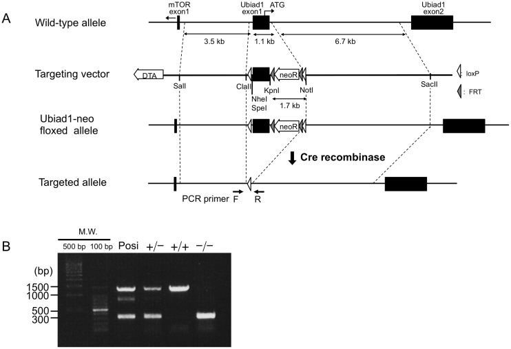
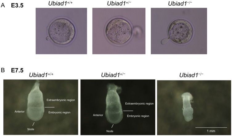
Mice Ubiad1 contains two exons. To disrupt Ubiad1, the targeting vector was designed to flank exon 1 with two loxP sequences, and a frameshift was generated by excision with Cre recombinase (Figure 1). Ubiad1 +/− mice having a C57BL/6 background identified by PCR were viable and fertile. In comparison to control littermates (Ubiad1 +/+), they did not exhibit any overt phenotype and had similar body size and weight (Figure S1). Male and female Ubiad1 +/− mice were intercrossed to obtain Ubiad1 −/− mice. However, no Ubiad1 −/− pup could be identified among over 150 analysed offspring, suggesting that the disruption of Ubiad1 leads to embryonic lethality. In contrast, Ubiad1 +/+ and Ubiad1 +/− mice were obtained at the Mendelian frequency (Table 1). To assess the time of death in utero, IVF were performed using sperm and eggs obtained from male and female Ubiad1 +/− mice, respectively and the embryos in utero of pseudopregnant Ubiad1 +/+ mice were dissected and genotyped at several gestation times from E3.5 to E10.5. At E3.5, Ubiad1 −/− blastocysts were microscopically indistinguishable from Ubiad1 +/+ and Ubiad1 +/− blastocysts (Figure 2A). However, at E7.5, both Ubiad1 +/+ and Ubiad1 +/− embryos showed elongation of the egg cylinder, which developed to a primitive streak with the formation of mesoderm between ectoderm and endoderm. In contrast, Ubiad1 −/− embryos were consistently smaller than their littermates and exhibited no signs of primitive streak formation. The constriction marking the extraembryonic-embryonic ectoderm border was absent, and mesoderm had not formed in Ubiad1 −/− embryos (Figure 2B). At E10.5, Ubiad1 −/− embryos were not found at all, similar to the results observed in the intercrosses of male and female Ubiad1 +/− mice in vivo.
Figure 1. Generation of Ubiad1 knockout mice.
(A) Schematic presentation of ubiad1 genome, targeting vector and disrupted Ubiad1 genome. (B) PCR genotyping of Ubiad1 +/+, Ubiad1 +/− and Ubiad1 −/− embryos. PCR genotyping of tail DNA of Ubiad1 +/+, Ubiad1 +/− and Ubiad1 −/− embryos. Lane 1, positive controls for Ubiad1 +/− allele. Lane 2, PCR bands of Ubiad1 +/− embryos. Lane 3, PCR bands of Ubiad1 +/+ embryos. Lane 4, PCR bands of Ubiad1 −/− embryos.
Table 1. Analysis of embryos and weaning from intercrosses of Ubiad1 +/− mice.
| Stage | Total offspring | Ubiad1 +/+ | Ubiad1 +/− | Ubiad1 −/− | Resorbed |
| E3.5 | 19 | 3 | 13 | 3 | 0 |
| E7.5 | 56 | 7 | 22 | 13 | 14 |
| E10.5 | 103 | 15 | 51 | 0 | 37 |
| E15.5 | 111 | 20 | 69 | 0 | 22 |
| Weanling | 151 | 49 | 102 | 0 |
Figure 2. Developmental arrest in Ubiad1 knockout embryos.
(A) Morphology of E3.5 blastocysts. Blastocysts were cultured from in vitro fertilised embryos of Ubiad1 +/+, Ubiad1 +/− and Ubiad1 −/− mice. (B) Morphology of E7.5 embryos.
Ubiad1 −/− ES cells were unable to synthesize MK-4, but able to synthesize CoQ9 similar to wild-type ES cells
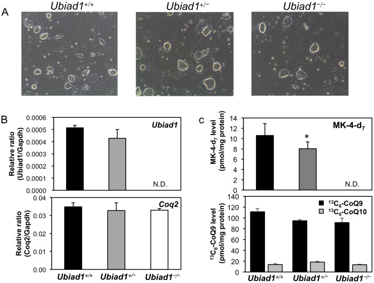
Ubiad1 −/− mice are embryonic lethal. For alternative confirmation of the successful ablation of Ubiad1, we established ES cells from fertilised ova of Ubiad1 +/− mice. Ubiad1 −/− ES cells exhibited neither Ubiad1 mRNA and protein expression nor MK-4 synthesis activity, whereas they exhibited CoQ9 or CoQ10 synthesis activity and coenzyme Q2 4-hydroxybenzoate-polyprenyltransferase (Coq2) mRNA expression similar to those of Ubiad1 +/+ and Ubiad1 +/− ES cells (Figure 3). These data exclude the presence of an additional biosynthesis pathway of MK-4 and indicate that UBIAD1 is the sole MK-4 biosynthetic enzyme in embryonic development of mice.
Figure 3. Ubiad1 and Coq2 expression and the biosynthesis of MK-4 and CoQ9 in ES cells derived from Ubiad1 +/+, Ubiad1 +/− and Ubiad1 −/− embryos.
(A) Morphology of Ubiad1 +/+, Ubiad1 +/− and Ubiad1 −/− ES cells. (B) Ubiad1 and Coq2 mRNA expression in Ubiad1 +/+, Ubiad1 +/− and Ubiad1 −/− ES cells. (C) The biosynthesis of MK-4-d7, 13C6-CoQ9 and 13C6-CoQ10 in Ubiad1 +/+, Ubiad1 +/− and Ubiad1 −/− ES cells. Mean ± s.e.m. Dunnett's test, *P<0.05. N.D.: not detected.
Neither MK-4 nor CoQ10 treatment to pregnant Ubiad1 +/− mice rescued their Ubiad1 −/− embryos from lethality, but extended the lifespan of many Ubiad1 −/− embryos to term
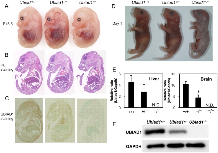
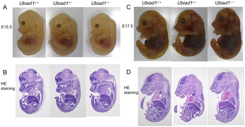
To examine whether MK-4 or CoQ10 treatment rescues the embryonic lethality of Ubiad1 −/− mice, either MK-4 or CoQ10 at a dose of 10 µmol/kg/day or 2.25 µmol/kg/day was orally administered to Ubiad1 +/− pregnant mice from 1 day before crossing and throughout pregnancy. As a result, approximately 1.8% of Ubiad1 −/− embryos survived by E15.5 and approximately 12.5% of Ubiad1 −/− embryos survived by day 1 in MK-4-treated Ubiad1 +/− pregnant mice. In CoQ10-treated Ubiad1 +/− pregnant mice, approximately 2.2% of Ubiad1 −/− embryos survived by E15.5 and approximately 1.8% of Ubiad1 −/− embryos survived by day 1, although no Ubiad1 −/− embryo was identified at E15.5 in the non-treated Ubiad1 +/− pregnant mice (Table 2). A Ubiad1 −/− embryo at E15.5 from the Ubiad1 +/− mice orally administered CoQ10 throughout pregnancy were indistinguishable from their Ubiad1 +/+ and Ubiad1 +/− littermates (Figure 4A–C). A weanling Ubiad1 −/− mouse on day 1 from the Ubiad1 +/− mice orally administered CoQ10 throughout pregnancy died immediately after birth or stillbirth (Figure 4D). The cause of death was not identified. In this Ubiad1 −/− mouse, we could find no corneal and hemorrhagic abnormalities that had been observed in SCD patients [15] or ubiad1-mutated zebrafish [12], [14]. The size and morphological features of Ubiad1 −/− embryo at E15.5 and E17.5 from the Ubiad1 +/− mice orally administered MK-4 throughout pregnancy did not differ from those of their Ubiad1 +/+ and Ubiad1 +/− littermates (Figure 5A–D). A weanling Ubiad1 −/− mouse on day 1 was not alive. UBIAD1 mRNA and protein expression in Ubiad1 −/− embryos at E15.5 was completely abolished by the targeting, as demonstrated by real-time RT-PCR and western blotting (Figure 4E, F). These results suggest that MK-4 or CoQ10 treatment would help to rescue the embryonic lethality of Ubiad1 −/− mice, although their contribution might be small and limited.
Table 2. Analysis of embryos and weaning from intercrosses of Ubiad1 +/− mice rescued by MK-4 or CoQ10.
| Stage | Total offspring | Ubiad1 +/+ | Ubiad1 +/− | Ubiad1 −/− | Resorbed |
| Rescured by MK-4 | |||||
| E15.5 | 113 | 25 | 70 | 2 | 16 |
| Day 1 | 24 | 6 | 15 | 3 leathal | |
| Rescured by CoQ10 | |||||
| E15.5 | 270 | 71 | 147 | 6 | 46 |
| Day 1 | 147 | 43 | 101 | 3 leathal | |
Figure 4. Morphological examination of Ubiad1 +/+, Ubiad1 +/− and Ubiad1 −/− embryos and weanling mice (postnatal day 1) from pregnant Ubiad1 +/− mice orally administered CoQ10.
(A) Morphology of Ubiad1 +/+, Ubiad1 +/− and Ubiad1 −/− embryos at E15.5 from CoQ10-supplemented pregnant Ubiad1 +/− mice. (B) HE staining of embryos at E15.5. (C) Immunohistochemical staining of UBIAD1 in Ubiad1 +/+, Ubiad1 +/− and Ubiad1 −/− embryos at E15.5. (D) Morphology of Ubiad1 +/+, Ubiad1 +/− and Ubiad1 −/− embryos at postnatal day 1. Ubiad1 +/+ and Ubiad1 +/− mice were born alive, but Ubiad1 −/− mice were stillborn. (E) Ubiad1 mRNA expression in the livers and brains of Ubiad1 +/+, Ubiad1 +/− and Ubiad1 −/− embryos at E15.5 from CoQ10-supplemented pregnant Ubiad1 +/− mice. Mean ± s.e.m. Dunnett's test, *P<0.05. N.D.: not detected. (F) UBIAD1 expression in the brains of Ubiad1 +/+, Ubiad1 +/− and Ubiad1 −/− embryos at E15.5 from CoQ10-supplemented pregnant Ubiad1 +/− mice.
Figure 5. Morphological examination of Ubiad1 +/+, Ubiad1 +/− and Ubiad1 −/− embryos from pregnant Ubiad1 +/− mice orally administered MK-4.
(A) Morphology of Ubiad1 +/+, Ubiad1 +/− and Ubiad1 −/− embryos at E15.5 from MK-4-supplemented pregnant Ubiad1 +/− mice. (B) HE staining of embryos at E15.5. (C) Morphology of Ubiad1 +/+, Ubiad1 +/− and Ubiad1 −/− embryos at E17.5 from MK-4-supplemented pregnant Ubiad1 +/− mice. (D) HE staining of embryos at E17.5.
Levels of MK-4 and its epoxide in tissues of Ubiad1 +/− mice are approximately half of those in tissues of Ubiad1 +/+ mice at 28 weeks of age
PK originating in a laboratory chow diet was found in all (n = 23) tissues measured, although at widely varying levels, whereas PK epoxide was found in only five and six tissues of Ubiad1 +/+ and Ubiad1 +/− mice, respectively. There was no significant difference between both groups in the levels of PK and its epoxide. In contrast, MK-4 was found in all tissues measured, although at widely varying levels. MK-4 epoxide was found in 17 and 12 tissues of Ubiad1 +/+ and Ubiad1 +/− mice, respectively. Again, levels of MK-4 in the tissues of Ubiad1 +/− mice were approximately half of those in tissues of the wild type (Table S1).
Levels of CoQ9 and CoQ10 in tissues of Ubiad1 +/− mice are similar to those of Ubiad1 +/+ mice at 28 weeks of age
In Ubiad1 +/+ and Ubiad1 +/− mice, both CoQ9 and CoQ10 were found in all tissues measured, although at widely varying levels; there was no significant difference in levels between the two groups (Table S2). We first measured the concentrations of MK-4, MK-4 epoxide, CoQ9 and CoQ10 in five tissues (cerebrum, heart, liver, kidney and small intestine) that are known to be important and representative tissues of vitamin K and CoQ functions. As a result, the concentrations of MK-4 and MK-4 epoxide in the above tissues of Ubiad1 +/− mice were found likely to be approximately half of those of Ubiad1 +/+ mice (Table S1), whereas the concentrations of CoQ9 and CoQ10 in the same tissues of Ubiad1 +/− mice were found likely to be similar to those of Ubiad1 +/+ mice (Table S2). To confirm the influence of Ubiad1 ablation, we further measured the concentrations of MK-4 and MK-4 epoxide in additional 18 tissues of both Ubiad1 +/− and Ubiad1 +/+ mice (Table S1). It is obvious that UBIAD1 is the enzyme responsible for the synthesis of MK-4, but not CoQ9 and CoQ10 in mice.
Serum levels of total cholesterol, free cholesterol and HDL-cholesterol in Ubiad1 +/− mice are higher than those in Ubiad1 +/+ mice at 28 weeks of age
In blood chemical analysis, values of total cholesterol, free cholesterol and HDL-cholesterol in Ubiad1 +/− mice were significantly higher than those in Ubiad1 +/+ mice. However, values of calcium, phosphorus, glucose, LDL-cholesterol and triglyceride were not significantly different between the two groups (Table S3). These results suggest that UBIAD1 deficiency may affect cholesterol metabolism in mice similarly in human SCD patients [15], [36].
UBIAD1 expression and MK-4 synthesis activity in the cerebrum of Ubiad1 +/− mice are at approximately half of wild-type levels at 28 weeks old
UBIAD1 mRNA and protein expression levels were evaluated by real-time RT-PCR and Western blot analysis, respectively. As expected, significant reductions in UBIAD1 mRNA and protein were observed compared to wild-type levels. Similar reductions in MK-4 biosynthetic activity in the cerebrum of the Ubiad1 +/− mice were observed compared to the wild-type mice (Figure S2).
Ubiad1 +/− MEF cells exhibit approximately half the MK-4 synthetic activity of the Ubiad1 +/+ MEF cells, but CoQ9 synthetic activity is similar between both genotypes of MEF cells
As Ubiad1 −/− embryos uniformly die beyond E7.5, we generated MEF cells from Ubiad1 +/+ and Ubiad1 +/− embryos at E15.5 and measured their MK-4 and CoQ9 synthetic activities. Both Ubiad1 +/+ and Ubiad1 +/− MEF cells grew normally and were indistinguishable from each other microscopically. Similarly to the results for the ES cells, Ubiad1 +/− MEF cells exhibited approximately half of the MK-4 synthetic activity of Ubiad1 +/+ MEF cells, whereas both genotypes of MEF cells exhibited similar CoQ9 synthetic activity, suggesting that UBIAD1 is a MK-4 synthetic enzyme, but may not be a CoQ9 synthetic enzyme in mice. (Figure S3).
Discussion
UBIAD1 is a recently identified MK-4 biosynthetic enzyme in mice and humans. UBIAD1 is expressed and coexists with vitamin K throughout the body, suggesting various physiological functions of vitamin K. We showed, for the first time to our knowledge, that Ubiad1 knockout mice uniformly failed to survive beyond E7.5, exhibiting a small-sized body and prominent gastrulation arrest. Oral administration of MK-4 or CoQ10 to Ubiad1 +/− pregnant mice crossed with male Ubiad1 +/− mice resulted in the appearance of a few Ubiad1 −/− embryos from E10.5 to term, but no pup was observed alive after birth. These results suggest that both MK-4 and CoQ10 can only partially rescue the Ubiad1 −/− embryonic lethal phenotype. Hitherto, there have been inconsistent reports with respect to the functions of Ubiad1 as a MK-4 and/or CoQ10 biosynthetic enzyme. Hegarty et al. reported that ubiad1-generated MK-4 rescued the zebrafish ubiad1 vascular integrity/maintenance mutant reddishS587 (reh), which develops a functional vasculature by 24 to 36 hours after fertilisation, but then displays cranial vascular haemorrhages because of vascular degeneration by 48 hours after fertilisation [14]. In contrast, CoQ10 was unable to rescue the reh vascular phenotype. Vos et al. reported that Drosophila ubiad1/heix is a modifier of pink 1, a gene mutated in Parkinson's disease with a defect of mitochondrial function, and that MK-4 but not CoQ10 rescued the ubiad1/heix mutant phenotype [7]. In contrast, Mugoni et al. recently reported that UBIAD1 is a non-mitochondrial CoQ10 synthetic enzyme with specific cardiovascular protective function via modulation of eNOS activity, and that loss of UBIAD1 induces cardiovascular failure in zebrafish embryos by increasing oxidative stress [12]. Though it remains uncertain whether UBIAD1 in zebrafish and Drosophila is able to synthesize MK-4 like humans and mice [10], it is obvious that mutations in ubiad1 lead to severe or lethal cardiovascular failure in these species. Considering these findings, complete loss of Ubiad1 function as observed in the present study may lead to a cardiovascular defect in a mouse embryo, leading in turn to foetal demise. To further elucidate the function of UBIAD1, it will be necessary to analyse the Ubiad1 knockout mouse phenotype, but such an analysis is currently made difficult by the uniform death of Ubiad1 knockout mice beyond E7.5 and the very low numbers able to survive from mid-embryonic stage to term with supplementation with MK-4 or CoQ10. To overcome this limitation, we are currently generating tissue-specific Ubiad1 knockout mice that will develop normally and will enable us to determine whether UBIAD1 regulates vascular integrity/maintenance in mice, as observed in zebrafish and Drosophila.
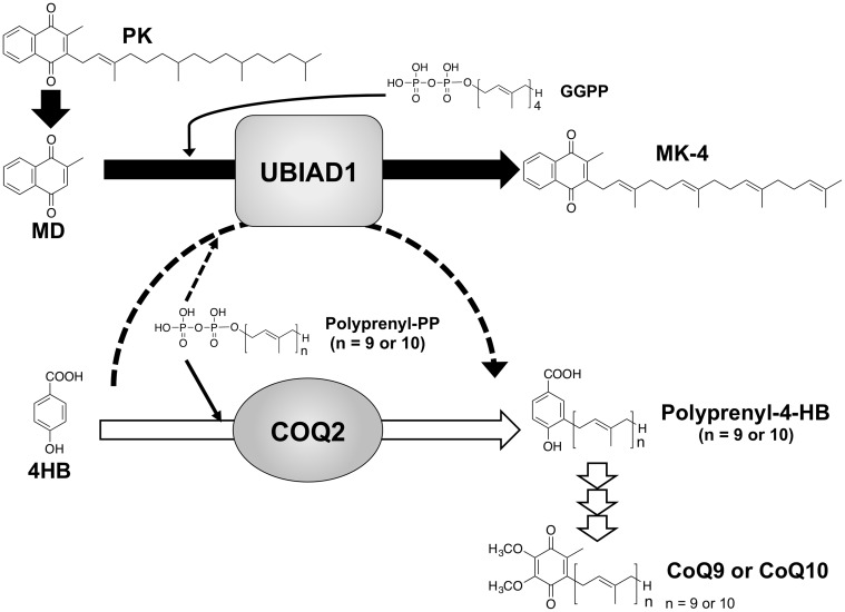
It remains unclear whether UBIAD1 can synthesize MK-4 and/or CoQ9 in mice. Mugoni et al. reported that zebrafish can synthesize CoQ10 but not MK-4 [12]. However, we previously reported that short interfering RNA treatment against the UBIAD1 gene and the transfection of UBIAD1 expression vector in human osteoblast-like MG-63 cells resulted in a marked reduction and a significant increase of the biosynthesis of MK-4, respectively. We further confirmed that microsomes prepared from human UBIAD1 baculovirus-infected Sf9 cells catalyse the biosynthesis of MK-4 in a dose-dependent manner [10]. In the present study, Ubiad1 −/− ES cells exhibited neither UBIAD1 mRNA and protein expression nor biosynthesis activity of MK-4 (Figure 3) and Ubiad1 +/− ES cells exhibited approximately half of the MK-4 synthetic activity of Ubiad1 +/+ ES cells. Ubiad1 +/− MEF cells exhibited approximately half of the MK-4 synthetic activity of Ubiad1 +/+ MEF cells; however, CoQ9 synthetic activity was similar among the three genotypes of ES cells and also between both genotypes of MEF cells. Levels of tissue MK-4 and MK-4 synthesis activity of the cerebrum of Ubiad1 +/− mice were approximately half of those of Ubiad1 +/+ mice at 28 weeks old (Table S1). These findings may exclude the existence of an MK-4 biosynthetic enzyme other than UBIAD1, and suggest that UBIAD1 is the sole MK-4 biosynthetic enzyme at least in embryonic development of mice. However, it is uncertain whether UBIAD1 is a CoQ9 biosynthetic enzyme in mice, given that we could observe no significant difference in concentrations of CoQ9 between the tissues of Ubiad1 +/+ and Ubiad1 +/− mice, and that Ubiad1 −/− ES cells exhibited CoQ9 synthesis activity similar to that of Ubiad1 +/+ ES cells. At present, we have no explanation for the inconsistency of our results with those reported by Mugoni et al. [12]. One possible explanation may be the difference in the production rate of CoQ9 in the mitochondrial and Golgi membrane compartments. CoQ9 is well known to be generated predominantly by a mitochondrial enzyme COQ2 that catalyses the conversion of 13C6-4HB to 3-solanesyl-4HB, the first and rate-limiting step in the biosynthesis pathway of CoQ9 (Figure 6). Mugoni et al. reported that UBIAD1 is a non-mitochondrial CoQ9 biosynthetic enzyme in the Golgi membrane compartment [12]. It is thus plausible that the amounts of CoQ9 generated by UBIAD1 in the Golgi membrane are too small, compared to the amounts generated by COQ2 in the mitochondria, to show significant differences at the tissue and cellular CoQ9 levels. To date there are several conflicting reports showing higher or lower concentrations of CoQ9 in the mitochondrial membrane compartment than in the Golgi membrane compartment. When we measured separately the concentrations of CoQ9 in the mitochondria and Golgi membrane compartments of Ubiad1 +/− and Ubiad1 +/+ mice, we observed no marked difference in the concentrations of CoQ9 between both genotypes of mice (data not shown). To determine whether UBIAD1 is responsible for the biosynthesis of CoQ9, it would be more effective to determine whether the microsomes prepared from mouse UBIAD1 baculovirus-infected Sf9 cells catalyse the conversion of 13C6-4HB to 3-solanesyl-4HB in vitro. However, an authentic preparation of 3-solanesyl-4HB, required for the evaluation of the enzyme reaction, is not currently commercially available, and chemical synthesis of this compound is currently being undertaken in our laboratory.
Figure 6. MK-4 and CoQ9/CoQ10 biosynthetic mechanisms of UBIAD1 and COQ2 in mammals.
MD is released from PK in the intestine and converted to MK-4. 13C64-HB is prenylated to polyprenyl-4-HB by COQ2. Polyprenyl-4-HB is finally converted to CoQ9/CoQ10 by several enzymes.
In the present study, oral supplementation with MK-4 or CoQ10 to pregnant Ubiad1 +/− mice only partially rescued their Ubiad1 −/− embryos in utero, but failed to rescue them by term, raising the possibility that UBIAD1 has additional roles beyond its role in synthesizing MK-4 and/or CoQ9. Hegarty et al. reported that mutations in ubiad1 cause severe vascular defects and cardiac defects in zebrafish and that endothelial/endocardial expression of wild-type ubiad1 in the mutants led to rescue of both vascular and cardiac functions; however, MK-4 treatment rescued vascular but not cardiac phenotype. Warfarin-treated zebrafish exhibited atretic cranial vasculature and haemorrhaging, owing to endothelial cell apoptosis [14]. Interestingly, these warfarin-treated zebrafish did not develop appreciable cardiac oedema, supporting the idea that UBIAD1 possesses additional functions besides the biosynthesis MK-4 that regulate endocardial and myocardial functions. In human studies, UBIAD1 has been implicated in SCD, a rare autosomal-dominant disease associated with at least one of 22 different heterozygous UBIAD1 missense mutations. Surprisingly, mutations in UBIAD1 in SCD patients are not necessarily associated with their MK-4 synthetic activity, given that we observed that some mutations have weak but others strong activity [36]. In this study, serum concentrations of total cholesterol, free cholesterol and HDL-cholesterol in Ubiad1 +/− mice were significantly higher than those of Ubiad1 +/+ mice (Table S3). SCD has been reported to be characterized by the abnormal deposition of cholesterol, phospholipids and HDL apolipoproteins in the corneas [37]. Although there is a difference between cornea in SCD patients and blood in Ubiad1 +/− mice, it is common that values of total cholesterol and HDL-cholesterol are consistently high on accounts of UBIAD1 mutation and deletion. Consequently, these results suggest that UBIAD1 may affect cholesterol metabolism in mice similarly in human SCD patients [15], [36].
UBIAD1 (also known as TERE1) message and protein expression is reduced in human bladder transitional cell carcinoma (TCC) and metastatic prostate cancer. TERE1/UBIAD1 overexpression inhibits the growth of TCC cell lines and prostate cancer cell lines, although no UBIAD1 mutations have been identified in individuals with TCC, suggesting that UBIAD1 synthesizes anti-tumour proliferating metabolites other than MK-4 or interacts directly with anti-cancer modulators [17]–[20]. To date, no homozygous UBIAD1 mutations have been reported in animals or humans. Considering our present results in mice, complete loss of UBIAD1 function may lead to severe phenotypes, such as severe vascular and cardiac defects, which may lead to embryonic lethality. We are uncertain whether phenotypes observed in Ubiad1 −/− mice can be linked to SCD family genetics because they are embryonic lethal. It would be interesting to investigate any abnormalities in corneas in corneal-specific Ubiad1 −/− mice, if they are alive after birth. Nickerson et al. have recently reported complex molecular links between enzymes such as UBIAD1 and HMGCR or SOAT1 catalysing vitamin K and cholesterol metabolism, respectively, and an interaction between UBIAD1 and the cholesterol transport protein, apoE [36]. Because UBIAD1 is considered to act in a complex with HMGCR, SOAT1 or ApoE on cholesterol metabolism [36], the phenotype of Ubiad1 −/− mice may serve to explain the role of multi-protein complexes in cholesterol metabolism and SCD genetics. Thus, future studies investigating additional functions of UBIAD1 beyond the canonical functions of vitamin K are warranted, with the aim of elucidating the physiological and pathophysiological roles of UBIAD1 in cardiovascular homeostasis.
There were several limitations to this study. First, the number of Ubiad1 −/− embryos observed beyond E10.5, even supplemented with supra-physiological doses of MK-4 or CoQ10 were quite few and no pup was observed alive after birth. For this reason we were unable to analyse in detail the Ubiad1 −/− embryo phenotype morphologically and histologically. In the present study, the Ubiad1 −/− embryo was partially rescued by not only MK-4 but also CoQ10, although UBIAD1 may not be a likely CoQ9 and/or CoQ10 synthetic enzyme. We are uncertain why the phenotype of Ubiad1 −/− embryo was alleviated by CoQ10 treatment. Hegarty et al. recently reported that the phenotype of ubiad1-mutant zebrafish was rescued by MK-4 but not CoQ10 treatment [14]. In contrast, Mugoni et al. reported that the phenotype of ubiad1-mutant zebrafish was rescued by CoQ10 but not MK-4 treatment [12]. Given that both MK-4 and CoQ10 have an anti-oxidative effect and function as electron carriers in cells, CoQ10 treatment may have served partially to compensate the roles of anti-oxidation and electron transport of MK-4, which was completely abolished in Ubiad1 −/− mice. Second, we were unable to obtain in vivo evidence as to whether UBIAD1 is a CoQ9 synthetic enzyme in mice. To confirm this possibility, it would be more effective to examine. whether microsomes prepared from UBIAD1 baculovirus-infected Sf9 cells catalyse the conversion of 13C6-4HB to 3-solanesyl-4HB, the first product in the biosynthesis pathway of CoQ9. However, 3-solanesyl-4HB is not commercially available, and chemical synthesis of this compound is currently being undertaken in our laboratory.
In summary, the present study shows for the first time that UBIAD1 is the sole enzyme responsible for the biosynthesis of MK-4 in the tissues of mice and the complete ablation of the Ubiad1 gene leads to embryonic lethality. Rescued only partially by oral supplementation with MK-4 or CoQ10, UBIAD1 may play a critical role in embryonic development through the biosynthesis of MK-4, but an alternative UBIAD1/vitamin K-independent pathway may be involved in the embryonic development of mice. Given that systemic Ubiad1 knockout mice uniformly die between E7.5 to E10.5, the elucidation of the physiological and pathophysiological roles of Ubiad1 will require the generation of mice exhibiting tissue-specific deficiency of Ubiad1 without embryonic and postnatal lethality.
Supporting Information
Body weight curves of male and female Ubiad1 +/+ and heterozygous Ubiad1 +/− mice. (A) Body weight curves of male Ubiad1 +/+ and Ubiad1 +/− mice (n = 10/genotype). (B) Body weight curves of female Ubiad1 +/+ and Ubiad1 +/− mice (n = 10/genotype). Mean ± s.e.m.
(TIF)
Ubiad1 expression and MK-4 synthesis activity in the cerebrum of Ubiad1 +/+ and Ubiad1 +/− mice. (A) Ubiad1 mRNA expression in the cerebrum of Ubiad1 +/+ and Ubiad1 +/− mice (28 weeks old). (B) UBIAD1 protein expression in the cerebrum of Ubiad1 +/+ and Ubiad1 +/− mice (28 weeks old). (C) The biosynthesis of MK-4-d7 from MD-d8 in the cerebrum of Ubiad1 +/+ and Ubiad1 +/− mice (10 weeks old). Mean ± s.e.m. Student's t test, *P<0.05 and **P<0.01. N.D.: not detected.
(TIF)
The biosynthesis of MK-4 from MD-d8 and that of CoQ9/CoQ10 from 13C6-4HB in MEF from Ubiad1 +/+ and Ubiad1 +/− mice embryo. (A) Morphology of Ubiad1 +/+ and Ubiad1 +/− MEF cells. (B) The biosynthesis of MK-4-d7 and its epoxide from MD-d8 in MEF cells. (C) The biosynthesis of 13C6-CoQ9 and 13C6-CoQ10 from 13C6-4HB in MEF cells. Mean ± s.e.m. N.D.: not detected. * P<0.05.
(TIF)
Concentrations of PK, MK-4 and their epoxides in the tissues of Ubiad1 +/+ and Ubiad1 +/− mice (28 weeks old).
(DOCX)
Concentrations of CoQ9 and CoQ10 in the tissues of Ubiad1 +/+ and Ubiad1 +/− mice (28 weeks old).
(DOCX)
Blood chemical values of Ubiad1 +/+ and Ubiad1 +/− mice (28 weeks old).
(DOCX)
Acknowledgments
We are grateful to Aya Sasaki, Hiroaki Tashiro, Ayaka Nakanishi, Kayo Marutani, Rie Suzaki, Yui Kawahito, Kazuki Oe and Ayako Nakanishi for technical assistance. We are grateful to Akihiko Shimono, Rie Fujikawa, Yoshimi Tokashiki for expert help with the IVF and ES cell derivation. The authors thank Enago (www.enago.jp) for English language review.
Data Availability
The authors confirm that all data underlying the findings are fully available without restriction. All relevant data are within the paper and its Supporting Information files.
Funding Statement
This work was supported in part by a Grant-in-aid for Scientific Research (B) [grant number 23390022] to TO, a Grant-in-aid for Scientific Research (Young Scientists-B) [grant number 23790110] to KN from JSPS, the Naito Foundation Subsidy for Female Researchers after Maternity Leave to KN from the Naito Foundation and a Grant-in-aid for JSPS Fellows [grant number 24-7941] to YH. The funders had no role in study design, data collection and analysis, decision to publish, or preparation of the manuscript.
References
- 1. Furie B, Bouchard BA, Furie BC (1999) Vitamin K-dependent biosynthesis of gamma-carboxyglutamic acid. Blood 93: 1798–1808. [PubMed] [Google Scholar]
- 2. Stanley TB, Wu SM, Houben RJ, Mutucumarana VP, Stafford DW (1998) Role of the propeptide and gamma-glutamic acid domain of factor IX for in vitro carboxylation by the vitamin K-dependent carboxylase. Biochemistry 37: 13262–13268. [DOI] [PubMed] [Google Scholar]
- 3. Azuma K, Urano T, Ouchi Y, Inoue S (2009) Vitamin K2 suppresses proliferation and motility of hepatocellular carcinoma cells by activating steroid and xenobiotic receptor. Endocrine J 56: 843–849. [DOI] [PubMed] [Google Scholar]
- 4. Ichikawa T, Horie-Inoue K, Ikeda K, Blumberg B, Inoue S (2006) Steroid and xenobiotic receptor SXR mediates vitamin K2-activated transcription of extracellular matrix-related genes and collagen accumulation in osteoblastic cells. J Biol Chem 281: 16927–16934. [DOI] [PubMed] [Google Scholar]
- 5. Tabb MM, Sun A, Zhou C, Grün F, Errandi J, et al. (2003) Vitamin K2 regulation of bone homeostasis is mediated by the steroid and xenobiotic receptor SXR. J Biol Chem 278: 43919–43927. [DOI] [PubMed] [Google Scholar]
- 6. Ichikawa T, Horie-Inoue K, Ikeda K, Blumberg B, Inoue S (2007) Vitamin K2 induces phosphorylation of protein kinase A and expression of novel target genes in osteoblastic cells. J Mol Endocrinol 39: 239–247. [DOI] [PubMed] [Google Scholar]
- 7. Vos M, Esposito G, Edirisinghe JN, Vilain S, Haddad DM, et al. (2012) Vitamin K2 is a mitochondrial electron carrier that rescues pink1 deficiency. Science 336: 1306–1310. [DOI] [PubMed] [Google Scholar]
- 8. Okano T, Shimomura Y, Yamane M, Suhara Y, Kamao M, et al. (2008) Conversion of phylloquinone (Vitamin K1) into menaquinone-4 (Vitamin K2) in mice: two possible routes for menaquinone-4 accumulation in cerebra of mice. J Biol Chem 283: 11270–11279. [DOI] [PubMed] [Google Scholar]
- 9. Hirota Y, Tsugawa N, Nakagawa K, Suhara Y, Tanaka K, et al. (2013) Menadione (vitamin K3) is a catabolic product of oral phylloquinone (vitamin K1) in the intestine and a circulating precursor of tissue menaquinone-4 (vitamin K2) in rats. J Biol Chem 288: 33071–33080. [DOI] [PMC free article] [PubMed] [Google Scholar]
- 10. Nakagawa K, Hirota Y, Sawada N, Yuge N, Watanabe M, et al. (2010) Identification of UBIAD1 as a novel human menaquinone-4 biosynthetic enzyme. Nature 468: 117–121. [DOI] [PubMed] [Google Scholar]
- 11. Wang X, Wang D, Jing P, Wu Y, Xia Y, et al. (2013) A novel Golgi retention signal RPWS for tumour suppressor UBIAD1. PLoS One 8: 72015. [DOI] [PMC free article] [PubMed] [Google Scholar]
- 12. Mugoni V, Postel R, Catanzaro V, De Luca E, Turco E, et al. (2013) Ubiad1 is an antioxidant enzyme that regulates eNOS activity by CoQ10 synthesis. Cell 152: 504–518. [DOI] [PMC free article] [PubMed] [Google Scholar]
- 13. Nickerson ML, Kostiha BN, Brandt W, Fredericks W, Xu KP, et al. (2010) UBIAD1 mutation alters a mitochondrial prenyltransferase to cause Schnyder corneal dystrophy. PLoS One 5: e10760. [DOI] [PMC free article] [PubMed] [Google Scholar]
- 14. Hegarty JM, Yang H, Chi NC (2013) UBIAD1-mediated vitamin K2 synthesis is required for vascular endothelial cell survival and development. Development 140: 1713–1719. [DOI] [PMC free article] [PubMed] [Google Scholar]
- 15. Orr A, Dubé MP, Marcadier J, Jiang H, Federico A, et al. (2007) Mutations in the UBIAD1 gene, encoding a potential prenyltransferase, are causal for Schnyder crystalline corneal dystrophy. PLoS One 2: e685. [DOI] [PMC free article] [PubMed] [Google Scholar]
- 16. McGarvey TW, Nguyen T, Puthiyaveettil R, Tomaszewski JE, Malkowicz SB (2003) TERE1, a novel gene affecting growth regulation in prostate carcinoma. Prostate 54: 144–155. [DOI] [PubMed] [Google Scholar]
- 17. Fredericks WJ, McGarvey T, Wang H, Lal P, Puthiyaveettil R, et al. (2011) The bladder tumor suppressor protein TERE1 (UBIAD1) modulates cell cholesterol: implications for tumor progression. DNA Cell Biol 30: 851–864. [DOI] [PMC free article] [PubMed] [Google Scholar]
- 18. Fredericks WJ, McGarvey T, Wang H, Zheng Y, Fredericks NJ, et al. (2013) The TERE1 protein interacts with mitochondrial TBL2: regulation of trans-membrane potential, ROS/RNS and SXR target genes. J Cell Biochem 114: 2170–2187. [DOI] [PubMed] [Google Scholar]
- 19. Fredericks WJ, Sepulveda J, Lai P, Tomaszewski JE, Lin MF, et al. (2013) The tumour suppressor TERE1 (UBIAD1) prenyltransferase regulates the elevated cholesterol phenotype in castration resistant prostate cancer by controlling a program of ligand dependent SXR target genes. Oncotarget 4: 1075–1092. [DOI] [PMC free article] [PubMed] [Google Scholar]
- 20. Fredericks WJ, Yin H, Lal P, Puthiyaveettil R, Malkowicz SB, et al. (2013) Ectopic expression of the TERE1 (UBIAD1) protein inhibits growth of renal clear cell carcinoma cells: altered metabolic phenotype associated with reactive oxygen species, nitric oxide and SXR target genes involved in cholesterol and lipid metabolism. Int J Oncol 43: 638–652. [DOI] [PubMed] [Google Scholar]
- 21. Suhara Y, Wada A, Okano T (2009) Elucidation of the mechanism producing menaquinone-4 in osteoblastic cells. Bioorg Med Chem Lett 19: 1054–1057. [DOI] [PubMed] [Google Scholar]
- 22. Nukui K, Yamagishi T, Miyawaki H, Kettawan A, Okamoto T, et al. (2007) Comparison of uptake between PureSorb-Q40 and regular hydrophobic coenzyme Q10 in rats and humans after single oral intake. J Nutr Sci Vitaminol (Tokyo) 53: 187–190. [DOI] [PubMed] [Google Scholar]
- 23. Iwaki T, Malinverno C, Smith D, Xu Z, Liang Z, et al. (2010) The generation and characterization of mice expressing a plasmin-inactivating active site mutation. J Thromb Haemost 8: 2341–2344. [DOI] [PMC free article] [PubMed] [Google Scholar]
- 24. Yanagawa Y, Kobayashi T, Ohnishi M, Kobayashi T, Tamura S, et al. (1999) Enrichment and efficient screening of ES cells containing a targeted mutation: the use of DT-A gene with the polyadenylation signal as a negative selection maker. Transgenic Res 8: 215–221. [DOI] [PubMed] [Google Scholar]
- 25.Robertson EJ editor (1987) Teratocarcinomas and embryonic stem cells: a practical approach. IRL Press. [Google Scholar]
- 26. Toyoda Y, Yokoyama M, Hosi T (1971) Studies on the fertilization of mouse eggs in vitro. In vitro fertilization of eggs by fresh epididymal sperm. Jap J Anim Reprod 16: 147–151. [Google Scholar]
- 27. Quinn P, Kerin JF, Warnes GM (1985) Improved pregnancy rate in human in vitro fertilization with the use of a medium based on the composition of human tubal fluid. Fert Steril 44: 493–498. [DOI] [PubMed] [Google Scholar]
- 28. Lawitts JA, Biggers JD (1993) Culture of preimplantation embryos. Methods Enzymol 225: 153–164. [DOI] [PubMed] [Google Scholar]
- 29. Nakao K, Nakagata N, Katsuki M (1997) Simple and effcient procedure for cryopreservation of mouse embryos by simple vitrification. Exp Anim 46: 231–234. [DOI] [PubMed] [Google Scholar]
- 30. Nakagata N (1992) Embryo transfer through the wall of the fallopian tube in mice. Exp Anim 41: 387–388. [DOI] [PubMed] [Google Scholar]
- 31. Teshima K, Kondo T (2005) Analytical method for ubiquinone-9 and ubiquinone-10 in rat tissues by liquid chromatography/turbo ion spray tandem mass spectrometry with 1-alkylamine as an additive to the mobile phase. Anal Biochem 338: 12–19. [DOI] [PubMed] [Google Scholar]
- 32. Zu Y, Zhao C, Li C, Zhang L (2006) A rapid and sensitive LC-MS/MS method for determination of coenzyme Q10 in tobacco (Nicotiana tabacum L.) leaves. J Sep Sci 29: 1607–1612. [DOI] [PubMed] [Google Scholar]
- 33. Mugoni V, Medana C, Santoro MM (2013) 13C-isotope-based protocol for prenyl lipid metabolic analysis in zebrafish embryos. Nat Protoc 8: 2337–2347. [DOI] [PubMed] [Google Scholar]
- 34. Nukui K, Yamagishi T, Miyawaki H, Kettawan A, Okamoto T, et al. (2008) Blood CoQ10 levels and safety profile after single-dose or chronic administration of PureSorb-Q40: animal and human studies. Biofactors 32: 209–219. [DOI] [PubMed] [Google Scholar]
- 35. Sato Y, Kanoko T, Satoh K, Iwamoto J (2005) Menatetrenone and vitamin D2 with calcium supplements prevent nonvertebral fracture in elderly women with Alzheimer's disease. Bone 36: 61–68. [DOI] [PubMed] [Google Scholar]
- 36. Nickerson ML, Bosley AD, Weiss JS, Kostiha BN, Hirota Y, et al. (2013) The UBIAD1 prenyltransferase links menaquinone-4 synthesis to cholesterol metabolic enzymes. Hum Mutat 34: 317–329. [DOI] [PMC free article] [PubMed] [Google Scholar]
- 37. Gaynor PM, Zhang WY, Weiss JS, Skarlatos SI, Rodrigues MM, et al. (1996) Accumulation of HDL apolipoproteins accompanies abnormal cholesterol accumulation in Schnyder's corneal dystrophy. Arterioscler Thromb Vasc Biol 16: 992–999. [DOI] [PubMed] [Google Scholar]
Associated Data
This section collects any data citations, data availability statements, or supplementary materials included in this article.
Supplementary Materials
Body weight curves of male and female Ubiad1 +/+ and heterozygous Ubiad1 +/− mice. (A) Body weight curves of male Ubiad1 +/+ and Ubiad1 +/− mice (n = 10/genotype). (B) Body weight curves of female Ubiad1 +/+ and Ubiad1 +/− mice (n = 10/genotype). Mean ± s.e.m.
(TIF)
Ubiad1 expression and MK-4 synthesis activity in the cerebrum of Ubiad1 +/+ and Ubiad1 +/− mice. (A) Ubiad1 mRNA expression in the cerebrum of Ubiad1 +/+ and Ubiad1 +/− mice (28 weeks old). (B) UBIAD1 protein expression in the cerebrum of Ubiad1 +/+ and Ubiad1 +/− mice (28 weeks old). (C) The biosynthesis of MK-4-d7 from MD-d8 in the cerebrum of Ubiad1 +/+ and Ubiad1 +/− mice (10 weeks old). Mean ± s.e.m. Student's t test, *P<0.05 and **P<0.01. N.D.: not detected.
(TIF)
The biosynthesis of MK-4 from MD-d8 and that of CoQ9/CoQ10 from 13C6-4HB in MEF from Ubiad1 +/+ and Ubiad1 +/− mice embryo. (A) Morphology of Ubiad1 +/+ and Ubiad1 +/− MEF cells. (B) The biosynthesis of MK-4-d7 and its epoxide from MD-d8 in MEF cells. (C) The biosynthesis of 13C6-CoQ9 and 13C6-CoQ10 from 13C6-4HB in MEF cells. Mean ± s.e.m. N.D.: not detected. * P<0.05.
(TIF)
Concentrations of PK, MK-4 and their epoxides in the tissues of Ubiad1 +/+ and Ubiad1 +/− mice (28 weeks old).
(DOCX)
Concentrations of CoQ9 and CoQ10 in the tissues of Ubiad1 +/+ and Ubiad1 +/− mice (28 weeks old).
(DOCX)
Blood chemical values of Ubiad1 +/+ and Ubiad1 +/− mice (28 weeks old).
(DOCX)
Data Availability Statement
The authors confirm that all data underlying the findings are fully available without restriction. All relevant data are within the paper and its Supporting Information files.


