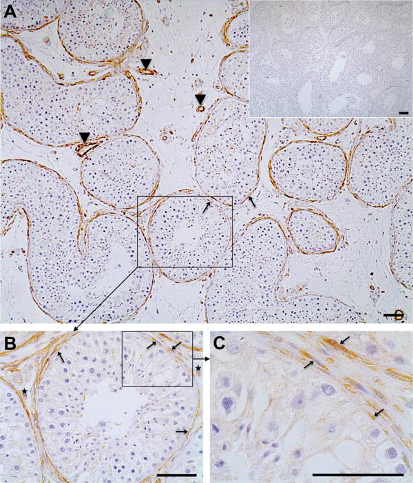
Figure 1. Immunohistochemistry of GPER in the human testis (A–C).
In the human testes smooth muscle-like peritubular cells (arrows; A–C) and smooth muscle cells of blood vessels (arrowheads; A, C) express GPER. Few interstitial cells (asterisks, B) also stain for GPER. Negative control (A, inset) was performed with IgG instead of the antibody. Haematoxylin was used to stain nuclei. Scale bars: 50 μm.

