Abstract
Persistent deficits in social behavior are among the major negative consequences associated with exposure to ethanol during prenatal development. Prior work from our laboratory has linked deficits in social behavior following moderate prenatal alcohol exposure (PAE) in the rat to functional alterations in the ventrolateral frontal cortex [21]. In addition to social behaviors, the regions comprising the ventrolateral frontal cortex are critical for diverse processes ranging from orofacial motor movements to flexible alteration of behavior in the face of changing consequences. The broader behavioral implications of altered ventrolateral frontal cortex function following moderate PAE have, however, not been examined. In the present study we evaluated the consequences of moderate PAE on social behavior, tongue protrusion, and flexibility in a variant of the Morris water task that required modification of a well-established spatial response. PAE rats displayed deficits in tongue protrusion, reduced flexibility in the spatial domain, increased wrestling, and decreased investigation, indicating that several behaviors associated with ventrolateral frontal cortex function are impaired following moderate PAE. A linear discriminant analysis revealed that measures of wrestling and tongue protrusion provided the best discrimination of PAE rats from saccharin-exposed control rats. We also evaluated all behaviors in young adult (4-5 mos.) or older (10-11 mos.) rats to address the persistence of behavioral deficits in adulthood and possible interactions between early ethanol exposure and advancing age. Behavioral deficits in each domain persisted well into adulthood (10-11 mos.), however, there was no evidence that age enhances the effects of moderate PAE within the age ranges that were studied.
Keywords: Fetal Alcohol Spectrum Disorders, Fetal Alcohol Syndrome, Prefrontal Cortex, Morris water task, Spatial Navigation, Aggression, Aging
1. Introduction
Deficits in behavior and cognition have long been identified as consequences of ethanol exposure during nervous system development [2, 23, 32, 39, 40, 47, 53, 55, 58, 72]. An estimated 1%-5% of children are diagnosed with fetal alcohol spectrum disorders (FASDs; [41]) which encompass, in decreasing order of severity, full-blown Fetal Alcohol Syndrome (FAS), partial FAS (pFAS), and alcohol-related neurodevelopmental disorders (ARNDs)[12]. Negative consequences are not limited to high levels of prenatal alcohol exposure (PAE), as moderate PAE that does not lead to the conspicuous morphological, behavioral and cognitive deficits characteristic of FAS can cause comparatively subtle, but nonetheless persistent, deficits in humans with FASDs [13, 59, 60] and non-human animals exposed to ethanol during brain development [66]. The importance of understanding the behavioral and corresponding neurobiological consequences of moderate PAE is underscored by current estimates indicating that the large majority of FASD cases fall within the less severe range of the spectrum [42]. Further, the incidence of less severe FASDs may be expected to increase as nearly 50% of women report drinking alcohol prior to recognition of pregnancy [11, 17, 19] and an alarmingly high percentage of women (ranging from 5% to >30%) report consumption of some ethanol while pregnant [10, 14, 49, 68] despite increased efforts to communicate the dangers of drinking for the developing fetus.
Deficits in social behavior and cognition are among the most common adverse outcomes observed in children with FASDs [15, 26, 29, 64]. Several independent laboratories have reported alterations in rodent social behavior related to ethanol exposure during brain development, including decreased investigation and interaction [21, 45, 65, 67], altered play [44, 45, 56], increased aggressive interactions [56], alterations in responsiveness to social stimuli [28, 36, 37], and deficits in socially acquired food preferences and social recognition memory [30]. Social behavior deficits have been observed following exposure to heavy (BECs ~3.0mg/dL, [30, 46]) or more moderate levels of ethanol (BECs ~0.80mg/dL, [21]), and across a broad range of parameters for other significant factors including exposure timing, duration of exposure, and age at the time of behavioral measurement. The neural mechanisms and circuits implicated in social behavior deficits are not well understood, however, because behavioral and cognitive processes in the social domain have been firmly associated with frontal cortex [1, 6, 16] this region is an obvious target for investigation. Prior work from our laboratory [21, 22] has linked alterations in social behavior following moderate PAE to alterations in the structure and function of frontal cortex neurons. Adult rats prenatally exposed to either moderate levels of ethanol or saccharin interacted with another rat for 10 minutes after which expression of the immediate early genes (IEGs) c-fos and Arc were quantified as a marker of neural activity in several regions of frontal cortex, including the ventrolateral frontal cortex (agranular insular cortex (Zilles’ area AID [73]) and the lateral orbital cortex (LO)), prelimbic cortex (Cg3) and the medial and lateral agranular cortices (Fr1 and Fr2). Social interaction in saccharin-exposed control rats yielded increases in c-fos expression in AID, LO, and Cg3 and increased Arc expression exclusively in AID. These social-experience-related increases in IEG expression were not observed in PAE rats. Further, exposure to novel cage-mates altered dendritic morphology on pyramidal neurons of AID in saccharin-exposed control rats but not following moderate PAE [21]. A prior lesion study by Kolb and Nonneman [33] revealed that normal adult rats with selective damage to ventrolateral frontal cortex that included the AID and LO regions resulted in increased aggression, whereas lesions of medial frontal cortex did not appear to affect social behavior, but instead led to increased general agitation or “touchiness”. Together with the IEG expression and structural plasticity data described above the available lesion data support the conclusion that PAE leads to alterations in behaviors associated with ventrolateral frontal cortex function. The broader behavioral and cognitive implications of alterations in ventrolateral frontal cortex function following PAE, however, have not been investigated.
The primary goal of the present study was to evaluate the effects of moderate PAE on social, perseverative, and motor behaviors associated with the ventrolateral frontal cortex. Pregnant Long-Evans rat dams voluntarily consumed moderate levels of ethanol or saccharin throughout pregnancy, and measures of social behavior, spatial response perseveration errors and tongue protrusion were obtained in adult male offspring (our prior studies have yielded robust social behavioral deficits in male but not female animals). For quantification of spatial response perseveration errors, we adopted a variant of the Morris water task in which rats were first given extensive training to navigate to single platform location after which the platform was moved to a novel position for an extended period of training. This allowed measurement of response perseveration errors for a well-learned spatial response over a much longer period of training than possible in the standard moving-platform variant of the task in which the platform is relocated to a new position every 4-8 trials [69]. Preliminary data revealed that pharmacological inactivation or blockade of metabotropic glutamate receptors selectively in AID but not Cg3 resulted in increased response perseveration errors in the absence of effects on initial learning or performance (unpublished observations). Deficits in spatial reversal learning, characterized by response perseveration errors, are observed in rodents following heavy ethanol exposure during brain development [54, 62, 63], however, general deficits of this form have not been investigated following moderate PAE. Tongue protrusion was examined using procedures previously described by Whishaw and colleagues [70, 71], who demonstrated that deficits in tongue protrusion are among the more robust outcomes following ventrolateral frontal cortex damage in the rat. These authors also found that near complete decortication that spared only the ventrolateral aspect of frontal cortex left tongue protrusion unimpaired; subsequent lesions to the remaining ventrolateral frontal cortical tissue resulted in profound deficits in tongue protrusion, suggesting that the ventrolateral frontal cortex circuitry targeted in these studies is both necessary and sufficient for this behavior. Related to our primary goal, we also sought to evaluate the relative utility of these behavioral measures for distinguishing PAE from non-exposed rats, particularly considering the need for model systems approaches tailored for investigating the frontocortical bases of PAE. To address this goal we performed a linear discriminant analysis that included all dependent measures obtained for social interaction, tongue protrusion, Morris water task learning and response perseveration errors.
A secondary goal of the present study was to evaluate the persistence of ethanol-related alterations in these behaviors. Prior studies by our laboratory and others have primarily evaluated social behavior in PAE offspring during the juvenile [44, 67], peri-adolescent [45, 67] or young adult stages ([21]; e.g., ~90-120 day old). We obtained each behavioral measure in PAE and saccharin-exposed rats at either 4-5 or 10-11 months of age. To our knowledge there are presently no reports that evaluate the interaction between early ethanol exposure and age in adulthood on the behaviors of interest. The possibility that these factors could be additive or synergistic, such that the more subtle consequences of moderate PAE may be enhanced and more readily detectable with age, provided additional motivation for evaluating behavioral outcomes at multiple ages in adulthood.
2. Method
2.1 Subjects
Subjects were 32 male Long-Evans rats obtained from the University of New Mexico Health Sciences Center Animal Resource Facility (see breeding protocol below). After weaning, all animals were housed with a single animal from the same prenatal treatment and different litter in standard plastic cages with water and food available ad libitum. All cage-mate pairs were matched for age and weight and animals were at least 4 months of age prior to behavioral testing. Groups of 6 saccharin-exposed and 6 prenatal-ethanol-exposed rats sampled from 12 litters were tested at 4-5 months of age. Groups of 10 saccharin-exposed and 10 prenatal-ethanol-exposed rats sampled from 12 litters were tested at 10-11 months of age. The sample size in the 10-11 month group was increased to allow for potential attrition with increasing age. Lights were maintained on a reverse 12h:12h light:dark cycle with lights on at 0900h. All procedures were approved by the Institutional Animal Care and Use Committee of either the main campus or Health Sciences Center at University of New Mexico.
2.2 Materials and Procedures
2.2.1 Breeding and Voluntary Ethanol Consumption During Gestation
All breeding procedures were conducted in the University of New Mexico HSC Animal Resource Facility (ARF). Three to four-month-old Long-Evans rat breeders (Harlan Industries, Indianapolis, IN) were single-housed in plastic cages at 22°C and kept on a reverse 12-hour light/dark schedule (lights on from 2100 to 0900 hours) with Purina Breeder Block rat chow and tap water ad libitum. After at least one week of acclimation to the animal facility, all female rats were provided 0.066% saccharin in tap water for four hours each day from 1000 to 1400 hours. On Days 1 and 2, the saccharin water contained 0% ethanol, on Days 3 and 4, saccharin water contained 2.5% ethanol (v/v). On Day 5 and thereafter, saccharin water contained 5% ethanol (v/v). Daily four-hour consumption of ethanol was monitored for at least two weeks and mean daily ethanol consumption was determined for each female breeder. Following two weeks of daily ethanol consumption females that drank less than one standard deviation below the mean of the entire group were removed from the study. The remainder of the females were assigned to either a saccharin control or 5% ethanol drinking group and matched such that the mean pre-pregnancy ethanol consumption by each group was comparable.
Subsequently, females were placed with proven male breeders until pregnant as evidenced by the presence of a vaginal plug. Female rats did not consume ethanol during the breeding procedure. Beginning on Gestational Day 1, rat dams were provided saccharin water containing either 0% or 5% ethanol for four hours a day, beginning precisely at 1000 hours (1 hour following the onset of the dark cycle). The volume of 0% ethanol saccharin water provided to the controls was matched to the mean volume of 5% ethanol in saccharin water consumed by the ethanol-drinking group, which has remained relatively consistent at about 16mL per four-hour drinking period over multiple breeding rounds. Rat chow was available ad libitum during both the drinking and non-drinking periods. Maternal weight gain during pregnancy and offspring birth-weight did not differ based on prenatal treatment group [21, 57]. Daily four-hour ethanol consumption was recorded for each dam. Ethanol consumption ceased at birth, and all litters were weighed and culled to 10 pups. Offspring were weaned at 24 days of age and transferred from the HSC-ARF to the Department of Psychology ARF. To minimize potential litter effects only 1-2 animals per each breeding pair were included in the present study.
2.2.2. Morris water task
A circular pool, 1.5 m in diameter and 48 cm in height, was filled to a depth of 26 cm with room temperature water (23° C) made opaque with a small amount (~200g) of non-toxic powdered paint. An escape platform (15 cm × 15 cm) was submerged ~1 cm below the surface of the water. A single room adjacent to the animal colony was used for all water task testing. A variety of distinctive, conspicuous items (posters, cabinets, equipment, etc.) on the walls of the room served as visual cues. All behavior was recorded via an overhead camera and digital camcorder. Videos were transferred to a computer for tracking and analysis using custom software developed in our laboratory. All trials were conducted in blocks of four in which each of four release points around the perimeter of the pool (NE,NW,SW,SE) were utilized with the order determined by pseudorandom selection without replacement. A trial ended when the animal either reached the platform or after 60 s had elapsed in which case the rat was placed on the platform by the experimenter. Rats were removed from the platform after ~5 s and were placed in holding cages between trials. The intertrial interval was ~ 5 min. All testing was conducted ~1-2 hours after the onset of the dark cycle.
The water task was conducted in three phases (see Figure 1). For all phases a total of three trial blocks were administered on each day of training. During Phase 1, rats were given 10 trial blocks; the first 9 blocks were distributed over days 1 through 3 and one additional block was conducted as the first block on day 4. Two possible platform locations were used (the center of the east or west quadrant), with each location used for half of the rats in each combination of prenatal treatment condition and age. Initial training was conducted in this manner to establish strong responding to the initial location and to minimize the effects of between-day reductions in performance on performance during the subsequent relocation trials. During Phase 2 the platform was repositioned to the other platform location (east or west) and 6 blocks of trials were conducted; the first two trial blocks were conducted on day 4 immediately following the last block of Phase 1, with three blocks on day 5 and one block at the beginning of day 6. The inclusion of one block of training on day 6 prior to the Phase 3 trials was based on the same rationale described for Phase 1. During Phase 3 the platform was returned to the original location and 5 additional blocks of training were conducted; two blocks were conducted on day 6 immediately following the final block of Phase 2 and three blocks were conducted on day 7. Mean latency and path length to reach the platform were measured for each trial block. For Phases 2 and 3, the number of trials per block that included a visit to the platform location used in the immediately preceding phase was also quantified. Separate repeated measures ANOVAs with prenatal treatment, age, and trial block as factors were conducted for each phase.
Figure 1.
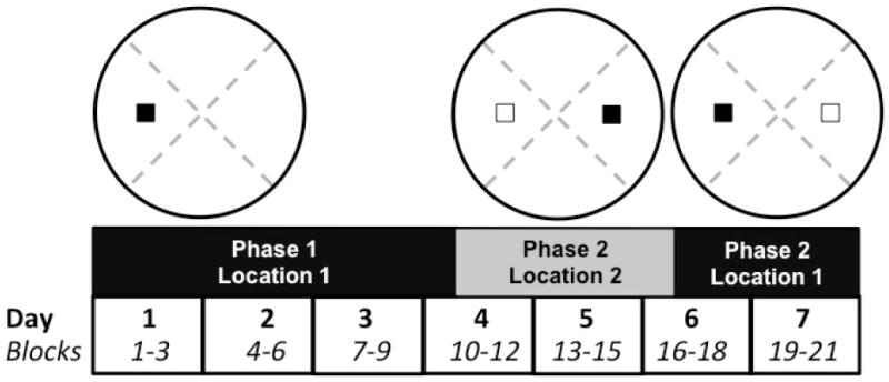
Diagram illustrating example platform locations and sequences of training in the Morris water task. Phase 1 included 10 training blocks of 4 trials spread over 4 days. The platform was either in the east or west (shown) for half of the rats for each combination of diet condition and age. The 6 blocks of Phase 2 were distributed over days 4-6. The platform was relocated to either the west or east (shown) depending on the location used in Phase 2. The number of trials per block that included visits to the former platform location (white rectangle, west as shown) was quantified. During the 5 blocks of Phase 3 the platform was moved back to the original location (west as shown). The number of trials per block that included visits to the platform location from Phase 2 (white rectangle, east as shown) was quantified.
2.2.3. Social Behavior
The procedures for housing, data collection and quantification of social behavior were similar to those utilized by [21]. Cagemate pairs were placed into a box (95 cm × 47 cm × 43 cm) with a Plexiglass front, opaque sides, and a mirrored back wall for 30 minutes on each of three consecutive days to habituate them to the apparatus. At the end of the final habituation session the animals were housed in isolation for 24 h. At the end of the isolation period the animals were placed into the test apparatus together and behavior was video-taped for 12 minutes. Videos were digitized for behavioral quantification using software developed in our laboratory. The duration, frequency and latency to first occurrence of the following behaviors were quantified: wrestling (including pinning), crawling (crossing) over/under the partner, anogenital sniffing, other sniffing of the partner’s body (body sniffing), allogrooming (grooming of the partner), rearing, and sniffing/digging in the bedding. We have previously quantified boxing [21], however, this behavior was only observed briefly in one pair of animals and is, therefore, not included in the analyses reported here. The six behaviors quantified here were selected based on a large body of extant literature [5] to target partner-directed (e.g., wrestling, investigation) behaviors, and have been quantified in prior work in our laboratory with PAE rats [21]. Separate analyses of variance (ANOVAs) were conducted for each measure with prenatal treatment and age as factors.
2.2.4. Tongue protrusion
Tongue protrusion measures were obtained in a single session in a room that was dimly lit by a red lamp. Rats were placed in a plastic hanging cage placed onto one side so that the wire mesh top was oriented vertically. To familiarize the animal with the food substrate used for measurement (chocolate syrup or creamy peanut butter) a small amount was placed inside the cage. Once it had been consumed an experimenter smeared a small amount of chocolate syrup or peanut butter onto one end of a thin piece of Plexiglass (2 mm × 5 cm × 7 cm). The end containing the food substrate was placed against the edge of the wire mesh. As the animal approached the food and attempted to lick, the Plexiglass was reoriented such that it was parallel to the animal’s mouth. After a lick attempt the Plexiglass was removed and an experimenter measured the length of the indentation left in the food substrate by the tongue. This was repeated until 5-12 measurements had been obtained for an individual animal in a 20 min. session. All attempts for a single animal were averaged for analysis. An ANOVA was conducted with prenatal treatment and age as factors.
2.2.5. Exploratory discriminant analysis
An additional, exploratory stepwise discriminant analysis [9] was conducted to evaluate which of the 75 dependent variables from the three behavioral tasks provide the best discrimination between PAE rats and prenatal saccharin-exposed control rats. The variables included tongue protrusion, latency and path length during each of the 21 blocks of the Morris water task, number of visits to the initial platform locations during each of the 6 blocks of Phase 2, number of visits to the Phase 2 platform location during each of the 5 blocks of Phase 3, and frequency, duration, and latency to first instance of wrestling, crossing over/under, anogenital sniffing, social sniffing, allogrooming, rearing, and digging.
3. Results
An alpha of p < 0.05 was adopted for all analyses. In addition to omnibus ANOVAs, the prenatal treatment factor was also evaluated within levels of the age factor for significant prenatal treatment main effects. Effect sizes (partial eta squared, η ) are provided for all significant effects.
3.1. Maternal ethanol consumption
During pregnancy, rat dams consumed a mean of 2.72 grams of ethanol during the daily four-hour drinking session. The mean peak serum ethanol concentration resulting from the voluntary consumption of 5% ethanol was reported previously as 84.0 + 5.5 mg/dL [57]. No significant differences in offspring birthweight, litter size or growth curves were observed using this moderate PAE paradigm (data not shown).
3.2. Morris water task
Mean escape latency and path length for each trial block during Phases 1-3 are shown in Figure 2.
Figure 2.
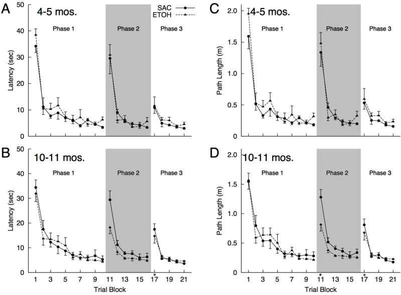
Mean (±SEM) latency (A-B) and path length (C-D) to navigate to the escape platform during each trial blocks of Phases 1-3 for prenatal treatment condition and age). [ * indicates a significant effect of Age at p < 0.05].
Phase 1. Initial training
There was a significant effect of trial block for latency [Greenhouse-Geisser corrected, F(4.35, 121.92) = 77.58, p < 0.001, η = 0.735] and path length [Greenhouse-Geisser corrected, F(4.32, 120.91) = 78.99, p < 0.001, η = 0.738] which resulted from a decrease in both measures across trial blocks. There were no significant prenatal treatment main effects, age main effects or interaction terms for either dependent measure [all ps > 0.23].
Phase 2. Platform relocation
For the 6 trial blocks during which the platform was placed in a new location there was a significant Trial Block X Age interaction for latency [Greenhouse-Geisser corrected, F(1.67, 46.70) = 3.53, p = 0.045, η = 0.112] and path length [Greenhouse-Geisser corrected, F(1.84, 51.51) = 5.62, p = 0.008, η = 0.167]. These interactions were due to significant age effects [10-11 mos. > 4-5 mos.] for path length during the first block of Phase 2 [day 4 trial block 2; F(1, 30) = 4.81, p = 0.036, η = 0.138] and for latency F(1, 30) = 5.90, p = 0.021, η = 0.164] and path length F(1, 30) = 5.69, p = 0.024, η = 0.159] during the second trial block of day 5, while all other age effects within trial blocks were not significant [all ps > 0.135]. All other interaction terms as well as the prenatal treatment and age main effects for the latency and path length measures were not significant [all ps > 0.096].
The mean numbers of visits to the original platform location during each of the 6 trial blocks of Phase 2 are shown in Figure 3. There was a significant block effect [F(5, 140) = 59.66, p < 0.001, η = 0.681] which resulted from a decrease in visits to the Phase 1 platform location across trial blocks. There was also a significant Trial Block X Prenatal Treatment interaction [F(5, 140) = 2.86, p = 0.017, η = 0.093], however, there were no other significant main effects or interactions [all ps > 0.13]. Inspection of individual swim paths suggested that PAE rats continued to visit the Phase 1 location more frequently than saccharin-exposed rats during the later stages of training, particularly during the final block of Phase 2. Example swim paths during the final Phase 2 trial block for each diet condition and age are shown in Figure 4. Analysis of simple effects revealed that PAE rats visited the Phase 1 platform location significantly more than saccharin-exposed rats during the final block of Phase 2 [F(1, 30) = 11.58, p = 0.002, η = 0.093], whereas no other prenatal treatment main effects within levels of the trial block factor were significant [all ps > 0.22]. The diet effect observed for the final trial block of Phase 2 was significant within each age group; 4-5 month old rats [ETOH > SAC; F(1, 10) = 5.98, p = 0.035, η = 0.374], 10-11 month old rats [ETOH > SAC; F(1, 18) = 5.23, p = 0.035, η = 0.225] .
Figure 3.
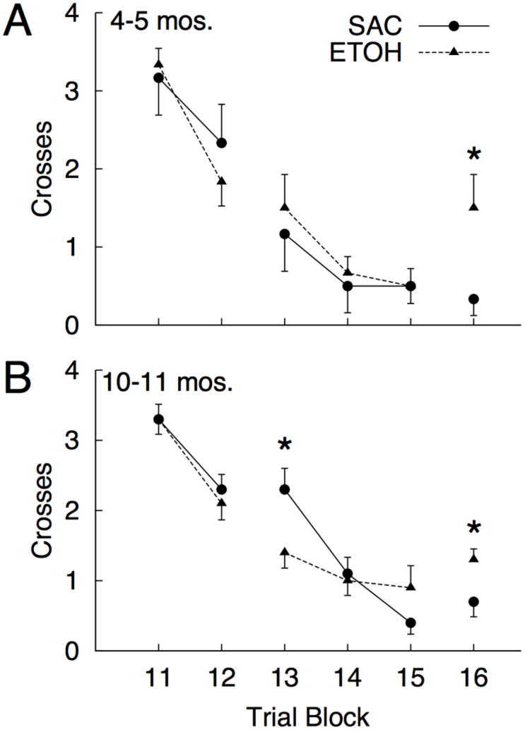
Mean (±SEM) visits to the Phase 1 (initial) platform location for each prenatal treatment and age group during the 6 trial blocks of Phase 2 training. During Phase 2, the platform was repositioned in the pool (see Figure 1). [ * p < 0.05].
Figure 4.
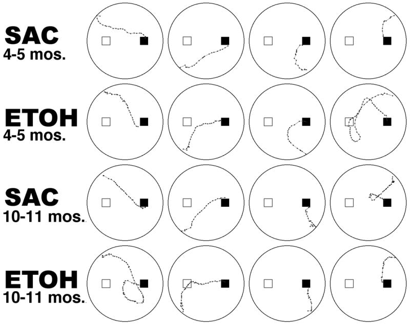
Representative swim paths during block 16 (Day 6, block 1; the final block of Phase 2) for each prenatal treatment and age group.
Phase 3. Original platform location
For the 5 trial blocks during which the platform was returned to the Phase 1 location there were significant reductions in latency [Greenhouse-Geisser corrected, F(1.29, 34.68) = 37.98, p < 0.001, η = 0.576] and path length [Greenhouse-Geisser corrected, F(1.29, 36.19) = 38.23, p < 0.001, η = 0.577] across trial blocks. No other main effects or interactions were observed [all ps > 0.068]. The mean numbers of visits to the Phase 2 platform location during each of the 5 trial blocks of Phase 3 are shown in Figure 5. There was a significant block effect [Greenhouse-Geisser corrected, F(3.06, 85.79) = 57.15, p < 0.001, η = 0.671] which resulted from a decrease in visits to the Phase 2 platform location across trial blocks. There was also a significant Trial Block X Prenatal Treatment interaction [Greenhouse-Geisser corrected, F(3,06, 85.79) = 2.85, p = 0.041, η = 0.092]. There were no other main effects or interactions [all ps > 0.39]. Analysis of simple effects revealed that PAE rats visited the Phase 2 platform location significantly less than saccharin-exposed rats during the first block of Phase 3 [F(1, 30) = 5.99, p = 0.021, η = 0.166], whereas no other diet main effects within levels of the trial block factor were significant [all ps > 0.17]. The prenatal treatment effect observed for the first trial block was significant for 10-11 month old rats [ETOH < SAC; F(1, 18) = 5.06, p = 0.037, η = 0.22], however, the effect for 4-5 month old rats was not significant [ETOH < SAC; F(1, 10) = 1.22, p = 0.29, η = 0.108].
Figure 5.
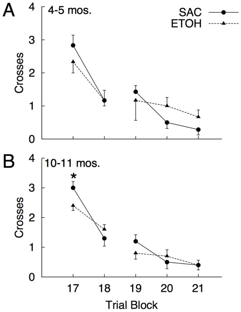
Mean (±SEM) visits to the Phase 2 (repositioned) platform location for each prenatal treatment and age group during the 5 trial blocks of Phase 3. During Phase 3, the platform returned to the initial (Phase 1) location in the pool (see Figure 1). [ * p < 0.05].
3.3. Social Behavior
Mean values for the frequency, duration and latency to first occurrence of each behavior quantified during the 12-minute social interaction session are shown in Table 1. For wrestling there were significant prenatal treatment main effects for the frequency [ETOH > SAC; F(1, 28) = 28.23, p < 0.001, η = 0.502], duration [ETOH > SAC; F(1, 28) = 22.43, p < 0.001, η = 0.445] and latency to first occurrence [ETOH < SAC; F(1, 28) = 7.25, p = 0.012, η = 0.206]. There were also significant main effects of age for duration [4-5 mos. > 10-11 mos.; F(1, 28) = 22.56, p < 0.001, η = 0.446] and latency to first occurrence of wrestling [4-5 mos. < 10-11 mos.; F(1, 28) = 5.46, p = 0.027, η = 0.163], and a Prenatal Treatment X Age interaction for duration [F(1, 28) = 8.59, p = 0.007, η = 0.235] of wrestling. Within the 4-5 month age group there were significant prenatal treatment comparisons for the frequency [ETOH > SAC; F(1, 10) = 60.50, p < 0.001, η = 0.858], duration [ETOH > SAC; F(1, 10) = 20.12, p < 0.001, η = 0.858], and latency to first occurrence [ETOH < SAC; F(1, 10) = 6.61, p = 0.028, η = 0.398] of wrestling. Within the 10-11 month age group there were significant effects for frequency [ETOH > SAC; F(1, 18) = 10.66, p = 0.004, η = 0.372 ] and latency to first occurrence [ETOH > SAC; F(1, 18) = 7.37, p = 0.014, η = 0.291] of wrestling. Although PAE rats wrestled more than saccharin-exposed rats, the duration effect was not significant for 10-11 month old rats [p > 0.13].
Table 1.
Mean (±SEM) frequency (1a), duration (1b) and latency to first occurrence (1c) for each behavior quantified during the 12 minute social interaction session for saccharin- (SAC) and prenatal ethanol-exposed (ETOH) rats from each age group. Significant (p < 0.05) main effects (p = prenatal treatment, a = age) and interactions (a*p) for separate ANOVAs conducted for each dependent measure are indicated in brackets.
| a. Frequency | 4-5 months | 10-11 months | ||
|---|---|---|---|---|
| SAC | ETOH | SAC | ETOH | |
| Wrestling [p] | 1.67 (0.21) | *5.33 (0.42) | 1.00 (0.37) | *4.00 (0.84) |
| Crossing over/under | 0.00 (0.00) | 0.33 (0.21) | 0.10 (0.10) | 0.30 (0.15) |
| Anogenital Sniffing | 4.83 (0.87) | 3.67 (1.33) | 2.00 (0.37) | 3.40 (1.13) |
| Body Sniffing | 15.17 (1.54) | 15.67 (3.59) | 9.40 (1.02) | 12.10 (2.91) |
| Allogrooming [a, a*p] | 5.00 (2.03) | 1.33 (0.56) | 0.60 (0.27) | 0.70 (0.50) |
| Rearing | 38.67 (4.70) | 50.00 (7.55) | 41.80 (3.02) | 44.00 (4.73) |
| Digging | 3.50 (0.76) | 5.17 (1.40) | 6.70 (2.03) | 11.90 (3.43) |
| b. Duration (sec) | 4-5 months | 10-11 months | ||
| SAC | ETOH | SAC | ETOH | |
|
| ||||
| Wrestling [p, a, a*p] | 16.21 (4.00) | *50.49 (6.51) | 8.08 (3.24) | 16.15 (4.08) |
| Crossing over/under [p] | 0.00 (0.00) | 0.65 (0.42) | 0.05 (0.05) | 0.21 (0.11) |
| Anogenital Sniffing | 9.59 (1.41) | 5.29 (1.75) | 4.56 (2.30) | 10.71 (6.43) |
| Body Sniffing [a] | 32.08 (3.55) | 29.46 (6.95) | 13.99 (2.77) | 20.10 (3.45) |
| Allogrooming [p, a] | 34.44 (10.81) | *8.10 (4.06) | 8.21 (5.29) | 3.97 (2.99) |
| Rearing | 101.04 (14.05) | 139.98 (21.34) | 117.70 (9.94) | 103.37 (10.68) |
| Digging | 12.49 (5.93) | 25.45 (9.34) | 16.87 (5.84) | 32.45 (9.10) |
| c. Latency (sec) | 4-5 months | 10-11 months | ||
| SAC | ETOH | SAC | ETOH | |
|
| ||||
| Wrestling [p, a] | 268.32 (32.15) | *174.51 (17.25) | 533.18 (64.96) | *243.03 (84.85) |
| Crossing over/under | 720.00 (0.00) | 661.17 (37.21) | 714.37 (5.63) | 593.26 (64.55) |
| Anogenital | 64.83 (36.08) | 181.62 (42.71) | 209.04 (41.81) | 214.29 (85.59) |
| Body Sniffing | 35.83 (13.64) | 17.89 (4.78) | 56.60 (12.36) | 45.25 (17.71) |
| Allogrooming [p] | 261.24 (109.00) | #549.10 (75.55) | 478.16 (99.50) | 618.95 (64.62) |
| Rearing | 17.08 (4.50) | 10.33 (2.60) | 126.50 (78.12) | 71.11 (60.03) |
| Digging | 187.81 (36.71) | 303.84 (69.57) | 323.03 (108.21) | 166.27 (65.42) |
indicates a significant prenatal treatment effect at p < 0.05 within levels of the age factor [ # p = 0.055].
There was a significant prenatal treatment main effect for duration of crossing over/under [ETOH > SAC; F(1, 28) = 5.07, p = 0.032, η = 0.153], and the prenatal treatment effect for latency to first occurrence of crossing over/under approached significance [ETOH < SAC; F(1, 28) = 4.05, p = 0.054, η = 0.126]. For allogrooming, there was a significant prenatal treatment main effect for duration [ETOH < SAC; F(1, 28) = 6.71, p = 0.015, η = 0.193] and latency to first occurrence [ETOH < SAC; F(1, 28) = 5.38, p = 0.028, η= 0.161]. The prenatal treatment effect for the frequency of allogrooming approached significance [ETOH < SAC; F(1, 28) = 4.13, p = 0.052, η = 0.128]. There was, however, a significant Prenatal Treatment X Age interaction for the frequency of allogrooming [F(1, 28) = 4.60, p = 0.041, η = 0.141], which resulted from a significant age effect in saccharin-exposed rats [4-5 mos. > 10-11 mos.; F(1, 14) = 7.79, p = 0.014, η = 0.358] that was absent in PAE rats [p > 0.42]. There were several significant effects of age, including main effects for the frequency [4-5 mos. > 10-11 mos; F(1, 28) = 8.22, p = 0.008, η = 0.227] and duration [4-5 mos. > 12 mos.; F(1, 28) = 6.61, p = 0.016, η = 0.191] of allogrooming.
There were no significant effects of prenatal treatment for anogenital sniffing, body sniffing, rearing, or digging [all ps > 0.19]. The only additional significant effect was an age main effect for duration of body sniffing [4-5 mos. > 10-11 mos.; F(1, 28) = 11.02, p = 0.003, η = 0.282]. All other effects of age failed to reach significance [all ps > 0.07].
3.4. Tongue Protrusion
Mean tongue protrusion values for each prenatal treatment and age group are shown in Figure 6. There were significant effects of age [4-5 mos. > 10-11 mos.; F(1, 28) = 72.16, p < 0.001, η = 0.72] and prenatal treatment [ETOH < SAC; F(1, 28) = 42.18, p < 0.001, η = 0.601]. Within age groups there were significant prenatal treatment effects at 4-5 months [ETOH < SAC; F(1, 10) = 18.14, p = 0.002, η = 0.645] and at 10-11 months of age [ETOH < SAC; F(1, 18) = 29.09, p < 0.001, η = 0.618]. The Prenatal Treatment X Age interaction was not significant [p = 0.64].
Figure 6.
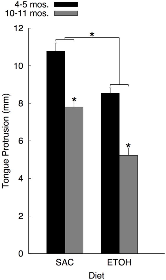
Mean (±SEM) tongue protrusion for saccharin (SAC) and prenatal ethanol-exposed (ETOH) rats taken at age 4-5 months (n = 6 per prenatal treatment condition) or 10-11 months (n = 10 per prenatal treatment condition). [ * p < 0.05].
3.4. Exploratory discriminant analysis
The stepwise discriminant analysis resulted in a single discriminant function that included the following 6 variables (and their standardized canonical discriminant function coefficients): wrestling frequency (1.360), tongue protrusion (-1.241), duration of body sniffing (0.835), anogenital sniffing frequency (0.664), rearing frequency (0.584), and the swim path length during the first block of Phase 2 (0.479). This discriminant function accounted for 86.68% of the between group variability, canonical R2=.87, [λ = 0.133, χ2 = 54.50, p < 0.001]. The cross-validated classification showed that 100 percent of cases were classified correctly. Inspection of the structure matrix revealed that the loading of two variables met criterion of magnitude greater than ±0.3, including frequency of wrestling (.377) and latency to first instance of wrestling (-.345), indicating that these variables provided the best discrimination between groups. We also note that several variables had loadings greater in magnitude than ±0.26, including tongue protrusion (-0.267), latency to the first instance of allogrooming (0.271), the number of visits to the initial platform location during the fifth block of Phase 2 (0.286), and the escape latency during the fourth block of Phase 3 (0.280).
4. General Discussion
Rats exposed to moderate levels of ethanol during prenatal brain development displayed sizeable deficits in tongue protrusion, alterations in social behavior including increased wrestling and decreased interaction in the form of reduced allogrooming, and increases in spatial response perseveration errors in a modified Morris water task. These observations indicate that the negative consequences of moderate PAE include several behavioral and cognitive processes that engage and depend upon ventrolateral frontal cortex, and expand the scope of the deficits beyond the social domain to include motor (tongue protrusion) and perseverative behavior. The effect sizes for tongue protrusion and specific social behaviors (e.g., wrestling) following moderate PAE were large in comparison to the more modest effect sizes observed for spatial response perseveration errors. The linear discriminant analysis revealed that two measures of social interaction (the frequency and latency to first instance of wrestling) provided the best discrimination of prenatal treatment conditions, followed by tongue protrusion, other measures of social behavior (latency to first instance of allogrooming) and finally spatial response perseveration errors. Thus, while each behavioral task described here may hold utility in future investigations of PAE on frontal cortex function and related behaviors for a range of levels of analysis, the effects of ethanol on social behavior appear to be among the most robust adverse outcomes associated with moderate PAE. Although there were clear deficits associated with increasing age in adulthood for many of the behavioral variables of interest, the effects of age did not appear to enhance or exacerbate the effects due to moderate PAE.
4.1. Social Behavior
The effects of moderate PAE on wrestling and social investigation in adult rats replicate our prior observations [21] and join the growing body of work indicating that negative consequences in the social domain are among the major outcomes associated with ethanol exposure during brain development. Alterations in social behaviors have been reported by several laboratories using a variety of doses, timing of exposure, duration of exposure and ages at the time of behavioral measurement [21, 27, 28, 30, 45, 46, 65]. To our knowledge, the present study is the first to demonstrate alterations in social behavior that persist well into adulthood (at 10-11 months of age). Considering that the typical lifespan of a Long-Evans rat housed under normal laboratory settings is approximately 24-36 months [25, 38] the age ranges used in the present study did not include advanced age beyond reproductive senescence. To address whether alterations in social behavior following PAE persist for the life of the individual will require future investigations that include additional time-points beyond 1-2 years of age.
The observation by Pellis and colleagues [50] that early lesions to the ventrolateral frontal cortex in rats result in increased play behavior prompted us to also consider the possibility that wrestling observed in PAE rats actually reflects play rather than genuine aggression. In our previous studies in young adult animals [21], and in the present study with a broader age range, we have failed to observe conspicuous behavioral indicators of aggressive behavior, such as fighting or biting. In the rat, play behavior peaks during post-weaning development around postnatal days 30-40 [43, 51] and declines as animals approach adulthood. Further, the topographies of play and adult agonistic behaviors are similar, so the observation of increased wrestling in PAE rats as old as 10-11 months of age would seem to score against the idea that these behavioral outcomes are merely manifestations of delayed maturation. In an attempt to arrive at a more definitive evaluation, we also quantified the frequency of attacks directed at the nape of the neck, the primary target of playful attacks, and attacks directed toward the rump, a target of non-playful attacks [7]. Attacks directed at the nape were rare, occurring only 3 times for two animals from the entire group of PAE rats. In contrast, a total of 22 attacks directed at the rump were observed in 10 of 14 PAE rats. Considering that almost all PAE rats (14 of 16) engaged in wrestling the discrepancy between the number of attacks directed toward the rump and nape suggests that PAE-related increases in wrestling reflect aggressive, agonistic encounters rather than play. To more firmly address the distinction between aggression and play in PAE rats future studies could also include quantification of ultrasonic vocalizations (USVs) during behavior to evaluate whether the USVs produced during wrestling are more similar to those that occasion play behavior as opposed to aggressive interactions.
Considering that the present results regarding the effects of PAE on wrestling in adulthood closely match our previous observations [21] it is important to note that the present study deviated methodologically from our prior study in that all rats in the present study were housed with non-littermates immediately post-weaning rather than with littermates. Housing with non-littermates might be expected to impact social behavior, however, the effects of PAE on wrestling were comparable in both studies. Further, preliminary studies in our laboratory on tongue protrusion and spatial response perseveration errors in PAE and saccharin-exposed rats in which all animals were housed with littermates yielded similar results to those observed here. These observations suggest that the effects of PAE on the behaviors of interest may not be sensitive to housing with non-littermates, at least when the animals are familiar with the cage-mate at the time of testing [21]. In the present study all rats were housed with their cage-mate for 90 to 270 days prior to testing, thus, the cage-mates should have been familiar at the time of testing which may have been an important determinant of the outcomes observed here and the similarities to previous observations using different housing procedures.
In the present study, we also observed evidence of PAE-related alterations in allogrooming, which may be considered a form of social investigation. Allogrooming was observed rarely in our prior work with animals of approximately 4 months of age. The increased occurrence of allogrooming in the present study may be partially related to the fact that the duration of social interaction was increased by 20% (to 12 minutes) compared to our prior work, especially considering that most instances of allogrooming occurred later in the session. In our previous work [21] we also observed alterations in social investigation (sniffing) when cage-mate novelty rather than isolation was used to motivate interaction. Thus, future work could evaluate whether changes in social investigation following moderate PAE are equally persistent when different procedures to motivate social interaction are utilized. This may, however, prove difficult to assess considering that investigation appears to decrease quite dramatically with age, making detection of PAE-related alterations in social investigation more difficult.
4.2. Tongue protrusion
Effects of PAE on motor behaviors including deficits in skilled forelimb movements in rats [24] and sensory guided reaching in humans with FASDs [72] have been examined by several laboratories. Because of the well-established neural pathways involved in motor behaviors, the use of motor tasks is likely to factor prominently in future efforts to understand the neural bases of specific deficits associated with PAE [20, 72]. To our knowledge the consequences of PAE during brain development on orofacial movements, including movements of the tongue, have not been examined. Although the primary neuromuscular and sensory aspects of tongue movement are controlled by afferents from primary motor cortex (M1) and coordinated signaling in primary somatosensory cortex (S1) [3, 4], the principal motivation for examining tongue protrusion in the present study was driven by evidence that ventrolateral frontal cortex circuitry is necessary and sufficient for this behavior. Whishaw and Kolb [70] measured tongue protrusion as well as eating and drinking in rats that were completely decorticated or received selective lesions of the ventrolateral frontal cortex, motor cortex, medial frontal cortex, parietal cortex or lateral hypothalamus. The most profound deficits in tongue protrusion were observed in decorticate rats and rats with lesions of the ventrolateral frontal cortex, with intermediate deficits observed in rats with motor cortex damage, and no deficits observed following parietal or medial frontal cortex damage. Rats with damage to the lateral hypothalamus never returned to normal levels of eating and drinking, but almost fully recovered the capacity for tongue protrusion. A subsequent study [71] demonstrated that tongue protrusion was spared in rats that had all neocortical tissue removed except for ventrolateral frontal cortex, and ablation of the remaining ventrolateral frontal cortex impaired tongue protrusion. Thus, the robust deficits in tongue protrusion following moderate PAE are consistent with alterations in the functional of this circuitry. Although the lesion data indicate that damage to the ventrolateral frontal cortex is necessary and sufficient for deficits in tongue protrusion, it is important to reiterate that tongue protrusion involves a distributed set of brain regions and pathways, including the aforementioned primary sensory and motor regions. Thus, the dependence of tongue protrusion on ventrolateral frontal cortex cannot be taken to indicate that alterations in other aspects of this distributed circuit are absent following PAE or that alterations in other aspects of this circuit do not contribute to the ethanol-related deficits observed here. Future studies on the sensory and motor pathways involved in tongue protrusion are needed to evaluate the potential role of these pathways in PAE-related deficits in tongue protrusion.
One possibility that must be considered concerns the potential for alterations in motivation to account for the effects of moderate PAE in tongue protrusion. Two observations are relevant to evaluating this possibility. There were no differences between PAE and saccharin control rats in the number of lick attempts within the session (both approximately 6 attempts per group; data not shown). Further, taste reactivity has been evaluated using quinine and sucrose solutions as part of a separate project in our laboratory using the same PAE paradigm. In both cases, PAE rats decreased (quinine) or increased (sucrose) consumption to levels comparable to saccharin-exposed control rats [52]. This observation is of further importance in the present context because frank damage to AID leads to clear deficits in flavor discrimination [31]. Moderate PAE does not appear to have adverse effects in this regard while other aspects of behavior (e.g., tongue protrusion) are sensitive to PAE. Collectively, these observations suggest that moderate PAE does not have a notable effect on motivation to consume appetitive or avoid aversive flavors, which would further suggest that deficits in tongue protrusion are not simply a result of reduced motivation for consumption of the food substrate.
4.3. Morris water task : Learning and flexibility in the spatial domain
In the initial phase of training in the Morris water task there were no detectable differences between groups in learning to navigate to an escape platform in a fixed location, which replicates prior observations on place learning following moderate PAE [57] and is further consistent with a lack of motivational differences based on PAE as discussed above. The effects of moderate PAE appear to be limited to an increase in erroneous visits to the initial platform location following platform relocation. Prior observations in Sprague-Dawley rats suggested that moderate PAE reduced the rate of learning in a moving platform variant of the water task [61], in which the platform is moved to a novel position every 4-8 trials [69]. Specifically, PAE rats displayed less of a reduction in escape latency from trial 1 to trial 2 when the platform was moved, which may indicate impaired learning in a more challenging variant of the task and/or increased persistence in visits to the former platform location as would be expected with a reduction in flexibility. To address the latter possibility more clearly, we utilized a set of procedures in which rats were first trained to navigate to a hidden platform in a fixed location over the course of 4 days (40 trials) in order to establish a strong place response to a single location. During the sequence of trials on day 4 the platform was relocated to the diametrically opposite quadrant of the pool where it remained for a total of 24 trials (Phase 2), which were distributed over the following three days. Both PAE and saccharin-exposed control rats displayed reductions in escape latency and path length over the course of Phase 2. Whereas both groups continued to visit the previous platform location early during Phase 2, the PAE group continued to visit the former platform location even at the very end of Phase 2 training. These visits were rapid (i.e., not occasioned by persistence in searching) and may be considered opportunistic in that they tended to occur when animals were released nearby to the old platform location. Together with the fact that even well-trained control animals will execute suboptimal (i.e., indirect) paths on most trials, the visits to the former platform location by PAE rats were not sufficient to result in detectable alterations in path length and escape latency, and were only apparent through verification of visits to the former platform location in the video record. Although PAE rats in both age groups displayed increased errors late in Phase 2 training it is important to note that 10-11 month saccharin-exposed rats displayed more errors, longer latencies and higher path length that PAE rats early during Phase 2 training. These differences did not persist and were attributable to increased searching at the former location during the initial trials following platform relocation. Additional data consistent with increased persistence in visiting the Phase 1 platform location by ethanol-exposed rats come from the initial block of Phase 3 in which the platform was returned to the original location. In this block saccharin-exposed rats visited the more recently used Phase 2 location more frequently, suggesting a preference for the Phase 1 location in PAE rats.
The overall pattern of findings from the Morris water task were less robust than those observed for social behavior and tongue protrusion, but were nonetheless consistent with the expected outcomes based on other examinations of spatial response perseveration errors [54, 62] or reduced flexibility in the spatial domain [61]. Importantly, Kolb and colleagues [34] utilized a similar method to evaluate the effects of damage to the ventrolateral frontal cortex on spatial learning and navigation in the Morris water task. Similar to the present data, escape latencies of rats with damage to the ventrolateral frontal cortex during the “reversal” phase were comparable to that of control animals, however, perseveration errors were not quantified so a direct comparison is not possible. In preliminary work for the current study we evaluated the effects of more restricted, subtotal neurotoxic lesions of AID and LO or reversible inactivation of AID via local infusions of lidocaine on performance in this variant of water task. Both produced increases in perseveration errors when the platform was moved in the absence of effects on initial learning or reversal performance indicated by latency and path length (unpublished observations). Thus, several manipulations of ventrolateral frontal cortex are sufficient to yield increased perseveration errors as described here.
4.5. Age and persistence of ethanol-related behavioral consequences
With respect to our secondary goal, the effects of age observed here prompt two general conclusions. First, the effects of moderate PAE on tongue protrusion, response perseveration and wrestling appear to persist well into adulthood, as they were observed in 4-5 month old and 10-11 month old animals. One exception was allogrooming, however, this behavior decreased to very low levels in both PAE and saccharin-exposed rats at 10-11 months of age. Second, the effects of moderate PAE did not appear to be modulated by increasing age in adulthood, at least for the behavioral measures obtained here. One exception was a modest enhancement of some aspects of spatial response errors in the Morris water task.
4.6. Limitations and future directions
Given that the age range in the present study was limited to animals under 1 year of age, future studies should more thoroughly evaluate the potential interaction of PAE and aging using a broader range of ages that extends beyond reproductive senescence and into the later stages of life. The present study was also limited to examinations in male rats, which was based on several prior replications by our group demonstrating that effects of moderate PAE in social behaviors are primarily limited to males. Preliminary work on tongue protrusion and spatial response perseveration errors also failed to yield compelling effects of PAE in female offspring, although we have detected effects in other domains such as contextual fear conditioning [57]. To provide greater focus and emphasis on the factors of age and behavioral outcomes, we opted to limit the present report to male rats. However, characterization of sex effects are critical for future work and we are currently completing a separate study to evaluate the effects of sex and moderate PAE on these behaviors. Finally, although deficits in social behavior have been well documented in children with FASDs and in rodents exposed to ethanol during prenatal development, we are unaware of extant data on tongue protrusion or related deficits in humans with FASDs. Alterations in the length, duration or topography of tongue protrusion behaviors have been identified in humans with other disorders [8, 18, 35, 48]. Future studies to assess tongue protrusion in children with FASDs will be important for establishing whether the present findings yield corresponding results in humans. If so, the measurement of tongue protrusion and other orofacial movements may hold considerable importance for understanding the neural mechanisms and circuits that underlie behavioral consequences of PAE.
Summary
With respect to the primary goal of the current study we found that rats exposed to moderate levels of ethanol prenatally displayed deficits or alterations on several aspects social behavior, tongue protrusion and flexible spatial behavior. Collectively, these outcomes suggest that moderate PAE results in persistent alterations in behaviors that depend upon the ventrolateral frontal cortex and identify these and functionally-related regions as important targets for future research on the functional consequences of PAE during brain development.
Prenatal ethanol exposure impaired social behavior and tongue protrusion
Behavioral deficits suggest alterations in ventrolateral frontal cortex function
Prenatal ethanol exposure effects were persistent well into adulthood
Prenatal ethanol exposure effects were not enhanced with aging in adulthood
Acknowledgments
SUPPORT : Support provided by grant AA019462 to DAH and AA019884 to DDS.
Footnotes
Publisher's Disclaimer: This is a PDF file of an unedited manuscript that has been accepted for publication. As a service to our customers we are providing this early version of the manuscript. The manuscript will undergo copyediting, typesetting, and review of the resulting proof before it is published in its final citable form. Please note that during the production process errors may be discovered which could affect the content, and all legal disclaimers that apply to the journal pertain.
References
- 1.Anderson SW, Bechara A, Damasio H, Tranel D, Damasio AR. Impairment of social and moral behavior related to early damage in human prefrontal cortex. Nat Neurosci. 1999;2:1032–7. doi: 10.1038/14833. [DOI] [PubMed] [Google Scholar]
- 2.Autti-Ramo I, Granstrom ML. The psychomotor development during the 1st year of life of infants exposed to intrauterine alcohol of various duration - fetal alcohol exposure and development. Neuropediatrics. 1991;22:59–64. doi: 10.1055/s-2008-1071418. [DOI] [PubMed] [Google Scholar]
- 3.Avivi-Arber L, Fung M, Bernfeld N, Perez M, Lee J-C, Sessle BJ. Organization of tongue motor representations within rat primary motor and somatosensory cortex. Society for Neuroscience Abstract Viewer and Itinerary Planner. 2012 14.02. [Google Scholar]
- 4.Avivi-Arber L, Martin R, Lee JC, Sessle BJ. Face sensorimotor cortex and its neuroplasticity related to orofacial sensorimotor functions. Archives of Oral Biology. 2011;56:1440–65. doi: 10.1016/j.archoralbio.2011.04.005. [DOI] [PubMed] [Google Scholar]
- 5.Barnett SA. A study in behaviour: Principles of ethology and behavioural physiology displayed mainly in the rat. London: 1963. [Google Scholar]
- 6.Berthoz S, Armony JL, Blair RJ, Dolan RJ. An fMRI study of intentional and unintentional (embarrassing) violations of social norms. Brain. 2002;125:1696–708. doi: 10.1093/brain/awf190. [DOI] [PubMed] [Google Scholar]
- 7.Blanchard RJ, Blanchard DC, Takahashi T, Kelley MJ. Attack and defensive behavior in albino-rat. Anim Behav. 1977;25:622–34. doi: 10.1016/0003-3472(77)90113-0. [DOI] [PubMed] [Google Scholar]
- 8.Buntinx IM, Hennekam RCM, Brouwer OF, Stroink H, Beuten J, Mangelschots K, et al. Clinical profile of Angelman syndrome at different ages. American Journal of Medical Genetics. 1995;56:176–83. doi: 10.1002/ajmg.1320560213. [DOI] [PubMed] [Google Scholar]
- 9.Burns R, Burns R. Business Research Methods and Statistics using SPSS. London: Sage Publications Ltd.; 2008. [Google Scholar]
- 10.Centers for Disease Control. Alcohol use among women of childbearing age: United States, 1994-1999. Morb Mortal Weekly Rep. 2002;51:273–6. [PubMed] [Google Scholar]
- 11.Centers for Disease Control. Alcohol use among pregnant and nonpregnant women of childbearing age-United States, 1991-2005. Morb Mortal Weekly Rep. 2009;58:529–32. [PubMed] [Google Scholar]
- 12.Chasnoff IJ, Wells AM, Telford E, Schmidt C, Messer G. Neurodevelopmental functioning in children with FAS, pFAS, and ARND. Journal of Developmental and Behavioral Pediatrics. 2010;31:192–201. doi: 10.1097/DBP.0b013e3181d5a4e2. [DOI] [PubMed] [Google Scholar]
- 13.Conry J. Neuropsychological deficits in Fetal Alcohol Syndrome and fetal alcohol effects. Alcohol Clin Exp Res. 1990;14:650–5. doi: 10.1111/j.1530-0277.1990.tb01222.x. [DOI] [PubMed] [Google Scholar]
- 14.Day NL, Cottreau CM, Richardson GA. The epidemiology of alcohol, marijuana, and cocaine use among women of childbearing age and pregnant-women. Clinical Obstetrics and Gynecology. 1993;36:232–45. doi: 10.1097/00003081-199306000-00005. [DOI] [PubMed] [Google Scholar]
- 15.Disney ER, Iacono W, McGue M, Tully E, Legrand L. Strengthening the case: Prenatal alcohol exposure is associated with increased risk for conduct disorder. Pediatrics. 2008;122:E1225–E30. doi: 10.1542/peds.2008-1380. [DOI] [PMC free article] [PubMed] [Google Scholar]
- 16.Eisenberger NI, Lieberman MD, Williams KD. Does rejection hurt? An fMRI study of social exclusion. Science. 2003;302:290–2. doi: 10.1126/science.1089134. [DOI] [PubMed] [Google Scholar]
- 17.Floyd RL, Decoufle P, Hungerford DW. Alcohol use prior to pregnancy recognition. Am J Prev Med. 1999;17:101–7. doi: 10.1016/s0749-3797(99)00059-8. [DOI] [PubMed] [Google Scholar]
- 18.Glatz-Noll E, Berg R. Oral dysfunction in children with Down’s Syndrome an evaluation of treatment effects by means of video registration. European Journal of Orthodontics. 1991;13:446–51. doi: 10.1093/ejo/13.6.446. [DOI] [PubMed] [Google Scholar]
- 19.Grant TM, Huggins JE, Sampson PD, Ernst CC, Barr HM, Streissguth AP. Alcohol use before and during pregnancy in western Washington, 1989-2004: implications for the prevention of Fetal Alcohol Spectrum Disorders. American Journal of Obstetrics and Gynecology. 2009;200 doi: 10.1016/j.ajog.2008.09.871. [DOI] [PMC free article] [PubMed] [Google Scholar]
- 20.Hamilton DA. The importance of measurement precision and behavioral homologies in evaluating the behavioral consequences of fetal-ethanol exposure: Commentary on Williams and colleagues (“Sensory-motor deficits in children with Fetal Alcohol Spectrum Disorder assessed using a robotic virtual reality platform“) Alcohol Clin Exp Res. 2014;38:40–3. doi: 10.1111/acer.12328. [DOI] [PMC free article] [PubMed] [Google Scholar]
- 21.Hamilton DA, Akers KG, Rice JP, Johnson TE, Candelaria-Cook FT, Maes LI, et al. Prenatal exposure to moderate levels of ethanol alters social behavior in adult rats: Relationship to structural plasticity and immediate early gene expression in frontal cortex. Behav Brain Res. 2010;207:290–304. doi: 10.1016/j.bbr.2009.10.012. [DOI] [PMC free article] [PubMed] [Google Scholar]
- 22.Hamilton DA, Candelaria-Cook FT, Akers KG, Rice JP, Maes LI, Rosenberg M. Patterns of social-experience-related c-fos and Arc expression in the frontal cortices of rats exposed to saccharin or moderate levels of ethanol during prenatal brain development. Behav Brain Res. 2010;214:66–74. doi: 10.1016/j.bbr.2010.05.048. [DOI] [PMC free article] [PubMed] [Google Scholar]
- 23.Hamilton DA, Kodituwakku P, Sutherland RJ, Savage DD. Children with Fetal Alcohol Syndrome are impaired at place learning but not cued-navigation in a virtual Morris water task. Behav Brain Res. 2003;143:85–94. doi: 10.1016/s0166-4328(03)00028-7. [DOI] [PubMed] [Google Scholar]
- 24.Heck DH, Roy S, Xie N, Waters RS. Prenatal alcohol exposure delays acquisition and use of skilled reaching movements in juvenile rats. Physiol Behav. 2008;94:540–4. doi: 10.1016/j.physbeh.2008.03.011. [DOI] [PubMed] [Google Scholar]
- 25.Holloszy JO. Mortality rate and longevity of food-restricted exercising male rats: A reevaluation. J Appl Physiol. 1997;82:399–403. doi: 10.1152/jappl.1997.82.2.399. [DOI] [PubMed] [Google Scholar]
- 26.Kelly SJ, Day N, Streissguth AP. Effects of prenatal alcohol exposure on social behavior in humans and other species. Neurotoxicol Teratol. 2000;22:143–9. doi: 10.1016/s0892-0362(99)00073-2. [DOI] [PMC free article] [PubMed] [Google Scholar]
- 27.Kelly SJ, Day N, Streissguth AP. Effects of prenatal alcohol exposure on social behavior in humans and other species. Neurotoxicol Teratol. 2000;22:143–9. doi: 10.1016/s0892-0362(99)00073-2. [DOI] [PMC free article] [PubMed] [Google Scholar]
- 28.Kelly SJ, Dillingham RR. Sexually dimorphic effects of perinatal alcohol exposure on social interactions and amygdala DNA and DOPAC concentrations. Neurotoxicol Teratol. 1994;16:377–84. doi: 10.1016/0892-0362(94)90026-4. [DOI] [PubMed] [Google Scholar]
- 29.Kelly SJ, Goodlett CR, Hannigan JH. Animal models of fetal alcohol spectrum disorders: Impact of the social environment. Dev Disabil Res Rev. 2009;15:200–8. doi: 10.1002/ddrr.69. [DOI] [PMC free article] [PubMed] [Google Scholar]
- 30.Kelly SJ, Tran TD. Alcohol exposure during development alters social recognition and social communication in rats. Neurotoxicol Teratol. 1997;19:383–9. doi: 10.1016/s0892-0362(97)00064-0. [DOI] [PubMed] [Google Scholar]
- 31.Kesner RP, Gilbert PE. The role of the agranular insular cortex in anticipation of reward contrast. Neurobiol Learn Mem. 2007;88:82–6. doi: 10.1016/j.nlm.2007.02.002. [DOI] [PMC free article] [PubMed] [Google Scholar]
- 32.Kodituwakku PW. Defining the behavioral phenotype in children with fetal alcohol spectrum disorders: A review. Neurosci Biobehav Rev. 2007;31:192–201. doi: 10.1016/j.neubiorev.2006.06.020. [DOI] [PubMed] [Google Scholar]
- 33.Kolb B, Nonneman AJ. Frontolimbic lesions and social-behavior in rat. Physiol Behav. 1974;13:637–43. doi: 10.1016/0031-9384(74)90234-0. [DOI] [PubMed] [Google Scholar]
- 34.Kolb B, Sutherland RJ, Whishaw IQ. A comparison of the contribution of frontal and parietal association cortex to spatial localization in rats. Behav Neurosci. 1983;97:13–27. doi: 10.1037//0735-7044.97.1.13. [DOI] [PubMed] [Google Scholar]
- 35.Kyllerman M. Angelman syndrome. Handbook of clinical neurology. 2013;111 doi: 10.1016/B978-0-444-52891-9.00032-4. [DOI] [PubMed] [Google Scholar]
- 36.Lugo JN, Marino MD, Cronise K, Kelly SJ. Effects of alcohol exposure during development on social behavior in rats. Physiology and Behavior. 2003;78:185–94. doi: 10.1016/s0031-9384(02)00971-x. [DOI] [PubMed] [Google Scholar]
- 37.Lugo JN, Marino MD, Gass JT, Wilson MA, Kelly SJ. Ethanol exposure during development reduces resident aggression and testosterone in rats. Physiology and Behavior. 2006;87:330–7. doi: 10.1016/j.physbeh.2005.10.005. [DOI] [PMC free article] [PubMed] [Google Scholar]
- 38.Masoro EJ. Mortality and growth-characteristics of rat strains commonly used in aging research. Experimental Aging Research. 1980;6:219–33. doi: 10.1080/03610738008258359. [DOI] [PubMed] [Google Scholar]
- 39.Mattson SN, Riley EP. Implicit and explicit memory functioning in children with heavy prenatal alcohol exposure. J Int Neuropsychol Soc. 1999;5:462–71. doi: 10.1017/s1355617799555082. [DOI] [PubMed] [Google Scholar]
- 40.Mattson SN, Roesch SC, Glass L, Deweese BN, Coles CD, Kable JA, et al. Further development of a neurobehavioral profile of Fetal Alcohol Spectrum Disorders. Alcohol Clin Exp Res. 2013;37:517–28. doi: 10.1111/j.1530-0277.2012.01952.x. [DOI] [PMC free article] [PubMed] [Google Scholar]
- 41.May PA, Blankenship J, Marais AS, Gossage JP, Kalberg WO, Barnard R, et al. Approaching the prevalence of the full spectrum of Fetal Alcohol Spectrum Disorders in a South African population-based study. Alcohol Clin Exp Res. 2013;37:818–30. doi: 10.1111/acer.12033. [DOI] [PMC free article] [PubMed] [Google Scholar]
- 42.May PA, Fiorentino D, Coriale G, Kalberg WO, Hoyme HE, Aragon AS, et al. Prevalence of children with severe Fetal Alcohol Spectrum Disorders in communities near Rome, Italy: New estimated rates are higher than previous estimates. Int J Env Res Public Health. 2011;8:2331–51. doi: 10.3390/ijerph8062331. [DOI] [PMC free article] [PubMed] [Google Scholar]
- 43.Meaney MJ, Stewart J. A descriptive study of social-development in the rat (Rattus-Norvegicus) Anim Behav. 1981;29:34–45. [Google Scholar]
- 44.Meyer LS, Riley EP. Social play in juvenile rats prenatally exposed to alcohol. Teratology. 1986;34:1–7. doi: 10.1002/tera.1420340102. [DOI] [PubMed] [Google Scholar]
- 45.Middleton FA, Varlinskaya EI, Mooney SM. Molecular substrates of social avoidance seen following prenatal ethanol exposure and its reversal by social enrichment. Dev Neurosci. 2012;34:115–28. doi: 10.1159/000337858. [DOI] [PMC free article] [PubMed] [Google Scholar]
- 46.Mooney SM, Varlinskaya EI. Acute prenatal exposure to ethanol and social behavior: Effects of age, sex, and timing of exposure. Behav Brain Res. 2011;216:358–64. doi: 10.1016/j.bbr.2010.08.014. [DOI] [PMC free article] [PubMed] [Google Scholar]
- 47.Nguyen TT, Ashrafi A, Thomas JD, Riley EP, Simmons RW. Children with heavy prenatal alcohol exposure have different frequency domain signal characteristics when producing isometric force. Neurotoxicol Teratol. 2013;35:14–20. doi: 10.1016/j.ntt.2012.11.003. [DOI] [PMC free article] [PubMed] [Google Scholar]
- 48.Panegyres PK, Goh JGS. The neurology and natural history of patients with indeterminate CAG repeat length mutations of the Huntington disease gene. J Neurol Sci. 2011;301:14–20. doi: 10.1016/j.jns.2010.11.015. [DOI] [PubMed] [Google Scholar]
- 49.Peadon E, Payne J, Henley N, D’Antoine H, Bartu A, O’Leary C, et al. Attitudes and behaviour predict women’s intention to drink alcohol during pregnancy: the challenge for health professionals. BMC Public Health. 2011;11 doi: 10.1186/1471-2458-11-584. [DOI] [PMC free article] [PubMed] [Google Scholar]
- 50.Pellis SM, Hastings E, Shimizu T, Kamitakahara H, Komorowska J, Forgie ML, et al. The effects of orbital frontal cortex damage on the modulation of defensive responses by rats in playful and nonplayful social contexts. Behav Neurosci. 2006;120:72–84. doi: 10.1037/0735-7044.120.1.72. [DOI] [PubMed] [Google Scholar]
- 51.Pellis SM, Pellis VC. The prejuvenile onset of play fighting in laboratory rats (Rattus norvegicus) Dev Psychobiol. 1997;31:193–205. [PubMed] [Google Scholar]
- 52.Rice JP, Peterson VL, Rice SL, Rosenberg M, Savage DD, Hamilton DA. Evaluating the relationship between moderate ethanol exposure during prenatal brain development, voluntary ethanol consumption in adulthood, and dendritic morphology in the nucleus accumbens. Society for Neuroscience Abstract Viewer and Itinerary Planner. 2012;42 [Google Scholar]
- 53.Riley EP, Infante MA, Warren KR. Fetal Alcohol Spectrum Disorders: An Overview. Neuropsychology Review. 2011;21:73–80. doi: 10.1007/s11065-011-9166-x. [DOI] [PMC free article] [PubMed] [Google Scholar]
- 54.Riley EP, Lochry EA, Shapiro NR, Baldwin J. Response perseveration in rats exposed to alcohol prenatally. Pharmacol Biochem Behav. 1979;10:255–9. doi: 10.1016/0091-3057(79)90097-2. [DOI] [PubMed] [Google Scholar]
- 55.Roebuck-Spencer TM, Mattson SN, Marion SD, Brown WS, Riley EP. Bimanual coordination in alcohol-exposed children: Role of the corpus callosum. Journal of the International Neuropsychological Society. 2004;10:536–48. doi: 10.1017/S1355617704104116. [DOI] [PubMed] [Google Scholar]
- 56.Royalty J. Effects of prenatal ethanol exposure on juvenile play-fighting and postpubertal aggression in rats. Psychol Rep. 1990;66:551–60. doi: 10.2466/pr0.1990.66.2.551. [DOI] [PubMed] [Google Scholar]
- 57.Savage DD, Rosenberg MJ, Wolff CR, Akers KG, El-Emawy A, Staples MC, et al. Effects of a Novel Cognition-Enhancing Agent on Fetal Ethanol-Induced Learning Deficits. Alcohol Clin Exp Res. 2010;34:1793–802. doi: 10.1111/j.1530-0277.2010.01266.x. [DOI] [PMC free article] [PubMed] [Google Scholar]
- 58.Simmons RW, Thomas JD, Levy SS, Riley EP. Motor response programming and movement time in children with heavy prenatal alcohol exposure. Alcohol. 2010;44:371–8. doi: 10.1016/j.alcohol.2010.02.013. [DOI] [PMC free article] [PubMed] [Google Scholar]
- 59.Streissguth AP, Aase JM, Clarren SK, Randels SP, LaDue RA, Smith DF. Fetal Alcohol Syndrome in adolescents and adults. JAMA-Journal of the American Medical Association. 1991;265:1961–7. [PubMed] [Google Scholar]
- 60.Streissguth AP, Barr HM, Sampson PD. Moderate prenatal alcohol exposure: effects on child IQ and learning problems at age 7 1/2 years. Alcoholism: Clinical and Experimental Research. 1990;14:662–9. doi: 10.1111/j.1530-0277.1990.tb01224.x. [DOI] [PubMed] [Google Scholar]
- 61.Sutherland RJ, McDonald RJ, Savage DD. Prenatal exposure to moderate levels of ethanol can have long-lasting effects on learning and memory in adult offspring. Psychobiology. 2000;28:532–9. doi: 10.1002/(SICI)1098-1063(1997)7:2<232::AID-HIPO9>3.0.CO;2-O. [DOI] [PubMed] [Google Scholar]
- 62.Thomas JD, Garcia GG, Dominguez HD, Riley EP. Administration of eliprodil during ethanol withdrawal in the neonatal rat attenuates ethanol-induced learning deficits. Psychopharmacology. 2004;175:189–95. doi: 10.1007/s00213-004-1806-x. [DOI] [PubMed] [Google Scholar]
- 63.Thomas JD, Garrison M, O’ Neill TM. Perinatal choline supplementation attenuates behavioral alterations associated with neonatal alcohol exposure in rats. Neurotoxicol Teratol. 2004;26:35–45. doi: 10.1016/j.ntt.2003.10.002. [DOI] [PubMed] [Google Scholar]
- 64.Thomas SE, Kelly SJ, Mattson SN, Riley EP. Comparison of social abilities of children with fetal alcohol syndrome to those of children with similar IQ scores and normal controls. Alcoholism: Clinical and Experimental Research. 1998;22:528–33. [PubMed] [Google Scholar]
- 65.Tunc-Ozcan E, Ullmann TM, Shukla PK, Redei EE. Low-dose thyroxine attenuates autism-associated adverse affects of fetal alcohol in male offspring’s social behavior and hippocampal gene expression. Alcohol Clin Exp Res. 2013;37:1986–95. doi: 10.1111/acer.12183. [DOI] [PMC free article] [PubMed] [Google Scholar]
- 66.Valenzuela CF, Morton RA, Diaz MR, Topper L. Does moderate drinking harm the fetal brain? Insights from animal models. Trends Neurosci. 2012;35:284–92. doi: 10.1016/j.tins.2012.01.006. [DOI] [PMC free article] [PubMed] [Google Scholar]
- 67.Varlinskaya EI, Mooney SM. Acute exposure to ethanol on gestational day 15 affects social motivation of female offspring. Behav Brain Res. 2014;261:106–9. doi: 10.1016/j.bbr.2013.12.016. [DOI] [PMC free article] [PubMed] [Google Scholar]
- 68.Walker MJ, Al-Sahab B, Islam F, Tamim H. The epidemiology of alcohol utilization during pregnancy: an analysis of the Canadian Maternity Experiences Survey (MES) Bmc Pregnancy and Childbirth. 2011;11 doi: 10.1186/1471-2393-11-52. [DOI] [PMC free article] [PubMed] [Google Scholar]
- 69.Whishaw IQ. Formation of a place learning-set by the rat: a new paradigm for neurobehavioral studies. Physiology and Behavior. 1985;35:139–43. doi: 10.1016/0031-9384(85)90186-6. [DOI] [PubMed] [Google Scholar]
- 70.Whishaw IQ, Kolb B. Stick out your tongue: Tongue protrusion in neocortex and hypothalamic damaged rats. Physiology and Behavior. 1983;30:471–80. doi: 10.1016/0031-9384(83)90154-3. [DOI] [PubMed] [Google Scholar]
- 71.Whishaw IQ, Kolb B. Tongue protrusion mediated by spared anterior ventrolateral neocortex in neonatally decorticate rats : Behavioral support for the neurogenetic hypothesis. Behav Brain Res. 1989;32:101–13. doi: 10.1016/s0166-4328(89)80078-6. [DOI] [PubMed] [Google Scholar]
- 72.Williams L, Jackson CPT, Choe N, Pelland L, Scott SH, Reynolds JN. Sensory-motor deficits in children with Fetal Alcohol Spectrum Disorder assessed using a robotic virtual reality platform. Alcohol Clin Exp Res. 2014;38:116–25. doi: 10.1111/acer.12225. [DOI] [PubMed] [Google Scholar]
- 73.Zilles K. The cortex of the rat: A stereotaxic atlas. Berlin: 1985. [Google Scholar]


