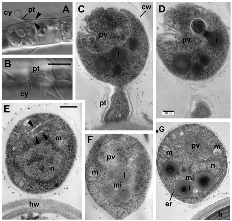
Figure 3. Cyst structure of Aphelidium aff. melosirae under light microscopy (LM) (A, B) and transmission electron microscopy (TEM) sections (C-G).
A – cyst (cy) with penetration tube (pt) between two halves of the host cell wall (arrowheads). B – empty cyst with penetrative tube on the surface of Tribonema filament. C-D and E-F – section pairs of cysts from two different series. Arrowheads on E show vesicles derived of the Golgi apparatus connected with posterior vacuole (pv) on F. Other abbreviations: cw – cyst wall, er-endoplasmic reticulum, h-host, hw-host cell wall, l-lipid globule, m-mitochondrion, mi-microbody, n-nucleus, pv-posterior vacuole. Scale bars: A, B – 10 μm, C, D – 200 nm, E-G – 500 nm.

