Abstract
Background
Melanocytes are present in both basal epidermis and hair follicles. Melanocyte stem cells have been found in hair follicle bulge. During embryogenesis, the outer cells of the bulge differentiate into the sebaceous gland (SG) and proliferate.
Objective
To identify and determine the distribution and morphological characteristics of melanocytes in human SGs.
Methods
A total of 171 biopsy specimens of face and scalp were studied. Of these specimens, 103 samples contained SGs. We conducted a retrospective review of slides stained with H&E, F-M, anti-S100, anti-c-kit, anti-HMB-45, anti-CD1a, anti-MITF, and anti-tyrosinase. The presence and distribution of melanocytes in human SGs was also evaluated by electron microscopy. In addition, melanocytes were isolated from SGs for primary culture.
Results
S-100-positive cells were observed mainly at the periphery of SGs in 34 of 54 specimens. We did not find F-M-positive and HMB-45-positive cells in SGs. CD1a-positve cells were identified in two specimens. We also found c-kit-, MITF-, and tyrosinase-positive cells in SGs. Electron micrograph showed the presence of melanocytes in the suprabasal portion of SGs. These melanocytes showed fewer melanin-containing granules than the melanocytes of basal epidermis. However, the individually distributed melanosomes in suprabasal melanocytes were larger than those in epidermal melanocytes. Primary culture of melanocytes derived from SGs showed morphologically homogeneous, slender cell bodies with few dendrites.
Conclusion
Our study confirms the presence of non-melanogenic melanocytes and Langerhans cells in human SGs. In addition, the characteristics of the melanocytes in SGs were found to be different from those of the epidermal melanocytes.
Keywords: Langerhans cells, Melanocytes, Sebaceous glands
INTRODUCTION
Melanocytes follow interesting and diverse developmental pathways. Most melanocytes originate in the neural crest and migrate to specific regions of the developing embryo, including the skin, hair follicles, inner ear, and the eye1. Melanocyte stem cells reside in the hair follicle bulge, located in the outer root sheath2. The sebaceous gland (SG) and hair follicles constitute the pilosebaceous unit of the skin. Therefore, it is possible that melanocytes are also present in SGs. However, little is known about the presence of melanocytes in human SGs.
The purpose of this study was to identify the presence of melanocytes and determine their distribution in human SGs. Determining the possible distribution of melanocytes and their functions within human SGs will offer several insights in the field of SG and melanocyte biology.
MATERIALS AND METHODS
Identification of the presence and distribution of melanocytes
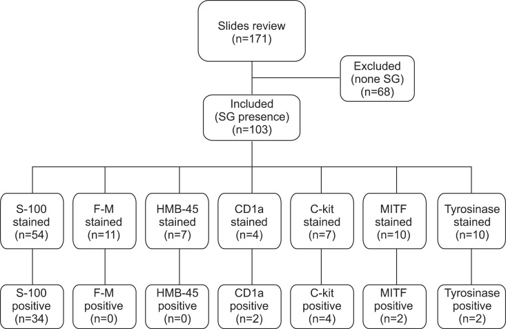
A total of 171 biopsy samples of the facial skin and the scalp which were randomly selected from the pathologic archives of the Department of Dermatology, Kyungpook National University Hospital, were used for the study. H&E staining of all samples (blocks of paraffin-embedded tissues) were reviewed retrospectively, and 103 samples with SGs were identified. Among them, 54 slides were already stained with the antiserum to S-100 (Dako, Ely, UK), 7 with antiserum to human melanoma black-45 (HMB-45; Dako), 11 with Fontana-Masson (F-M) stain (Junsei Chemical Co., Ltd., Tokyo, Japan), 4 with antiserum to CD1a (Novocastra, Newcastle, UK), 7 with antiserum to c-kit (Dako, Glostrup, Denmark), 10 with antiserum to microphthalmia-associated transcription factor (MITF; Leica Biosystems, Newcastle, UK), and 10 with antiserum to tyrosinase (Thermo Scientific, Fremont, CA, USA). All tissue sections contained epidermal melanocytes and Langerhans cells, which served as internal positive controls (Fig. 1).
Fig. 1.
We retrospectively reviewed 171 H&E slides taken from the scalp and facial skin. Among them, 103 slides containing sebaceous gland (SG) tissues were finally included in the study. Of these, 54 were stained with the antiserum to S-100, 11 with Fontana-Masson (F-M) stain, 7 with antiserum to human melanoma black-45 (HMB-45), 4 with antiserum to CD1a, 7 with antiserum to c-kit, 10 with antiserum to microphthalmia-associated transcription factor (MITF), and 10 with antiserum to tyrosinase. Out of the 54 specimens of human SGs, 34 slides showed S-100-positive cells. All specimens showed negative staining for F-M (11 specimens) and HMB-45 (7 specimens). In addition, 2 specimens were found to be CD1a-positive, and 4 were c-kit-positive. Out of 10 specimens of human SGs, 2 showed MITF-positive cells, and 2 others showed positive staining for tyrosinase.
Identification of melanocyte morphology by transmission electron microscopy
Facial skin and scalp biopsy specimens were obtained from two adult Korean males and post-fixed for 2 hours at 4℃ in 2% osmium tetroxide, and then dehydrated in graded alcohol and embedded in epoxy-embedding resin for transmission electron microscopy (TEM). Semi-thin sections of approximately 0.5-µm thickness were obtained using an ultramicrotome and stained with 0.75% toluidine blue; they were then examined under a light microscope. Ultra-thin sections were stained using a conventional method involving staining with 4% uranyl acetate for 10 to 15 minutes, followed by Reynold's lead citrate for 10 to 15 minutes. The specimens were observed under TEM (LEO/Zeiss, Oberkochen, Germany).
Isolation of primary melanocytes from human SGs in vitro
Human SGs were isolated from dissected hair follicles under a binocular microscope, and transferred to a tissue culture dish. The cells were maintained in Dulbecco modified Eagle medium at 37℃ in a humidified atmosphere of 5% CO2. Five days later, the medium was discarded and melanocyte growth medium (MGM) was added. The MGM was composed of MCDB-154 medium supplemented with 0.2% (v/v) bovine pituitary extract, 0.5% (v/v) fetal bovine serum, 5 µg ml-1 bovine insulin, 5 µg ml-1 bovine transferrin, 3 ng ml-1 basic fibroblast growth factor, 0.18 µg ml-1 hydrocortisone, 3 µg ml-1 heparin, 10 ng ml-1 phorbol 12-myristate 13-acetate (Cascade Biologics, Portland, OR, USA), 10 nmol L-1stem cell factor (Sigma, St. Louis, MO, USA), 100 U ml-1 penicillin (Gibco BRL, Grand Island, NY, USA), and 100 µg ml-1 streptomycin (Gibco BRL), 0.25 µg ml-1 fungizone (Gibco BRL). The medium was changed once every three days.
RESULTS
Identification of the presence and distribution of melanocytes by histochemical staining and immunohistochemical staining
S-100-positive cells were observed mainly at the periphery of SGs in 34 out of 54 specimens (63%) (Fig. 2A). Several cells, including Langerhans cells and melanocytes, showed a positive reaction to S-100. Therefore, immunohistochemistry with an antibody to HMB-45 and F-M stain was performed in order to prove melanogenic activity and melanin production in S-100-positive cells. All specimens showed negative staining for HMB-45 (seven specimens) and F-M (11 specimens). Two specimens, however, stained positively for CD1a, which is a specific indicator of Langerhans cells (Fig. 2B). In addition, c-kit-positive cells were observed in SGs in 4 specimens (Fig. 2C), and MITF-positive cells (Fig. 2D) and tyrosinase positive cells were observed in 2 specimens (Fig. 2E). These findings suggest that both differentiated melanocytes and Langerhans cells exist in human SGs.
Fig. 2.

(A) S-100-positive cells are mainly located in the periphery of the sebaceous glands (black arrow; ×200). (B) CD1a-positive Langerhans cells are seen in the sebaceous glands (black arrow; ×200). (C) C-kit-positive melanocytes are seen in the sebaceous glands (black arrows; ×40). (D) Microphthalmia-associated transcription factor-positive melanocytes are seen in the sebaceous glands (black arrows; ×200). (E) Tyrosinase-positive melanocytes are also shown in the sebaceous glands (black arrow; ×200).
Identification of melanocyte morphology by TEM
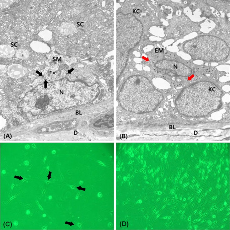
TEM images representing a series of observations made from different sections demonstrated the presence of melanocytes in the suprabasal portion of SGs. Sebaceous melanocytes were large and triangular or polygonal in shape, and the plasma membrane was devoid of tonofilaments and desmosomes. They were located between undifferentiated peripheral cells containing no lipid droplets. The nuclei had a slightly irregular contour with shallow indentations. These melanocytes showed fewer melanin-containing granules than the melanocytes in the basal surface of the epidermis. However, the individually distributed melanosomes in the suprabasal melanocytes were larger compared to those of the epidermal melanocytes. The melanin granules did not migrate to the surrounding sebocytes (Fig. 3A, B).
Fig. 3.
The specimen is from the facial skin of a Korean male (A, B: ×5,000. C, D: ×200). Electron micrograph shows a SM (A) in the suprabasal portion of the sebaceous gland (SG). The SM shows fewer melanin-containing granules (black arrows) compared to the EM (B). Evenly distributed melanosomes of SM (black arrows) are larger than those of EMs (red arrows). (C, D) Melanocytes cultured from SGs (black arrows) are seen in an early stage. The SMs have morphologically homogeneous, slender cell bodies with few-dendrites in the later stage. SC: sebocyte, SM: sebaceous melanocyte, N: nucleus, BL: basal lamina, D: dermis, KC: keratinocyte, EM: epidermal melanocyte.
Melanocyte culture from human SGs
We isolated melanocytes from SGs through cell culture. The sebaceous melanocytes appeared to be triangular or polygonal in shape. In cells cultured with MGM, the sebaceous melanocytes spread, developed long processes, and became bipolar, tripolar, or dendritic. The sebaceous melanocytes had morphologically homogeneous, slender cell bodies with few-dendrites (less than 4 dendrites per cell) (Fig. 3C, D).
DISCUSSION
Development of SGs begins from cells residing in the hair follicle bulge3. Previous studies have demonstrated that bulge cells possess stem cell properties, including high proliferative capacity and multipotency for regeneration of hair follicles, SGs, and the epidermis4,5. The results of this study show that sebaceous melanocytes may be used as a source of melanocyte stem cells.
In the current study, we identified melanocyte distribution in human SGs using specimens stained with antibodies against S-100, F-M, HMB-45, CD1a, c-kit, MITF, and tyrosinase. Melanocytes were mainly distributed in a peripheral layer of poorly differentiated sebocytes.
S-100 is a highly sensitive marker for melanocytes6. It is also a sensitive marker for Langerhans cells. C-kit-positive cells and CD1a-positive cells were also observed in SGs. This implies that both melanocytes and Langerhans cells are present in SGs. However, melanocytes in SGs showed negative reactivity to antibody against HMB-45 and F-M stain. MITF and tyrosinase-positive cells were also observed in SGs. We think that melanocytes in SGs are differentiated melanocytes with low melanogenic activity.
SG melanocytes are likely to have migrated from melanocyte stem cells in the hair follicle bulge. Thus, it is highly probable that the melanocytes are located within the periphery of the fibrous sheath. In our study, SG melanocytes showed negative staining for HMB-45 and F-M. Observation under TEM also revealed that they have smaller melanosomes than the epidermal melanocytes. This suggests that melanin production is lower in SG melanocytes.
We believe that the function of SG melanocytes needs to be further investigated. In addition, the role of Langerhans cells of SGs in the pathogenesis of acne should also be evaluated. Although previous reports have discussed the presence of melanocytes in human SGs7, only electron micrograph was used to study them. Our findings differed from the previous findings in two aspects: (i) the individually distributed melanosomes in SG melanocytes were larger than those in epidermal melanocytes and (ii) the melanin granules did not migrate to the surrounding sebocytes.
Recent evidence has shown that melanocytes have functions in the skin other than melanin production8. They secrete a wide range of signaling molecules, including cytokines, pro-opiomelanocortin peptides, catecholamines, and nitric oxide in response to ultraviolet irradiation and other stimuli9. Potential targets of these secretory products include keratinocytes, lymphocytes, fibroblasts, mast cells, and endothelial cells. Sebocytes may also be the target of secretions from sebaceous melanocytes.
Previous studies report that ectopic synthesis of melanin is observed in the human adipose tissue10,11. This implies that melanocytes and melanin can exist in various components of the skin.
Melanoblast/melanocyte proliferation and differentiation are regulated by the tissue environment, especially by keratinocytes12. Melanocyte differentiation is also stimulated by α-melanocyte-stimulating hormone (MSH).13 α-MSH is known to exert sebotrophic and immunomodulatory effects on human sebocytes, presumably via melanocortin receptors (MC-Rs)14,15. Therefore, we suppose that α-MSH in SGs may be a key factor in the coordination of MCRs-mediated sebum secretion and melanocyte function.
The limitations of this study were as follows: (1) The sample size was small. (2) F-M and immunohistochemical staining could not be performed again, so we only retrospectively reviewed already stained slides. (3) Finally, while two-color staining can increase the specificity of the data, we could not perform this. Therefore, only histochemical and immunohistochemical staining could be performed, which can be non-specific.
The morphology and characteristics of sebaceous melanocytes were found to differ from those of epidermal melanocytes. These differences are likely to be related to the functions of melanocytes in SGs other than melanin production. In this study, we isolated melanocytes from human SGs. We carefully separated the SGs from hair follicles by removing keratinocytes and fibroblasts. However, the possibility of contamination cannot be ignored. Dermal stem cells in the dermis are thought to be able to differentiate into melanocytes. Therefore, the possibility of stem cells differentiating into melanocytes in melanocyte culture media is not likely, but it cannot be ruled out. Further test of our hypothesis is required. The isolation of sebaceous melanocytes provides the opportunity to study their function and structure in vitro.
ACKNOWLEDGMENT
This research was supported by the Basic Science Research Program through the National Research Foundation of Korea (NRF) funded by the Ministry of Education, Science and Technology (NRF-2012R1A1A2007017).
References
- 1.Steel KP, Davidson DR, Jackson IJ. TRP-2/DT, a new early melanoblast marker, shows that steel growth factor (c-kit ligand) is a survival factor. Development. 1992;115:1111–1119. doi: 10.1242/dev.115.4.1111. [DOI] [PubMed] [Google Scholar]
- 2.Nishimura EK, Granter SR, Fisher DE. Mechanisms of hair graying: incomplete melanocyte stem cell maintenance in the niche. Science. 2005;307:720–724. doi: 10.1126/science.1099593. [DOI] [PubMed] [Google Scholar]
- 3.Amoh Y, Li L, Katsuoka K, Hoffman RM. Embryonic development of hair follicle pluripotent stem (hfPS) cells. Med Mol Morphol. 2010;43:123–127. doi: 10.1007/s00795-010-0498-z. [DOI] [PubMed] [Google Scholar]
- 4.Ohyama M. Hair follicle bulge: a fascinating reservoir of epithelial stem cells. J Dermatol Sci. 2007;46:81–89. doi: 10.1016/j.jdermsci.2006.12.002. [DOI] [PubMed] [Google Scholar]
- 5.Blanpain C, Lowry WE, Geoghegan A, Polak L, Fuchs E. Self-renewal, multipotency, and the existence of two cell populations within an epithelial stem cell niche. Cell. 2004;118:635–648. doi: 10.1016/j.cell.2004.08.012. [DOI] [PubMed] [Google Scholar]
- 6.Tímár J, Udvarhelyi N, Bánfalvi T, Gilde K, Orosz Z. Accuracy of the determination of S100B protein expression in malignant melanoma using polyclonal or monoclonal antibodies. Histopathology. 2004;44:180–184. doi: 10.1111/j.1365-2559.2004.01800.x. [DOI] [PubMed] [Google Scholar]
- 7.Ito K, Sato S, Nishijima A, Hiraga K, Hidano A. Melanogenic melanocytes in human sebaceous glands. Experientia. 1976;32:511–512. doi: 10.1007/BF01920827. [DOI] [PubMed] [Google Scholar]
- 8.Bellei B, Pitisci A, Catricalà C, Larue L, Picardo M. Wnt/β-catenin signaling is stimulated by α-melanocyte-stimulating hormone in melanoma and melanocyte cells: implication in cell differentiation. Pigment Cell Melanoma Res. 2011;24:309–325. doi: 10.1111/j.1755-148X.2010.00800.x. [DOI] [PubMed] [Google Scholar]
- 9.Luger TA, Brzoska T, Böhm M, Schiller M, Fisbeck T, Scholzen T. The effect of ultraviolet light onthe production and processing of proopiomelanocortin in the skin. In: Ortonne JP, Ballotti R, editors. Mechanisms of suntanning. London: Martin Dunitz; 2002. pp. 135–142. [Google Scholar]
- 10.Ito S. Melanins seem to be everywhere in the body, but for what? Pigment Cell Melanoma Res. 2009;22:12–13. doi: 10.1111/j.1755-148X.2008.00538.x. [DOI] [PubMed] [Google Scholar]
- 11.Randhawa M, Huff T, Valencia JC, Younossi Z, Chandhoke V, Hearing VJ, et al. Evidence for the ectopic synthesis of melanin in human adipose tissue. FASEB J. 2009;23:835–843. doi: 10.1096/fj.08-116327. [DOI] [PMC free article] [PubMed] [Google Scholar]
- 12.Hirobe T. How are proliferation and differentiation of melanocytes regulated? Pigment Cell Melanoma Res. 2011;24:462–478. doi: 10.1111/j.1755-148X.2011.00845.x. [DOI] [PubMed] [Google Scholar]
- 13.Tsatmali M, Ancans J, Thody AJ. Melanocyte function and its control by melanocortin peptides. J Histochem Cytochem. 2002;50:125–133. doi: 10.1177/002215540205000201. [DOI] [PubMed] [Google Scholar]
- 14.Whang SW, Lee SE, Kim JM, Kim HJ, Jeong SK, Zouboulis CC, et al. Effects of α-melanocyte-stimulating hormone on calcium concentration in SZ95 sebocytes. Exp Dermatol. 2011;20:19–23. doi: 10.1111/j.1600-0625.2010.01199.x. [DOI] [PubMed] [Google Scholar]
- 15.Böhm M, Schiller M, Ständer S, Seltmann H, Li Z, Brzoska T, et al. Evidence for expression of melanocortin-1 receptor in human sebocytes in vitro and in situ. J Invest Dermatol. 2002;118:533–539. doi: 10.1046/j.0022-202x.2001.01704.x. [DOI] [PubMed] [Google Scholar]


