The wireless revolution and medicine
The digital revolution and the rapid development of smart phones, mobile connectivity and social networking have changed the way we live. The average American is constantly connected via high bandwidth to a vast network of data and sophisticated digital platforms with over 90% of American adults owning a cell phone and 55% having a smart phone.1 The digital revolution has transformed virtually every industry as well as every facet of our personal lives but has been conspicuously absent from the world of medicine. Physicians and health care networks have been slow to adopt electronic medical records and integrate medical data with the ubiquitous mobile device. Recently however, novel devices for wireless monitoring have emerged and begun to be integrated with the care of the cardiac patient. We believe that the evolution of these wireless cardiac monitoring devices will mark a new era in medicine and a transition from population level health care to individualized medicine where suitable patients are equipped with advanced biosensors that, in turn, have their data processed through sophisticated algorithms to predict events before they occur. This review is meant to be a comprehensive overview of the novel wireless cardiac monitoring devices that are available as well as the technologies that are currently under development and poised to revolutionize the way we practice cardiology. A comprehensive list of a number of devices that are currently available or under development is available in Table 1 for reference.
Table 1.
Comprehensive overview of existing wireless cardiac monitoring devices
| NAME | COMPANY | LINK | BRIEF DESCRIPTION |
|---|---|---|---|
| COMPREHENSIVE VITAL SIGN MONITORING | |||
| VitalSigns Camera | Phillips | http://www.vitalsignscamera.com/index.html | Skin micro-blush change in capillary filling to measure heart rate and chest movement to measure respiratory rate |
| Scout | Scanadu | http://www.scanadu.com/ | Measures temperature, pulse, oximetry, ECG, heart rate variability, pulse wave transit time |
| BioPatch | Zephyr | http://www.zephyranywhere.com/healthcare/biopatch/ | Adhesive patch transmits wirelessly pulse, R-R interval, respiratory rate, activity, respirations, ECG, position and posture. |
| Hexoskin | Hexoskin Wearable Body Metrics | http://www.hexoskin.com/en?utm_campaign=Listly&utm_medium=list&utm_source=listly | Shirt measures HR, HRV, respiratory rate and volume, activity and estimates VO2 max. |
| OMSignal | OMSignal | http://www.omsignal.com/ | Washable shirt that monitors 3-lead ECG, respirations, stress, temperature |
| Sensor Bra | Microsoft | http://www.cs.rochester.edu/hci/pubs/pdfs/FoodMood.pdf | Sensors built into bra: heart rate, respiration, EDA, 3-axis accelerometer, 2-axis gyroscope. Designed to track emotions and study of emotional eating. |
| Intermittent ECG | |||
| Alivecor System | Alivecor | http://www.alivecor.com/ | With app able to analyze and print ECGs as PDFs. ECG data syncs between the app and online ECG hub. Rx only. |
| ECG Check | CardiacDesigns | http://cardiacdesigns.com/ | With app able to analyze and print ECGs as PDFs. ECG data syncs between the app and online ECG hub. |
| EPI Mini (also EPI Life) | EPI Mobile Health Solutions | http://epimhealth.com.sg/ | Separate device that transmits ECG to smartphone, which can forward it to a “Health Concierge” service that can send back a reading. FDA cleared for consumer use. |
| 12-lead ECG | MobilECG | http://mobilecg.hu/ | USB-based open source 12-lead clinical ECG. |
| Prolonged ECG Monitoring | |||
| eMotion ECG Mobile | Mega Electronics | http://www.megaemg.com/products/emotion-ecg/ | 3-lead ECG data is transmitted from the wearable ECG sensor to mobile phone via Bluetooth. Phone forwards the data over mobile network to server which stores the data. The data can be monitored in real time or a specialist can investigate and analyze the stored ECG data. |
| BodyGuardian | Preventice | http://www.preventice.com/ | Patch monitor of ECG, activity, respirations and body position. |
| Zio XT Patch | iRhythm | http://www.irhythmtech.com/?utm_campaign=Listly&utm_medium=list&utm_source=listly | 14 day continuous cardiac rhythm monitoring with a single adhesive chest wall device. Once completed is mailed for analysis |
| NUVANT Mobile Cardiac Telemetry System | Corventis | http://www.corventis.com/ | Automatic and patient triggered 30-day cardiac rhythm monitoring. Arrhythmia detection the device transmits information via a wireless data transmission device, zLink® to the Corventis Monitoring Center |
| Ambulatory ECG | iHealth | http://ces.cnet.com/8301-35284_1-57616620/at-ces-2014-health-monitors-join-the-wearables-parade/ | Sensor attaches to chest and transmits ECG to smartphone. |
| Heart Failure | |||
| CoVa necklace | Perminova | http://www.perminova.com/sensor/ | Heart rate, respiratory rate, fluid levels |
| VitaLink | vg-bio | http://www.vgbio.com/vitalink-remote-patient-monitoring/ | Measures pulse, heart rate variability, transthoracic impedance, activity via head band and chest strap. |
| AVIVO Mobile Patient Monitoring System | Corventis | http://corventis.com/us/avivo.asp | Monitors thoracic impedance, HR, HRV, RR, Posture and heart rhythm with wireless transmission to Coreventis Monitoring Center |
| Telescale | Cardiocom | http://www.cardiocom.com/telescale.asp | For daily weights with automated verbal/feedback and communication to patient and provider. |
| Blood Pressure | |||
| Visi Mobile | Sotera Wireless | http://www.visimobile.com | Wireless vital sign monitoring with non-invasive continuous blood pressure monitor |
| Wireless Wrist Blood Pressure | iHealth | http://www.ihealthlabs.com/wireless-blood-pressure-wrist-monitor-feature_33.htm | Wireless wrist blood pressure measurement and heart rate transmitted to a mobile application |
| iPhone-connected BP cuff | Withings | http://www.withings.com/bloodpressuremonitor | Plugs into iPhone or iPad and tracks and displays all results. Also available in 2014 with Bluetooth connection between cuff and smartphone. |
| Continuous BP watch | Quanttus | http://www.technologyreview.com/news/524376/this-fitness-wristband-wants-to-play-doctor/ | Continuous blood pressure, heart rate and respirations. |
| BPro radial artery pressure monitor | HealthStats | http://www.healthstats.com | Watch-like device that samples radial artery waveforms via tonometry at regular time intervals, over a24-hour period. For assessment of ambulatory blood pressure. |
| Wearable, wireless ambulatory BP Monitor | iHealth | http://ces.cnet.com/8301-35284_1-57616620/at-ces-2014-health-monitors-join-the-wearables-parade/ | Vest-like device that allows BP to be measured as frequently as every 15 minutes throughout day. |
| ULTRASOUND | |||
| VScan | GE | http://www3.gehealthcare.com/en/Products/Categories/Ultrasound/Vscan | Stand-alone ultrasound imaging device that can download ant transmit images. |
| MobiUS SPI | Mobisante | http://www.mobisante.com/product-overview/ | Smartphone-based ultrasound |
| Terason USmart 3200T | Terason | http://www.terason.com/index.asp | Comprehensive ultrasound |
| Nanomaxx | Sonosite | http://www.sonosite.com/products/nanomaxx | Stand-alone ultrasound imaging device that can download and transmit images. |
Arrhythmia Detection
Cardiac arrhythmias, such as atrial fibrillation, are common and can be associated with adverse outcomes such as embolic stroke. Less common but more malignant rhythm disorders such as ventricular tachycardia can herald sudden cardiac death (SCD).2 The identification and management of arrhythmias in the patient with palpitations, syncope, a history of arrhythmia or high risk for SCD often relies on conventional Holter monitoring, event monitoring or continuous remote telemetry.3 These technologies, while effective, are burdensome to the patient, expensive (up to $750) and can often miss arrhythmias if the patient doesn’t activate the event monitor at the time of their symptoms.3, 4 A number of new and effective technologies for wireless monitoring of arrhythmias have been developed and validated and are currently available for patient care.
Real-time smart phone monitoring
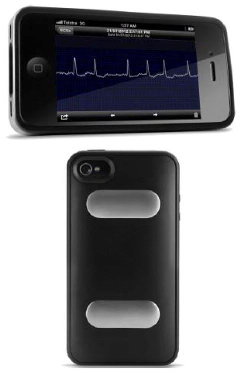
Using a novel smart phone adapter, patients are now able to capture and transmit single lead ECG data to their health care providers. The Alivecor (Figure 1) and ECG Check (Cardiac Designs) systems are FDA-approved single lead (Lead I) ECG monitoring systems that consist of a case that snaps on the back of a smart phone. Finger contact on the case of the smart phone activates ECG recording of bipolar lead I and is transmitted to the smart phone from the case using frequency modulation of an ultrasound or Bluetooth signal that is received in the smart phone’s speaker. Once captured digitally in the smart phone it can be viewed in real-time and transmitted to a secure server for the patient’s provider to review in PDF format. The system has been validated in a number of settings including event monitoring and screening of atrial fibrillation.5
Figure 1.
Alive Cor real-time monitoring of ECG. Finger contact on the case activates ECG recording of bipolar lead I and is transmitted to the smart phone.
ECG Patch monitoring
As opposed to the bulky and burdensome devices currently available for ambulatory ECG monitoring, the ECG patch monitor is an adhesive, single-lead ECG monitor that is applied to the left pectoral region. The typical ECG patch monitor consists of a System on a Chip (SoC) that converts analog ECG signals to digital format, an accelerometer to assist with artifact removal, a low-power Blue Tooth low energy processor that transmits the data, and a lithium polymer battery. There are several FDA-approved ECG patch-monitoring devices currently available.
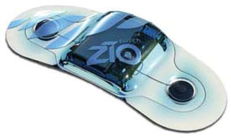
One example of this technology is the Zio® Patch (Figure 2- iRhythm technologies, San Francisco, CA), a waterproof, single-use, continuous ECG monitor that can be worn for up to 14 days. It lacks any leads or wires and its low profile design allows the patient to wear it with minimal interruption of daily activities. The device is mailed to the patient’s home in an envelope, worn for up to 14 days and returned in a prepaid envelope for analysis and reporting to the patient’s provider. A recent trial of the 14 day Zio Patch monitor compared to conventional 24-Holter monitor showed that the Zio Patch detected 57% more (96 events vs. 61 events, p < 0.001) significant clinical events than the conventional Holter.6 The majority (81%) of study participants preferred the Zio patch to conventional holter monitoring; 94% said they found the device comfortable to wear compared to only 52% for the Holter. There are several limitations to this technology however as the Zio patch offers only one lead as opposed to the multiple channels available with conventional Holter monitoring. This may have contributed to the Holter monitor being significantly more sensitive for the detection of events (61 events detected/24 hrs with Holter vs. 52 events/24 hrs with Zio, p < 0.013) during the 24 hours of dual monitoring. The extended duration of monitoring and comfort of use likely outweighs these limitations and makes this a meaningful device for use in patients with arrhythmias.
Figure 2.

Zio Patch – adhesive ECG patch.
Another example is the NUVANT® Mobile Cardiac Telemetry (MCT) system, which is a wireless-enabled arrhythmia event monitor. It consists of a wearable monitoring device and a portable data transmission device. Unlike the Zio Patch that records and stores all ECG data allowing for arrhythmia detection only in retrospect the NUVANT system performs real-time analysis and transmission. Once applied to the body the sensor automatically activates and begins monitoring and transmitting ECGs when a select list of rhythm abnormalities is encountered (i.e. 3 second pause, ventricular tachycardia).7 There is also a patient trigger magnet that can be used to collect and transmit symptomatic episodes. All events are transmitted to a cloud-based application where it can be analyzed by certified electrocardiographic technicians.
Injectable loop recorders and device-tailored anticoagulation
The miniaturization of cardiac monitoring devices as well as advances in remote data transmission capabilities has led to the development of extremely small injectable loop recorders and novel therapeutic strategies for anticoagulation in atrial fibrillation. Devices like the FDA-approved Medtronic REVEAL LINQ™ has provided clinicians with a long term monitoring device (up to 3 years) that can be injected under the skin and is one-third the size of a AAA battery. These miniaturized loop recorders build on the utility of the previously mentioned patch monitors and offer a long-term monitoring period for patients with clinical conditions such as unexplained syncope and cryptogenic stroke. In fact, the recently released data from the CRYSTAL-AF trial8 randomized patients with cryptogenic stroke to monitoring with an implantable REVEAL XT™ device vs. conventional monitoring and found a dramatic increase in the detection rate (30% vs. 3%) of atrial fibrillation at 36 months.
Remote monitoring of implantable cardiac defibrillators (ICD’s) and pacemakers is commonly utilized in current practice but there is emerging interest into use of remote atrial tachycardia (AT) monitoring for tailored anti-coagulation in patients with atrial fibrillation. The recently released data from the IMPACT trial9 randomized participants to device-tailored anticoagulation based on AT alerts from existing ICD’s or pacemakers vs. conventional office-based monitoring. While the study did not meet it’s primary endpoint for a reduction in stroke, systemic embolism or major bleed it was underpowered and there were a low rate of compliance with anti-coagulation in the intervention group.
The NIH has recently funded a novel trial using the implantable REVEAL XT monitor for device-tailored anticoagulation. The device performs daily monitoring of the patient for episodes of atrial fibrillation lasting more than one hour. The data is manually downloaded to the internet and reviewed by the patient’s health care provider for initiation of oral anticoagulation if needed.10 The recently approved LINQ monitor, however, will allow automated nightly downloads and smartphone alerts to streamline the process. This type of device-tailored anticoagulation is a novel method to minimize the complications associated with anticoagulation in atrial fibrillation while maximizing its benefits. Moreover, this type of intervention is a natural translation of biosensor driven data from a diagnostic to a therapeutic role and has the potential to transform the way we manage chronic diseases.
Wireless vital sign monitoring
Cardiac patients on the inpatient telemetry wards and intensive care units require close observation with frequent vital sign measurement and ECG monitoring. Even the most critically ill patients, however, have their complete vital signs measured and reported to their physicians only every hour in the ICU and every 4–6 hours on the telemetry units. Recent advances in non-invasive blood pressure monitoring and wireless delivery systems has made continuous vital sign monitoring available for all inpatients in the form of a small monitor slightly larger than a wrist watch. The Sotera Visi mobile® system is a patient worn monitor that is connected to a series of small biosensors including a pulse oximeter, skin temperature sensor, telemetry leads and a non-invasive continuous blood pressure monitor that uses pulse arrival time (PAT) and cuff calibration to continuously monitor the patient. The PAT is defined as the time required for the arterial pulse pressure wave to travel from the aortic valve to the periphery and can be estimated as the delay between the peak of the R wave on the ECG and the arrival of the corresponding pulse wave on pulse oximetry (photoplethysmography). It has been validated in a number of settings11, 12 as an accurate method for non-invasive assessment of blood pressure. The Sotera system wirelessly transmits data via secure encryption through existing wifi networks to the electronic medical record and remote alarm systems allowing the physician 24/7 access to the patient’s vital signs. Significant alteration in a patient’s vital signs alerts the provider immediately via their smart phone. Further advances in predictive analytics will build upon this technology by establishing algorithms for preventing clinical events such as impending circulatory disorders before they occur in the inpatient setting. Moreover, the continuous wireless monitoring of vital signs can be seamlessly translated into the outpatient setting upon hospital discharge, or even for use in prevention and real-time management in appropriate individuals.
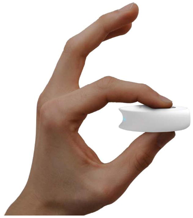
One such device designed for individual home use is the Scanadu Scout (Figure 3) that will soon enter FDA trials. It is a hockey puck shaped device that his held between two fingers and directed at a patients forehead. The device is designed to generate a complete set of vital signs including heart rate, blood pressure, temperature, respiratory rate and oxygen saturation in less than 10 seconds and wirelessly transmit the data to the patient’s smart phone where it can be stored, tracked, analyzed, and transmitted to a provider if desired. The technology underlying devices like the Scanadu Scout is based on photoplethysmography, which relies upon an infrared light source directed at the skin surface and the relationship of the backscattered light as detected by a photodetector to the variation in blood volume with each heartbeat. There are numerous applications of this technology as the reflected light can be calibrated to detect many important physiologic measures such as photoelectric activity (the shift in wavelength between oxygenated and deoxygenated hemoglobin that measures oxygen saturation), cardiac output and blood pressure. Similar but wearable devices such as watches and necklaces are also currently under development and in validation phases. Like the Scout, these wearable devices will transmit data to the patient’s smart phone and be continuously analyzed and interpreted, relying in large part on machine learning. In addition to conventional vital signs, many of these wearable devices in development can also monitor novel parameters like autonomic nervous system activity using heart rate variability and electro dermal tracking (using the changes in the conductance of the skin as a surrogate for sympathetic tone) as well as sleep quality measurements. This technology is still in its infancy but has the potential to revolutionize healthcare and bring the doctor’s office to the patient’s smart phone.
Figure 3.

Scanadu Scout – wireless vital sign monitor.
Handheld ultrasound
Echocardiography is a critical tool for the diagnosis and management of heart disease and over 20 million echocardiographic procedures are performed each year in the Unites States.13 Over 22% of these procedures, however, are currently deemed inappropriate14 and the rising costs of health care in the United states coupled with the increasing complexity of cardiac patients demands a more efficient and streamlined way to integrate echocardiography into our daily clinical practice. The recent development of high-resolution pocket mobile echocardiography (PME) devices has the potential to revolutionize bedside and outpatient management of cardiac patients and alleviate the burden of healthcare costs associated with cardiac imaging. These small devices consist of a small phased-array probe connected to a handheld viewer the size of a smart phone. Most models easily fit into a white coat pocket and have advanced imaging capabilities with high-resolution 2D imaging, color Doppler and measurement capabilities. There have been numerous studies that document the similar diagnostic accuracy of bedside evaluation with a PME and conventional transthoracic echocardiography (TTE) in clinical scenarios such as valvular heart disease, ejection fraction assessment and volume status assessment using IVC diameter. 15–18 Moreover, recent data suggests that PME not only has similar diagnostic accuracy as conventional TTE but it is also more cost-effective.19 Barriers to wide scale implementation of PME into the clinical arena include the significant costs of each device (up to $8000) as well as the logistics of physician re-imbursement for performing and interpreting bedside scans on daily rounds. As opposed to handheld viewing, next generations of these devices that are already available (Mobisante, INC – Redmond, WA) consist of a simple phased-array probe that wirelessly transmits data via WIFI or Bluetooth to a tablet-based tool and central EMR for image storage and subsequent interpretation and advanced diagnostics. It is possible that in the future patients may be able to acquire and transmit their own images to their health care providers for wireless monitoring of chronic cardiac conditions such as valvular heart disease.
Heart Failure
Despite the availability of a multitude of evidence-based therapies for the treatment of heart failure, the burden of heart failure on the US population remains unacceptably high with an estimated 1 million admissions per year. 20 Moreover, readmission rates for heart failure, the majority of which are for congestion,21 are a marker of worse prognosis and represent a significant health care expenditure as well as a performance measure for payers.22 Efforts to reduce the burden of rehospitalization have been largely ineffective given the poor sensitivity and specificity of conventional markers such as weight and symptoms.23, 24
Multiple parameter testing
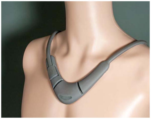
While the relative sensitivity and specificity of conventional stand alone markers of congestion is poor there has been intense interest in using remote monitoring of multiple markers for the assessment of impending congestive episodes and readmission. Anand et al.25, 26 recently developed and validated an algorithm for prediction of impending heart failure using a combination of physiologic signals obtained from an external device adhered to the chest. The MUSIC trial studied this predictive algorithm using a combination of bioimpedance, heart rate, respiratory rate, activity duration and body posture that was tailored to each participant. 543 participants with an ejection fraction of ≤ 40% and a recent admission for heart failure were remotely monitored for 90 days. The predictive algorithm had a sensitivity of 63% and a specificity of 92% for prediction of rehospitalization in the validation cohort – numbers that far exceed conventional performance characteristics of single parameter testing, although still far from ideal. Other wearable monitoring devices, such as the Perminova CoVa necklace (Figure 4), are under development for the long-term management of heart failure and have the capacity to monitor complex hemodynamics such as stroke volume and cardiac output non-invasively. The CoVa necklace, worn just a few minutes a day, measures thoracic bioimpedance and electrocardiographic waveforms to determine patients thoracic fluid index, heart rate, heart rate variability and respiratory rate. Newer versions of the device’s firmware, using the same hardware, will be able to monitor stroke volume, cardiac output and blood pressure. As the fidelity of biosensors improves and predictive analytics become more powerful multiple parameter testing has the potential to drastically improve the management of heart failure as well as many other chronic conditions.
Figure 4.
Perminova CoVa necklace uses thoracic bioimpedance and ECG monitoring to determine stroke volume, blood pressure and cardiac output in patients with chronic cardiac conditions.
Breath analysis
Breath metabolomics is the study of the complex mixture of volatile organic compounds in exhaled breath and has the potential to become a safe and noninvasive method of identifying patients with impending decompensation in a point of care fashion. Previous studies have demonstrated that elevated levels of acetone, pentane and nitric oxide levels in exhaled breath are correlated with heart failure disease severity.27–29 A recent analysis from a group at the Cleveland Clinic demonstrated that a “breathprint” derived from a select group of volatile organic compounds could accurately discriminate between HF patients and controls (Wilks’ Lambda 0.109, p < 0.0001).30 Breath analysis platforms using Bluetooth-enabled nanosensors are already under development (Vantage™) with the planned capability of transmitting “breathprint” data to a smart phone for analysis and upload.31 Breath metabolomics is a rapidly developing field and has the potential to offer patients a non-invasive diagnostic tool for complex diseases. Wireless invasive-pressure monitoring. It has been well documented that left ventricular filling pressures begin to rise in the days and weeks preceding a hospitalization for heart failure,24, 32 independent of weight, and a number of novel implantable devices have been developed to identify and intervene upon this rise in filling pressures before hospitalization. CardioMEMS (Atlanta, GA) is a passive, wireless, radiofrequency sensor implanted into the pulmonary artery that continuously monitors pulmonary artery pressure. This device was tested in the 2011 CHAMPION trial33 wherein 550 participants with Class III heart failure symptoms and a previous hospitalization for HF were randomized to implantation with continuous monitoring vs. standard of care and followed for the 6 month endpoint of HF hospitalization. At 6 months there were 84 HF-related hospitalizations in the sensor group vs. 120 in the usual care group (HR 0.72, 95% CI 0.60 – 0.85, p < 0.0002). At 15 months there was an impressive 39% reduction in HF hospitalizations in the sensor group with a significant improvement in quality of life compared to usual care. Importantly, physicians were given a standardized algorithm for the management of patients in the monitoring group and the benefits were independent of left ventricular ejection fraction. The initial FDA review of the device in 2011 was met with concern regarding the detailed recommendations of study sponsor nurses for management of PA pressure alarms in the treatment arm. Subsequent analyses of PA alerts in the absence of these detailed recommendations have been met by a favorable review by the FDA advisory committee in 2013.
Coronary Artery Disease
Secondary prevention with continuous ST monitoring
Acute myocardial infarction is a major cause of death and morbidity in the United States and despite improving door-to-balloon times among US hospitals the symptom onset to arrival at hospital times remain at 2.5 to 3 hours.2, 34–36 This delay in treatment results in potentially irreversible myocardial infarction and lethal arrhythmias that may have been otherwise avoided if the patient came to medical attention earlier. Continuous ST monitoring is able to detect “supply-related” ST segment shifts that occur in the absence of increased heart rates (“demand-related”) and are a highly sensitive and specific marker of ischemia and impending infarction. The Angelmed Guardian implantable cardiac device is a single right ventricle lead that is currently in clinical testing among patients with a recent (< 6 months) ACS event and at high risk for recurrence. The device detects ST shifts ≥ 3 standard deviations beyond a patients daily ST variation and alerts them to seek medical care. In the first in man trial37 37 patients were followed for 1.5 yrs and the device alerted 4 of these patients to seek medical attention, all of whom had angiographic evidence of coronary occlusion or ruptured plaques. The average alarm to door time was 19.5 minutes and there were 2 false positive findings due to arrhythmia.
Embedded nanosensors
In addition to electrical markers, biologic markers of impending plaque rupture are critically important for the prevention and early identification of myocardial infarction. One such biological marker currently being studied for use in the early identification of plaque rupture is circulating endothelial cells (CECs). Recent investigation has confirmed that CECs, identified by multiple surface markers using a fluorescence microassay, are significantly elevated in patients presenting with acute myocardial infarction vs. controls with an AUC of 0.95.38 Integration of a highly sensitive and endothelial specific genomic markers with blood-embedded nanosensors has the potential to make the new diagnosis of an imminent myocardial infarction before the event has initiated, representing the true prevention capability of wireless embedded sensing technology.
Wireless Cardiac Rehabilitation
Cardiac rehabilitation is a critical component to the secondary prevention of coronary artery disease and is well-established to improve outcomes in patients with CAD.39,40 Despite the clear benefits of cardiac rehabilitation, enrollment rates are still less than 30% and there are many patients without the resources or availability to attend three days a week of supervised exercise and education. 41 The utility of home-based programs for cardiac rehab hold the promise of improved compliance rates but still requires monitoring by a nurse or trained technician.42 Home-based cardiac rehab in conjunction with wireless ECG, blood pressure and heart rate monitoring may provide a means of improving compliance with cardiac rehab while maximizing safety and better individualizing management strategies and education arising in real-world activities. A study conducted in Korea43 randomized 50 patients to conventional care vs. home-based cardiac rehab for 12 weeks and monitored echocardiographic measures of left ventricular function and regional wall motion abnormalities. After 12 weeks, the group in the home-based cardiac rehab group showed significantly improved ejection fraction and a reduced number of regional wall motion abnormalities. This type of home-based exercise training can be further extrapolated to other chronic conditions where exercise has shown beneficial effects such as peripheral arterial disease and CHF.
Challenges that lie ahead
Need for validation, cost-effectiveness
The rapid development of the hardware and software involved in the new generation of wireless cardiac monitoring devices has outpaced its real world validation and large-scale, pragmatic studies are needed to validate the enormous amounts of data generated from these monitors. Ongoing clinical trials will be critical to determine the safety, efficacy and cost-effectiveness of this new technology relative to conventional methods of monitoring patients. Moreover, a much greater understanding of individual variability in the acceptance, engagement, and sustainability of these technologies as well as the most appropriate balance of patient and provider involvement are critically important areas of study.
Data Security
Personal health information (PHI) is heavily protected under the health insurance portability and accountability act (HIPAA) and the dissemination of electronic medical data to mobile and cloud-based technology requires encrypted and secure networks. The investment in online security for the mobile monitoring devices of the future must be a major priority as a data breach could be catastrophic for the future of this industry and expose patients to both an invasion of their privacy as well as the potential for personal harm.
Reimbursement and medico legal issues
The deluge of data that comes from wireless cardiac monitoring devices needs to be analyzed, as it is still the job of the health care provider to react to the information and provide appropriate care to the patient in the appropriate turn around time. Whereas improved data analytics with automated decision support will be a critical component of any successful widespread implementation of home monitoring, the product of these analyses will still require provider oversight that needs to be reimbursed; directly in the fee-for-service environment or better, indirectly through incentives to keep individuals well. Moreover, tasks such as performing a bedside echocardiographic assessment with a PME, which is financially dis-incentivized from multiple directions – not reimbursed, extends the duration of a poorly reimbursed physical exam, and lost revenues from a formal echocardiographic study not done – need to become part of a sustainable system of care. This new technology demands that we reexamine how physicians are paid in the current environment and efforts are underway in many states to re-imbursement physicians for electronic services such as returning emails and conducting “tele-visits.” The rapid adoption of accountable care organizations will also make wireless cardiac monitoring devices more relevant by reducing hospitalization and empowering patients and providers to focus on improving health.
Predictive analytics, machine learning, algorithms
The rapid development of hardware like biosensors has overshadowed the slower development of the software that will be needed to manage the enormous amount of data that these biosensors can generate. Mobile technologies will go well beyond providing data physicians already know how to treat (e.g. blood pressure at an office visit) and instead provide brand new data streams (e.g. continuous blood pressure during daily activities) that will require tremendous bioinformatics capabilities to eventually understand. As discussed in previous segments of this review, the capacity for technological advances in the way we are able to process big data using predictive analytics and network biology will usher in the era of personalized medicine and the ability to predict clinically significant events well before they occur.
The cardiovascular patient of the future
In light of the technological advances we’ve reviewed it is likely that the cardiac patient of the future, “wired” with a personalized network of biosensors, will be extremely different from the usual patient today. These biosensors will transmit a wealth of data about the patient’s genomic, metabolomic and clinical responses to daily stimuli to their smartphone peripheral brain. Once received the smart phone will process that information using sophisticated and personalized modeling software and direct the patient to take action; whether to contact their physician, adjusting a medication, or alerting EMS of an impending myocardial infarction. These devices have the potential to revolutionize medicine. The eventual integration and study of these devices in the coming years must carefully examine not only the relative safety and efficacy of this technology relative to conventional care, but also the impact of this technology on changing the costs of healthcare by preventing rehospitalization and empowering patients.
Acknowledgments
Funding Sources: This work was supported, in part through the Institutes of Health (NIH) /National Center for Advancing Translational Sciences NIH/NCATS 8 UL1 TR001114
Footnotes
Conflict of Interest Disclosures: JAW has nothing to disclose. SRS is supported by the National Institutes of Health (NIH) /National Center for Advancing Translational Sciences grant UL1TR001114 and a grant from the Qualcomm Foundation, and is an Advisory Board member for Agile Edge Technologies, Inc. EJT reports research funding from the Qualcomm Foundation and NIH/National Center for Advancing Translational Sciences grant UL1TR001114; currently serving on the boards of directors of Dexcom and Volcano and having served on the board of directors of SoteraWireless. He is serving as editor in chief of Medscape (WebMD) and is Chief Medical Advisor for AT&T.
References
- 1.Pew Internet fact sheet. Available at: http://www.pewinternet.org/fact-sheets/mobile-technology-fact-sheet/
- 2.Lloyd-Jones D, Adams R, Carnethon M, De Simone G, Ferguson TB, Flegal K, Ford E, Furie K, Go A, Greenlund K, Haase N, Hailpern S, Ho M, Howard V, Kissela B, Kittner S, Lackland D, Lisabeth L, Marelli A, McDermott M, Meigs J, Mozaffarian D, Nichol G, O'Donnell C, Roger V, Rosamond W, Sacco R, Sorlie P, Stafford R, Steinberger J, Thom T, Wasserthiel-Smoller S, Wong N, Wylie-Rosett J, Hong Y. Heart disease and stroke statistics--2009 update: a report from the American Heart Association Statistics Committee and Stroke Statistics Subcommittee. Circulation. 2009;119:480–486. doi: 10.1161/CIRCULATIONAHA.108.191259. [DOI] [PubMed] [Google Scholar]
- 3.Zimetbaum P, Goldman A. Ambulatory arrhythmia monitoring: choosing the right device. Circulation. 122:1629–1636. doi: 10.1161/CIRCULATIONAHA.109.925610. [DOI] [PubMed] [Google Scholar]
- 4.Reiffel JA, Schwarzberg R, Murry M. Comparison of autotriggered memory loop recorders versus standard loop recorders versus 24-hour Holter monitors for arrhythmia detection. Am J Cardiol. 2005;95:1055–1059. doi: 10.1016/j.amjcard.2005.01.025. [DOI] [PubMed] [Google Scholar]
- 5.Lau JK, Lowres N, Neubeck L, Brieger DB, Sy RW, Galloway CD, Albert DE, Freedman SB. iPhone ECG application for community screening to detect silent atrial fibrillation: a novel technology to prevent stroke. Int J Cardiol. 165:193–194. doi: 10.1016/j.ijcard.2013.01.220. [DOI] [PubMed] [Google Scholar]
- 6.Barrett PM, Komatireddy R, Haaser S, Topol S, Sheard J, Encinas J, Fought AJ, Topol EJ. Comparison of 24-hour Holter Monitoring with 14-day Novel Adhesive Patch Electrocardiographic Monitoring. Am J Med. 127:95 e11–97. doi: 10.1016/j.amjmed.2013.10.003. [DOI] [PMC free article] [PubMed] [Google Scholar]
- 7.Engel JM, Mehta V, Fogoros R, Chavan A. Study of arrhythmia prevalence in NUVANT Mobile Cardiac Telemetry system patients. Conf Proc IEEE Eng Med Biol Soc. 2012:2440–2443. doi: 10.1109/EMBC.2012.6346457. [DOI] [PubMed] [Google Scholar]
- 8.Passman R. American College of Cardiology 2014. San Francisco, CA: 2014. CRYptogenic STroke and underlying AtriaL Fibrillation (CRYSTAL AF): Long-Term Follow-Up Results. [Google Scholar]
- 9.Martin D. American College of Cardiology. San Francisco, CA: 2014. Randomized Trial of Anticoagulation Guided by Remote Rhythm Monitoring In Patients with Implanted Cardioverter-Defibrillator and Resynchronization Devices. [Google Scholar]
- 10.Zimetbaum P, Ellis ER, Waks JW, Passman RS. The importance of atrial fibrillation burden and the origin of device-tailored anticoagulation. Pacing Clin Electrophysiol. 36:1319–1324. doi: 10.1111/pace.12275. [DOI] [PubMed] [Google Scholar]
- 11.Ahlstrom C, Johansson A, Uhlin F, Lanne T, Ask P. Noninvasive investigation of blood pressure changes using the pulse wave transit time: a novel approach in the monitoring of hemodialysis patients. J Artif Organs. 2005;8:192–197. doi: 10.1007/s10047-005-0301-4. [DOI] [PubMed] [Google Scholar]
- 12.Schmalgemeier H, Bitter T, Bartsch S, Bullert K, Fischbach T, Eckert S, Horstkotte D, Oldenburg O. Pulse transit time: validation of blood pressure measurement under positive airway pressure ventilation. Sleep Breath. 16:1105–1112. doi: 10.1007/s11325-011-0609-7. [DOI] [PubMed] [Google Scholar]
- 13.Pearlman AS, Ryan T, Picard MH, Douglas PS. Evolving trends in the use of echocardiography: a study of Medicare beneficiaries. J Am Coll Cardiol. 2007;49:2283–2291. doi: 10.1016/j.jacc.2007.02.048. [DOI] [PubMed] [Google Scholar]
- 14.Bhatia RS, Carne DM, Picard MH, Weiner RB. Comparison of the 2007 and 2011 appropriate use criteria for transthoracic echocardiography in various clinical settings. J Am Soc Echocardiogr. 25:1162–1169. doi: 10.1016/j.echo.2012.07.018. [DOI] [PubMed] [Google Scholar]
- 15.Testuz A, Muller H, Keller PF, Meyer P, Stampfli T, Sekoranja L, Vuille C, Burri H. Diagnostic accuracy of pocket-size handheld echocardiographs used by cardiologists in the acute care setting. Eur Heart J Cardiovasc Imaging. 14:38–42. doi: 10.1093/ehjci/jes085. [DOI] [PubMed] [Google Scholar]
- 16.Cardim N, Fernandez Golfin C, Ferreira D, Aubele A, Toste J, Cobos MA, Carmelo V, Nunes I, Oliveira AG, Zamorano J. Usefulness of a new miniaturized echocardiographic system in outpatient cardiology consultations as an extension of physical examination. J Am Soc Echocardiogr. 24:117–124. doi: 10.1016/j.echo.2010.09.017. [DOI] [PubMed] [Google Scholar]
- 17.Prinz C, Voigt JU. Diagnostic accuracy of a hand-held ultrasound scanner in routine patients referred for echocardiography. J Am Soc Echocardiogr. 24:111–116. doi: 10.1016/j.echo.2010.10.017. [DOI] [PubMed] [Google Scholar]
- 18.Liebo MJ, Israel RL, Lillie EO, Smith MR, Rubenson DS, Topol EJ. Is pocket mobile echocardiography the next-generation stethoscope? A cross-sectional comparison of rapidly acquired images with standard transthoracic echocardiography. Ann Intern Med. 155:33–38. doi: 10.7326/0003-4819-155-1-201107050-00005. [DOI] [PMC free article] [PubMed] [Google Scholar]
- 19.Kitada R, Fukuda S, Watanabe H, Oe H, Abe Y, Yoshiyama M, Song JM, Sitges M, Shiota T, Ito H, Yoshikawa J. Diagnostic accuracy and cost-effectiveness of a pocket-sized transthoracic echocardiographic imaging device. Clin Cardiol. 36:603–610. doi: 10.1002/clc.22171. [DOI] [PMC free article] [PubMed] [Google Scholar]
- 20.Lloyd-Jones D, Adams RJ, Brown TM, Carnethon M, Dai S, De Simone G, Ferguson TB, Ford E, Furie K, Gillespie C, Go A, Greenlund K, Haase N, Hailpern S, Ho PM, Howard V, Kissela B, Kittner S, Lackland D, Lisabeth L, Marelli A, McDermott MM, Meigs J, Mozaffarian D, Mussolino M, Nichol G, Roger VL, Rosamond W, Sacco R, Sorlie P, Thom T, Wasserthiel-Smoller S, Wong ND, Wylie-Rosett J. Heart disease and stroke statistics--2010 update: a report from the American Heart Association. Circulation. 121:e46–e215. doi: 10.1161/CIRCULATIONAHA.109.192667. [DOI] [PubMed] [Google Scholar]
- 21.Adams KF, Jr, Fonarow GC, Emerman CL, LeJemtel TH, Costanzo MR, Abraham WT, Berkowitz RL, Galvao M, Horton DP. Characteristics and outcomes of patients hospitalized for heart failure in the United States: rationale, design, and preliminary observations from the first 100,000 cases in the Acute Decompensated Heart Failure National Registry (ADHERE) Am Heart J. 2005;149:209–216. doi: 10.1016/j.ahj.2004.08.005. [DOI] [PubMed] [Google Scholar]
- 22.Hunt SA, Abraham WT, Chin MH, Feldman AM, Francis GS, Ganiats TG, Jessup M, Konstam MA, Mancini DM, Michl K, Oates JA, Rahko PS, Silver MA, Stevenson LW, Yancy CW. 2009 focused update incorporated into the ACC/AHA 2005 Guidelines for the Diagnosis and Management of Heart Failure in Adults: a report of the American College of Cardiology Foundation/American Heart Association Task Force on Practice Guidelines: developed in collaboration with the International Society for Heart and Lung Transplantation. Circulation. 2009;119:e391–479. doi: 10.1161/CIRCULATIONAHA.109.192065. [DOI] [PubMed] [Google Scholar]
- 23.Chakko S, Woska D, Martinez H, de Marchena E, Futterman L, Kessler KM, Myerberg RJ. Clinical, radiographic, and hemodynamic correlations in chronic congestive heart failure: conflicting results may lead to inappropriate care. Am J Med. 1991;90:353–359. doi: 10.1016/0002-9343(91)80016-f. [DOI] [PubMed] [Google Scholar]
- 24.Adamson PB, Magalski A, Braunschweig F, Bohm M, Reynolds D, Steinhaus D, Luby A, Linde C, Ryden L, Cremers B, Takle T, Bennett T. Ongoing right ventricular hemodynamics in heart failure: clinical value of measurements derived from an implantable monitoring system. J Am Coll Cardiol. 2003;41:565–571. doi: 10.1016/s0735-1097(02)02896-6. [DOI] [PubMed] [Google Scholar]
- 25.Anand IS, Greenberg BH, Fogoros RN, Libbus I, Katra RP. Design of the Multi-Sensor Monitoring in Congestive Heart Failure (MUSIC) study: prospective trial to assess the utility of continuous wireless physiologic monitoring in heart failure. J Card Fail. 17:11–16. doi: 10.1016/j.cardfail.2010.08.001. [DOI] [PubMed] [Google Scholar]
- 26.Anand IS, Tang WH, Greenberg BH, Chakravarthy N, Libbus I, Katra RP. Design and performance of a multisensor heart failure monitoring algorithm: results from the multisensor monitoring in congestive heart failure (MUSIC) study. J Card Fail. 18:289–295. doi: 10.1016/j.cardfail.2012.01.009. [DOI] [PubMed] [Google Scholar]
- 27.Marcondes-Braga FG, Gutz IG, Batista GL, Saldiva PH, Ayub-Ferreira SM, Issa VS, Mangini S, Bocchi EA, Bacal F. Exhaled acetone as a new biomaker of heart failure severity. Chest. 142:457–466. doi: 10.1378/chest.11-2892. [DOI] [PubMed] [Google Scholar]
- 28.Sobotka PA, Brottman MD, Weitz Z, Birnbaum AJ, Skosey JL, Zarling EJ. Elevated breath pentane in heart failure reduced by free radical scavenger. Free Radic Biol Med. 1993;14:643–647. doi: 10.1016/0891-5849(93)90145-k. [DOI] [PubMed] [Google Scholar]
- 29.Schuster A, Thakur A, Wang Z, Borowski AG, Thomas JD, Tang WH. Increased exhaled nitric oxide levels after exercise in patients with chronic systolic heart failure with pulmonary venous hypertension. J Card Fail. 18:799–803. doi: 10.1016/j.cardfail.2012.08.356. [DOI] [PMC free article] [PubMed] [Google Scholar]
- 30.Samara MA, Tang WH, Cikach F, Jr, Gul Z, Tranchito L, Paschke KM, Viterna J, Wu Y, Laskowski D, Dweik RA. Single exhaled breath metabolomic analysis identifies unique breathprint in patients with acute decompensated heart failure. J Am Coll Cardiol. 61:1463–1464. doi: 10.1016/j.jacc.2012.12.033. [DOI] [PMC free article] [PubMed] [Google Scholar]
- 31.Nanobeak system. Available at: http://www.vantagehealthinc.com/
- 32.Zile MR, Bennett TD, St John Sutton M, Cho YK, Adamson PB, Aaron MF, Aranda JM, Jr, Abraham WT, Smart FW, Stevenson LW, Kueffer FJ, Bourge RC. Transition from chronic compensated to acute decompensated heart failure: pathophysiological insights obtained from continuous monitoring of intracardiac pressures. Circulation. 2008;118:1433–1441. doi: 10.1161/CIRCULATIONAHA.108.783910. [DOI] [PubMed] [Google Scholar]
- 33.Abraham WT, Adamson PB, Bourge RC, Aaron MF, Costanzo MR, Stevenson LW, Strickland W, Neelagaru S, Raval N, Krueger S, Weiner S, Shavelle D, Jeffries B, Yadav JS. Wireless pulmonary artery haemodynamic monitoring in chronic heart failure: a randomised controlled trial. Lancet. 377:658–666. doi: 10.1016/S0140-6736(11)60101-3. [DOI] [PubMed] [Google Scholar]
- 34.De Luca G, Suryapranata H, Ottervanger JP, Antman EM. Time delay to treatment and mortality in primary angioplasty for acute myocardial infarction: every minute of delay counts. Circulation. 2004;109:1223–1225. doi: 10.1161/01.CIR.0000121424.76486.20. [DOI] [PubMed] [Google Scholar]
- 35.Meischke H, Ho MT, Eisenberg MS, Schaeffer SM, Larsen MP. Reasons patients with chest pain delay or do not call 911. Ann Emerg Med. 1995;25:193–197. doi: 10.1016/s0196-0644(95)70323-3. [DOI] [PubMed] [Google Scholar]
- 36.Chew DP, Moliterno DJ, Herrmann HC. Present and potential future paradigms for the treatment of ST-segment elevation acute myocardial infarction. J Invasive Cardiol. 2002;14 (Suppl A):3A–20A. [PubMed] [Google Scholar]
- 37.Fischell TA, Fischell DR, Avezum A, John MS, Holmes D, Foster M, 3rd, Kovach R, Medeiros P, Piegas L, Guimaraes H, Gibson CM. Initial clinical results using intracardiac electrogram monitoring to detect and alert patients during coronary plaque rupture and ischemia. J Am Coll Cardiol. 56:1089–1098. doi: 10.1016/j.jacc.2010.04.053. [DOI] [PubMed] [Google Scholar]
- 38.Damani S, Bacconi A, Libiger O, Chourasia AH, Serry R, Gollapudi R, Goldberg R, Rapeport K, Haaser S, Topol S, Knowlton S, Bethel K, Kuhn P, Wood M, Carragher B, Schork NJ, Jiang J, Rao C, Connelly M, Fowler VM, Topol EJ. Characterization of circulating endothelial cells in acute myocardial infarction. Sci Transl Med. 4:126ra133. doi: 10.1126/scitranslmed.3003451. [DOI] [PMC free article] [PubMed] [Google Scholar]
- 39.Heran BS, Chen JM, Ebrahim S, Moxham T, Oldridge N, Rees K, Thompson DR, Taylor RS. Exercise-based cardiac rehabilitation for coronary heart disease. Cochrane Database Syst Rev. :CD001800. doi: 10.1002/14651858.CD001800.pub2. [DOI] [PMC free article] [PubMed] [Google Scholar]
- 40.Smith SC, Jr, Benjamin EJ, Bonow RO, Braun LT, Creager MA, Franklin BA, Gibbons RJ, Grundy SM, Hiratzka LF, Jones DW, Lloyd-Jones DM, Minissian M, Mosca L, Peterson ED, Sacco RL, Spertus J, Stein JH, Taubert KA. AHA/ACCF Secondary Prevention and Risk Reduction Therapy for Patients with Coronary and other Atherosclerotic Vascular Disease: 2011 update: a guideline from the American Heart Association and American College of Cardiology Foundation. Circulation. 124:2458–2473. doi: 10.1161/CIR.0b013e318235eb4d. [DOI] [PubMed] [Google Scholar]
- 41.Suaya JA, Shepard DS, Normand SL, Ades PA, Prottas J, Stason WB. Use of cardiac rehabilitation by Medicare beneficiaries after myocardial infarction or coronary bypass surgery. Circulation. 2007;116:1653–1662. doi: 10.1161/CIRCULATIONAHA.107.701466. [DOI] [PubMed] [Google Scholar]
- 42.Beatty AL, Fukuoka Y, Whooley MA. Using mobile technology for cardiac rehabilitation: a review and framework for development and evaluation. J Am Heart Assoc. 2:e000568. doi: 10.1161/JAHA.113.000568. [DOI] [PMC free article] [PubMed] [Google Scholar]
- 43.Lee Y, Lee J, Seo H, Kim K, Min D, Choi J. Effects of Home-based Exercise Training with Wireless Monitoring on the Left Ventricular Function of Acute Coronary Syndrome Patients. J Phys Ther Sci. 25:631–633. doi: 10.1589/jpts.25.631. [DOI] [PMC free article] [PubMed] [Google Scholar]


